Mastering PCR Cloning: 8 Critical Error Reduction Strategies for High-Fidelity Results
This comprehensive guide addresses PCR cloning error reduction for researchers and drug development professionals by covering four critical intents: 1) establishing foundational knowledge about PCR error sources and their downstream...
Mastering PCR Cloning: 8 Critical Error Reduction Strategies for High-Fidelity Results
Abstract
This comprehensive guide addresses PCR cloning error reduction for researchers and drug development professionals by covering four critical intents: 1) establishing foundational knowledge about PCR error sources and their downstream impacts, 2) presenting practical methodologies and validated protocols for error minimization, 3) providing systematic troubleshooting and optimization workflows for common issues, and 4) outlining robust validation techniques and comparative analysis of different approaches. The article synthesizes current best practices to improve cloning efficiency, sequence accuracy, and experimental reproducibility in molecular biology and therapeutic development workflows.
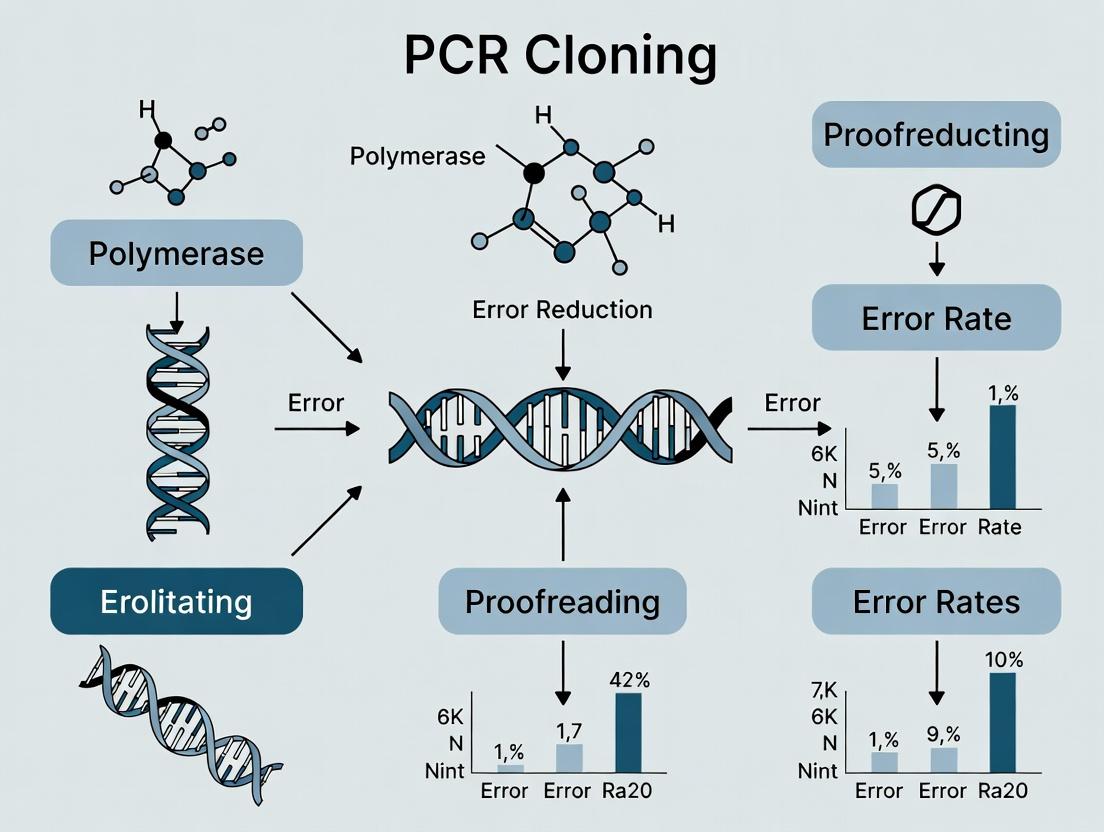
Understanding PCR Cloning Errors: Sources, Consequences, and Error-Prone Hotspots
Technical Support Center: Troubleshooting PCR Fidelity Issues
Support Context: This resource is part of a broader research thesis on PCR Cloning Error Reduction Strategies. The following guides address common, fidelity-related experimental failures in cloning, sequencing, and functional assays.
Troubleshooting Guides & FAQs
Q1: My cloned sequences after TA/Blunt-end cloning frequently contain unexpected point mutations. What is the likely cause and how can I fix it?
A: This is a classic symptom of polymerase infidelity during PCR. Standard Taq polymerase lacks proofreading (3'→5' exonuclease) activity, leading to misincorporation rates of ~1 x 10⁻⁴ errors per base per duplication.
- Solution: Switch to a high-fidelity (Hi-Fi) polymerase blend, which incorporates a proofreading enzyme (e.g., Pfu, Deep Vent). These can reduce error rates by 5-50 fold.
- Protocol: For critical cloning, use the following modified protocol:
- Polymerase: Use a certified high-fidelity polymerase (see Reagent Table).
- Cycling: Reduce the number of amplification cycles to the minimum required (e.g., 25 instead of 35).
- Template: Use high-quality, minimal-passage DNA template.
- Post-PCR: Always sequence multiple clones (at least 3-5) to identify consensus sequence.
Q2: I am getting a high percentage of non-functional clones in my protein expression assay. The gene sequence is correct by Sanger sequencing. What could be wrong?
A: Sanger sequencing from a pool of plasmids can miss low-frequency errors present in only a subset of your cloned DNA. A single PCR error early in amplification can be propagated, resulting in a plasmid sub-population expressing non-functional protein.
- Solution: Implement post-cloning sequence verification of single colonies and functional screening.
- Protocol: Colony PCR & Sequencing Workflow:
- Pick 8-12 individual E. coli colonies from your transformation plate.
- Perform colony PCR using vector-specific primers flanking the insert.
- Purify PCR products and submit each for Sanger sequencing.
- Only proceed with clones showing perfect sequence alignment for protein expression.
Q3: My NGS results for PCR-amplified libraries show an abnormally high rate of singletons and variant errors. How do I minimize PCR artifacts for sensitive applications like variant detection?
A: For NGS and rare variant detection, PCR errors are indistinguishable from true biological variants. Duplex sequencing or unique molecular identifiers (UMIs) are required, but wet-lab fidelity is the first defense.
- Solution: Use ultra-high-fidelity polymerases and limit amplification cycles.
- Protocol: Library Amplification for NGS:
- Use a polymerase with an error rate ≤ 2.0 x 10⁻⁶ errors/bp (see data table).
- Keep PCR cycles during library prep to ≤ 18 cycles.
- Perform technical replicates of the PCR step and compare variant calls; true variants should appear in both, while PCR errors will be stochastic.
Table 1: Error Rates and Characteristics of Common PCR Polymerases
| Polymerase Type | Example Enzymes | Proofreading Activity | Estimated Error Rate (errors per bp per duplication) | Best For |
|---|---|---|---|---|
| Standard Taq | Basic Taq | No | ~1.0 x 10⁻⁴ | Routine PCR, genotyping |
| High-Fidelity Blends | Q5, Phusion, KAPA HiFi | Yes | ~2.0 x 10⁻⁶ to 5.0 x 10⁻⁶ | Cloning, site-directed mutagenesis, library prep |
| Ultra-Hi-Fi / Next-Gen | Platinum SuperFi II, Q5 U | Yes (enhanced) | ≤ 1.0 x 10⁻⁶ | NGS, long amplicons, difficult templates |
Experimental Protocol: Validating Polymerase Fidelity (LacIα Complementation Assay)
This is a standard method for empirically determining polymerase error frequency.
Objective: To quantify the mutation frequency introduced by a PCR polymerase using a functional reporter gene (lacZα).
Materials: See "Research Reagent Solutions" below.
Methodology:
- Amplification: PCR-amplify the lacZα gene (~300 bp) from a control plasmid (e.g., pUC19) using the polymerase system under test. Use ≥ 30 cycles to amplify errors.
- Cloning: Purify the PCR product. Clone it into a prepared, linearized vector backbone that allows blue-white screening (requires complementation of lacZα).
- Transformation: Transform the ligation product into an appropriate E. coli strain. Plate onto LB agar containing X-Gal, IPTG, and the appropriate antibiotic.
- Analysis: Incubate plates overnight. Count the total colonies and the number of white (mutant) colonies. Blue colonies contain functional lacZα.
- Calculation: Error Rate = (Number of white colonies / Total colonies) / (Length of lacZα amplicon in bp).
Diagrams
Title: Downstream Impact Pathway of PCR Errors
Title: LacIα Assay Workflow for Error Rate Calculation
The Scientist's Toolkit: Research Reagent Solutions
Table 2: Essential Reagents for PCR Fidelity Research & Troubleshooting
| Reagent / Material | Function & Rationale |
|---|---|
| High-Fidelity Polymerase Blend (e.g., Q5, Phusion) | Contains a proofreading enzyme to excise misincorporated nucleotides, drastically reducing error rates for cloning. |
| dNTP Mix (Balanced, High-Purity) | Imbalanced dNTP concentrations can increase misincorporation by polymerase. Use high-quality, pH-verified mixes. |
| Template DNA (Low-Passage, High-Quality) | Damaged or contaminated template can cause polymerase stalling and increase error incorporation. |
| Cloning Vector for Blue-White Screening (e.g., pUC19) | Essential for the lacZα complementation assay to visually screen for functional (blue) vs. mutant (white) clones. |
| Competent E. coli (High-Efficiency) | Ensures transformation efficiency is not a bottleneck in cloning-based fidelity assays. |
| Sanger Sequencing Services/Primers | Required for definitive confirmation of nucleotide sequence in individual clones post-PCR and cloning. |
| Unique Molecular Identifiers (UMIs) | Short random nucleotide tags added to each template molecule before PCR to bioinformatically trace and remove PCR errors in NGS. |
Troubleshooting Guides & FAQs
FAQ 1: Why is my cloned sequence showing single base pair substitutions, and how can I determine if the error is from polymerase misincorporation or template damage?
- Answer: Single base pair substitutions are a common cloning error. To determine the source:
- Polymerase Misincorporation: These errors are introduced during the amplification cycle and are often stochastic. They may appear randomly across different clones and are not consistently reproducible from the same template. Using a high-fidelity polymerase (with 3'→5' exonuclease proofreading activity) significantly reduces their frequency.
- Template Damage: Errors from template damage (e.g., deamination of cytosine to uracil, oxidation of guanine to 8-oxo-guanine) are sequence context-dependent and reproducible. The same error will appear in multiple independent clones derived from the same damaged template stock, even with high-fidelity polymerases. Source template purification and limiting freeze-thaw cycles are critical.
FAQ 2: My high-fidelity PCR still yields clones with errors. Could template damage be the culprit, and how do I test for it?
- Answer: Yes. High-fidelity polymerases correct replication errors but cannot correct pre-existing lesions in the template DNA. To test:
- Perform parallel PCRs using the same polymerase but with different template preparations (e.g., a fresh aliquot vs. an old, repeatedly thawed one).
- Clone and sequence multiple colonies from each reaction.
- If the same non-consensus mutation appears in multiple clones from the old template but not from the fresh one, template damage is the likely source. Protocols like enzymatic treatment of template DNA with repair mixes (e.g., PreCR) can be attempted to mitigate this.
FAQ 3: What are the best practices to minimize both types of errors in a single experiment for critical cloning applications?
- Answer: Implement a combined strategy:
- For Polymerase Errors: Select a proven high-fidelity polymerase blend (see toolkit below). Minimize PCR cycle number. Use sufficient template to avoid late-cycle "jackpot" errors.
- For Template Damage: Use high-quality, freshly prepared template DNA. Aliquot template stocks to avoid freeze-thaw cycles. For valuable or damaged archives, consider pre-treatment with DNA repair enzymes. Always sequence multiple clones (at least 3-5) to establish consensus.
Table 1: Error Rate Comparison of Common PCR Polymerases
| Polymerase Type | Example Enzymes | Approximate Error Rate (mutations/bp/duplication) | Primary Error Reduction Mechanism |
|---|---|---|---|
| Standard Taq | Wild-type Taq | 1.0 x 10⁻⁴ | None (lacks proofreading) |
| Proofreading Enzymes | Pfu, Phusion, Q5 | 1.0 x 10⁻⁶ to 4.4 x 10⁻⁷ | 3'→5' exonuclease activity |
| High-Fidelity Blends | Mixes with proofreading + processivity enhancers | ~1.5 x 10⁻⁶ | Proofreading + optimized buffer |
Table 2: Impact of Template Damage on Cloning Fidelity
| Template Condition | Common Lesions | Resulting Cloning Mutation (after PCR & cloning) | Approximate Fold-Increase in Error Frequency* |
|---|---|---|---|
| Fresh, High-Quality | Minimal | Baseline (polymerase error rate only) | 1x |
| Multiple Freeze-Thaws | Single-strand breaks, base deamination | C→T / G→A transitions | 3-10x |
| Oxidized | 8-oxo-guanine | G→T transversions | 5-15x |
| UV-Damaged | Pyrimidine dimers | Deletions, complex errors | Variable, high |
*Compared to fresh template with high-fidelity polymerase. Data synthesized from current literature.
Experimental Protocols
Protocol 1: Assessing Polymerase Misincorporation Fidelity Objective: Quantify the intrinsic error rate of a PCR polymerase. Method:
- Template: Use a well-characterized, undamaged plasmid (e.g., plasmid with a lacZα gene for blue-white screening or a known antibiotic resistance gene).
- PCR Amplification: Amplify a 1-2 kb target using the test polymerase. Use a minimum of 30 cycles to potentially amplify errors.
- Cloning: Clone the PCR product into a linearized vector using a high-efficiency, blunt- or sticky-end ligation method. Transform into competent cells.
- Screening: For lacZα, plate on X-Gal/IPTG. White colonies indicate potential mutations. For other genes, perform functional screening (e.g., loss of antibiotic resistance).
- Analysis: Sequence the insert from a statistically significant number of colonies (e.g., 20-50). Calculate error rate using the formula: Error Rate = Total Mutations / (Total bp Sequenced × Number of PCR Duplications). The number of duplications is estimated as log2(DNA yield / template amount).
Protocol 2: Detecting Error Hotspots from Template Damage Objective: Identify reproducible mutations arising from a specific template stock. Method:
- Parallel Amplification: Set up two identical PCR reactions using a high-fidelity polymerase. Reaction A: "Old" template stock. Reaction B: "Fresh" template preparation of the same sequence.
- Independent Cloning: Purify PCR products A and B separately. Clone each into the same vector system using a high-yield assembly method (e.g., Gibson Assembly, Golden Gate).
- Colony Picking & Sequencing: Pick at least 10-15 colonies from each transformation plate. Ensure they are from independent E. coli colonies. Sequence the entire insert.
- Data Comparison: Align all sequences to the reference. Tabulate all mutations. A mutation signature (e.g., C→T at specific cytosines) present in multiple clones from Condition A but absent in all clones from Condition B is strong evidence of template damage (e.g., cytosine deamination).
Visualizations
Title: Decision Tree for Diagnosing PCR Cloning Errors
Title: Mechanisms of Error Propagation from Two Sources
The Scientist's Toolkit: Research Reagent Solutions
Table 3: Essential Reagents for PCR Cloning Fidelity Research
| Reagent / Material | Function & Rationale |
|---|---|
| High-Fidelity DNA Polymerase (e.g., Q5, Phusion, KAPA HiFi) | Contains proofreading (3'→5' exonuclease) activity to correct polymerase misincorporations during amplification, lowering error rates 50-100x compared to standard Taq. |
| dNTP Mix (Balanced, High-Purity) | Provides equimolar, uncontaminated nucleotides to prevent misincorporations due to substrate imbalance or impurities. |
| Template DNA Preparation/Purification Kit (e.g., column-based, magnetic bead) | Ensures template is free of contaminants (salts, organics, nucleases) that can promote damage or inhibit polymerase fidelity. |
| DNA Repair Enzyme Mix (e.g., PreCR Repair Mix) | Contains enzymes like Endonuclease IV, Fpg, and UDG to repair common template lesions (nicks, oxidized bases, uracil) before PCR. |
| High-Efficiency Cloning Kit (e.g., Gibson Assembly, Golden Gate) | Reduces the need for multiple PCR cycles (which can amplify errors) and minimizes background, increasing the probability of analyzing correct clones. |
| Chemically Competent E. coli (High-Efficiency, >1x10⁹ cfu/µg) | Ensures faithful replication of the plasmid post-cloning and allows for analysis of a large number of independent colonies to assess error frequency. |
| Next-Generation Sequencing (NGS) Service/Library Prep Kit | For deep sequencing of pooled clones or PCR products to quantitatively assess error rates and hotspots at high throughput. |
Technical Support Center
Troubleshooting Guides & FAQs
Q1: Why is my error-prone PCR (epPCR) reaction yielding no product or very low yield? A: This is often due to suboptimal polymerase activity. Ensure you are using a polymerase deficient in 3'→5' exonuclease (proofreading) activity, such as Taq DNA polymerase. Low yield can also result from excessive manganese concentration, which inhibits polymerization. Troubleshoot by:
- Titrating MnCl₂ (0.1-0.8 mM) while keeping MgCl₂ constant.
- Verifying template quality and concentration (10-100 ng for plasmid DNA).
- Testing a gradient annealing temperature (45-60°C) to find the optimal for your primers.
Q2: How can I achieve a more consistent and predictable mutation rate across different epPCR experiments? A: Inconsistent mutation rates are typically tied to variable concentrations of mutagenic agents. For reproducible results:
- Use a standardized master mix. Pre-mix dNTPs, Mg²⁺, Mn²⁺, buffer, and polymerase for multiple reactions.
- Precisely control the number of amplification cycles. More cycles increase mutation load.
- Use high-fidelity thermocyclers with accurate block temperature uniformity. Refer to Table 1 for parameter optimization.
Q3: My mutation rate is too low for effective directed evolution. Which parameters should I adjust first? A: To increase the mutation frequency, systematically adjust the following parameters in order:
- Increase MnCl₂ concentration within the 0.2-0.8 mM range.
- Increase MgCl₂ concentration (2-7 mM), as it is a cofactor for Taq polymerase. Maintain a total divalent cation (Mg²⁺ + Mn²⁺) concentration below 8-10 mM to prevent precipitation.
- Adjust dNTP ratios. Increasing dATP and dTTP relative to dCTP and dGTP can bias mutations toward A/T → G/C transitions.
- Increase the number of thermal cycles (e.g., from 25 to 35).
Q4: The amplified product contains too many double-stop codons or deleterious mutations. How can I bias toward more favorable mutations? A: This involves biasing the nucleotide misincorporation. Use nucleotide analogs or unbalanced dNTP pools.
- Unbalanced dNTPs: Use a commercial kit or prepare a mix with, for example, a 5-10X excess of two dNTPs relative to the other two.
- Nucleotide Analogs: Incorporate analogs like 8-oxo-dGTP or dPTP at low concentrations (e.g., 10-50 µM) to promote specific mispairing.
Q5: How do I quantify the actual mutation rate/kb from my epPCR library? A: You must sequence a representative sample. The standard method is:
- Clone the epPCR product into a vector.
- Pick 20-50 random colonies for Sanger sequencing.
- Align sequences to the original template.
- Calculate the mutation rate using the formula: Mutation rate (mutations/kb) = (Total number of mutations observed) / (Total number of base pairs sequenced).
Data Presentation
Table 1: Key Parameters Influencing Mutation Rate in Standard Taq-Based epPCR
| Parameter | Typical Standard PCR Range | Error-Prone PCR Optimization Range | Effect on Mutation Rate |
|---|---|---|---|
| MnCl₂ Concentration | 0 mM | 0.1 - 0.8 mM | Most critical. Increases misincorporation by reducing fidelity. >0.5 mM can drastically lower yield. |
| MgCl₂ Concentration | 1.5 - 2.5 mM | 2.0 - 7.0 mM | Higher concentrations increase polymerase processivity but decrease fidelity. Balance with Mn²⁺. |
| dNTP Concentration | 0.2 mM each | 0.2 - 1.0 mM each | Higher [dNTP] increases misincorporation. Unbalancing ratios (e.g., 1:1:8:8) is a key strategy. |
| Polymerase | Proofreading (e.g., Pfu) | Non-proofreading (e.g., Taq) | Mandatory. Proofreading enzymes correct errors, negating mutagenesis. |
| Thermal Cycles | 25 - 30 | 30 - 40 | Linear increase with cycle number. More replication events accumulate errors. |
| Template Amount | High (ng) | Low (pg-ng) | Lower template reduces template competition, enriching mutant sequences. |
Table 2: Comparison of Common epPCR Methodologies
| Method | Core Mechanism | Typical Mutation Rate (mutations/kb) | Key Advantage | Key Limitation |
|---|---|---|---|---|
| Standard Taq/Mn²⁺ | Mn²⁺-induced misincorporation + unbalanced dNTPs | 0.5 - 8 | Simple, inexpensive, adjustable. | Bias towards transitions (A/TG/C). |
| Nucleotide Analog | Incorporation of mutagenic base analogs (e.g., dPTP) | 1 - 15 | Can achieve higher rates, different bias. | Analogs can be expensive and toxic. |
| Commercial Kits | Optimized proprietary blends of mutagens | 1 - 20 (varies by kit) | Reproducible, user-friendly, high efficiency. | Costly, "black box" reagent composition. |
Experimental Protocols
Protocol 1: Standard Taq Polymerase-Based Error-Prone PCR This protocol is designed to generate a library with a moderate mutation rate (~2-4 mutations/kb).
Reagents:
- Template DNA (100-500 bp target, 10-50 ng)
- Forward and Reverse Primers (20-25 nt, 0.2-0.5 µM final)
- 10X Taq Buffer (no MgCl₂)
- MgCl₂ (25 mM stock)
- MnCl₂ (5 mM stock)
- dNTP mix (10 mM total, unbalanced e.g., 1mM dCTP, 1mM dGTP, 8mM dATP, 8mM dTTP)
- Taq DNA Polymerase (5 U/µL)
- Nuclease-free water
Procedure:
- Prepare a 50 µL reaction mix on ice:
- Nuclease-free water: to 50 µL
- 10X Taq Buffer: 5 µL
- MgCl₂ (25 mM): 3 µL (Final: 1.5 mM)
- MnCl₂ (5 mM): 5 µL (Final: 0.5 mM)
- Unbalanced dNTP mix (10 mM total): 5 µL (Final: 1 mM total dNTPs)
- Forward Primer (10 µM): 1 µL (Final: 0.2 µM)
- Reverse Primer (10 µM): 1 µL (Final: 0.2 µM)
- Template DNA: 1 µL (~20 ng)
- Taq DNA Polymerase: 0.25 µL (1.25 U)
- Mix gently and centrifuge briefly.
- Run PCR:
- Initial Denaturation: 95°C for 3 min.
- 30 Cycles:
- Denature: 95°C for 30 sec.
- Anneal: 55°C for 30 sec.
- Extend: 72°C for 1 min/kb.
- Final Extension: 72°C for 5 min.
- Hold: 4°C.
- Analyze 5 µL by agarose gel electrophoresis.
- Purify the remaining product using a PCR cleanup kit before downstream cloning.
Protocol 2: Mutation Rate Determination by Sequencing Procedure:
- Ligate the purified epPCR product into a blunt-end or TA-cloning vector according to manufacturer instructions.
- Transform the ligation into competent E. coli and plate on selective media.
- Randomly pick at least 20-30 colonies for colony PCR or plasmid purification.
- Submit samples for Sanger sequencing with an appropriate primer.
- Analyze sequences:
- Align each sequence to the wild-type template using software (e.g., Geneious, SnapGene, CLUSTAL Omega).
- Count all point mutations (ignore the vector sequence).
- Calculate: Total bp sequenced = (sequence read length) x (number of clones).
- Mutation Rate = (Total mutations observed) / (Total kb sequenced).
Mandatory Visualization
Title: Error-Prone PCR Experimental Workflow
Title: Parameter Comparison: Standard vs. Error-Prone PCR
The Scientist's Toolkit: Research Reagent Solutions
| Item | Function in epPCR | Key Consideration |
|---|---|---|
| Non-Proofreading DNA Polymerase (e.g., Taq) | Catalyzes DNA synthesis with low inherent fidelity, allowing misincorporated nucleotides to remain. | Must lack 3'→5' exonuclease activity. Do not use polymerases like Pfu or Phusion. |
| Manganese Chloride (MnCl₂) | The primary mutagen. Mn²⁺ substitutes for Mg²⁺ in the polymerase active site, promoting misincorporation of dNTPs. | Highly concentration-sensitive. Titrate carefully (0.1-0.8 mM). Store as small aliquots. |
| Unbalanced dNTP Mix | Biases the nucleotide pool to favor misincorporation of specific nucleotides, controlling mutation spectrum. | Can be prepared manually or purchased. Common ratio: 1:1:8:8 (dCTP:dGTP:dATP:dTTP). |
| High-Purity Template DNA | The sequence to be diversified. Minimizes background from pre-existing template errors. | Purify by gel extraction or column. Use minimal amount (pg-ng) to avoid wild-type carryover. |
| TA or Blunt-End Cloning Vector | For efficient ligation and cloning of the mutagenized PCR product for sequencing and library creation. | Taq-amplified products have 3'-A overhangs, making them compatible with TA vectors. |
| Commercial epPCR Kit | Provides optimized, pre-formulated buffers and nucleotide mixes for reproducible mutation rates. | Ideal for standardized workflows but offers less user control over individual parameters. |
The Impact of Amplicon Length and GC Content on Cloning Fidelity
Troubleshooting Guides & FAQs
Q1: My cloned insert has unexpected mutations after transformation and sequencing. Could this be related to the length of my PCR amplicon? A: Yes, amplicon length is a significant factor. Longer amplicons (>1kb) require more polymerase extension cycles, increasing the probability of polymerase misincorporation per full-length product. This is a core focus of thesis research on PCR cloning error reduction. For high-fidelity cloning of long fragments (>3kb), consider using a polymerase blend with proofreading activity and optimizing extension times.
Q2: I am trying to clone a high-GC (>70%) region. My PCR yield is low, and the clones I get often have deletions. What is the link, and how can I fix this? A: High GC content promotes secondary structure formation (e.g., hairpins), which can cause polymerase stalling, dissociation, and incomplete synthesis, leading to truncation artifacts. Within the thesis framework, this is addressed by employing PCR enhancers. A recommended protocol is below.
Q3: My sequencing results show point mutations (A->G, T->C) scattered throughout the clone, even with a "high-fidelity" polymerase. What's happening? A: This pattern often indicates residual PCR errors that escaped proofreading. All polymerases have a base misincorporation rate. The thesis strategy emphasizes that fidelity is a product of both polymerase choice and cycling conditions. Reducing cycle number and using sufficient template minimize error propagation.
Q4: How do I balance the need for high yield with the need for high fidelity when amplifying difficult templates? A: This is a key optimization challenge. The thesis proposes a tiered strategy: prioritize fidelity for cloning by using high-fidelity enzymes and minimal cycles. If yield is insufficient, scale up reaction volume rather than cycle number. For problematic GC-rich templates, use adjunct reagents (see Table 2) before increasing cycles.
Experimental Protocols
Protocol 1: Assessing Fidelity by Amplicon Length
- Design: Design primers to amplify regions of the same gene at 500bp, 1.5kb, and 3kb.
- PCR Setup: Use a high-fidelity polymerase (e.g., Q5, Phusion). Perform triplicate 50µL reactions for each length with identical template (10ng), cycling conditions (adjusted for length), and cycle number (25).
- Cloning: Gel-purify each amplicon. Clone into a blunt-end or TA vector (as appropriate). Transform into competent E. coli.
- Analysis: Pick 10-20 colonies per length. Perform colony PCR and Sanger sequence the inserts.
- Data Collection: Align sequences to the reference. Record total errors (mismatches, indels) per clone.
Protocol 2: Optimizing Cloning of High-GC Amplicons
- Standard Reaction: Set up a control PCR with standard buffer.
- Enhanced Reactions: Set up parallel reactions supplemented with:
- Condition A: 1M Betaine.
- Condition B: 5% DMSO.
- Condition C: 1M Betaine + 5% DMSO.
- Condition D: Commercial GC enhancer.
- PCR: Use a touch-down or slow-ramping cycling protocol (e.g., 0.5°C/sec ramp) to aid denaturation of secondary structures.
- Analysis: Compare yield via gel electrophoresis, then clone and sequence products from highest-yield condition as in Protocol 1 to assess fidelity impact.
Data Presentation
Table 1: Error Frequency vs. Amplicon Length (Representative Data)
| Amplicon Length | Average Error Rate (errors/kb)* | Percentage of Perfect Clones (%) | Most Common Error Type |
|---|---|---|---|
| 500 bp | 0.8 | 85 | Single bp substitution |
| 1500 bp | 1.5 | 65 | Single bp substitution |
| 3000 bp | 3.2 | 30 | Deletions > 5bp |
*Data based on sequencing of 20 clones per group using a common high-fidelity polymerase.
Table 2: Effect of GC-Enhancers on Yield and Fidelity of a 72% GC Template
| Condition | Relative Yield (%) | Average Error Rate (errors/kb) | Notes |
|---|---|---|---|
| Standard Buffer | 100 (baseline) | 2.1 | Low yield, high error |
| 1M Betaine | 450 | 1.8 | Greatly improved yield |
| 5% DMSO | 300 | 2.5 | Good yield, slightly higher error |
| Betaine + DMSO | 600 | 1.9 | Highest yield |
| Commercial Enhancer | 500 | 1.7 | Balanced performance |
Visualizations
Title: PCR Cloning Fidelity Optimization Workflow
Title: How GC & Length Lead to Cloning Errors
The Scientist's Toolkit: Research Reagent Solutions
| Item | Function in Cloning Fidelity Context |
|---|---|
| High-Fidelity DNA Polymerase (e.g., Q5, Phusion, KAPA HiFi) | Contains a proofreading (3'→5' exonuclease) domain to correct misincorporated nucleotides during synthesis, drastically reducing error rates. |
| PCR Enhancers (Betaine) | Equalizes the stability of AT and GC base pairing, reducing secondary structure formation in GC-rich templates and improving yield and accuracy. |
| PCR Enhancers (DMSO) | A destabilizing agent that helps denature DNA secondary structures by interfering with base pairing, facilitating polymerase progression. |
| dNTP Mix (Balanced, High-Quality) | Provides equimolar, pure nucleotides to prevent misincorporation due to substrate imbalance or contaminants. |
| Cloning Vector with Blue-White Screening (e.g., pUC-based) | Allows rapid visual screening of successful ligations, reducing the number of clones needing sequencing to find correct ones. |
| Competent E. coli (High-Efficiency) | Ensures high transformation efficiency, which is critical when working with low-yield or difficult amplicons to obtain sufficient clones for screening. |
| Gel Extraction Kit | Removes primer dimers, non-specific products, and enzyme inhibitors prior to ligation, improving cloning success of the intended amplicon. |
| Next-Generation Sequencing (NGS) Service | For comprehensive fidelity assessment beyond Sanger, enabling detection of low-frequency errors within a pooled clone population. |
Technical Support Center
Troubleshooting Guides & FAQs
FAQ 1: What are the main post-PCR artifacts that can compromise my cloning results?
- Chimeras: Hybrid molecules formed from two or more different parent sequences during amplification, especially in multi-template reactions. They cause recombinant clones.
- Heteroduplexes: Mismatched double-stranded DNA molecules formed by the annealing of similar but not identical strands (e.g., from different alleles or paralogs). They cause mixed sequencing signals and are a major source of base-calling errors in Sanger sequencing of cloned products.
- Primer-Dimer Carryover: Small, non-specific amplification products formed by primer self-annealing. When present in a cloning ligation, they compete with the insert, drastically reducing the fraction of recombinant clones containing your target.
FAQ 2: How can I specifically detect and quantify heteroduplexes in my PCR product before cloning? Method: Heteroduplex Mobility Assay (HMA) using Polyacrylamide Gel Electrophoresis (PAGE).
- Protocol:
- Run your standard PCR amplification.
- Denature the PCR product: Heat to 95°C for 5 minutes.
- Re-anneal: Rapidly cool on ice for 5 minutes, then incubate at 4°C for 60 minutes. This step promotes heteroduplex formation between polymorphic strands.
- Prepare an 8-12% non-denaturing polyacrylamide gel.
- Load the re-annealed product alongside the native PCR product and a homoduplex control (a known homozygous sample).
- Run electrophoresis at a constant voltage (e.g., 100-150V) at 4°C for optimal resolution.
- Stain with SYBR Gold or Ethidium Bromide and visualize. Heteroduplexes will appear as additional, slower-migrating bands compared to the homoduplex band(s).
FAQ 3: What is the most effective wet-lab strategy to eliminate chimeras and heteroduplexes prior to cloning? Method: Post-PCR Treatment with a Nuclease Specific for Heteroduplex/Mismatched DNA.
- Protocol: Using a DNA Mismatch-Specific Endonuclease (e.g., Surveyor Nuclease S, Cel I, or T7 Endonuclease I):
- PCR Amplification: Generate your product from a mixed template pool.
- Heteroduplex Formation: Denature and re-anneal as in the HMA protocol (steps 2-3 above).
- Nuclease Digestion: Set up a reaction with the re-annealed DNA (e.g., 100-200 ng), the provided nuclease buffer, and 1 µL of the mismatch-specific endonuclease. Incubate at 42°C for 20-60 minutes.
- Purification: Purify the digested product using a standard PCR clean-up kit or column. The nuclease cleaves at mismatch sites, linearizing heteroduplexes and chimeras, while leaving perfect homoduplexes intact.
- Cloning: Proceed to clone the purified product. The cleaved artifacts will not ligate efficiently, enriching your clone library for correct, homoduplex products.
Table 1: Comparison of Post-PCR Artifact Reduction Methods
| Method | Target Artifact(s) | Mechanism | Efficiency Reduction (Quantitative) | Downstream Impact |
|---|---|---|---|---|
| Reconditioning PCR | Heteroduplexes, Primer-Dimer | Limited-cycle re-amplification of pure product | Reduces heteroduplexes by ~50-70% | Moderate improvement in sequencing clarity |
| Gel Extraction | Primer-Dimers, Non-specific Products | Physical size separation and excision | ~90-99% removal of visible primer-dimer | High increase in recombinant clone ratio |
| Mismatch-specific Nuclease | Heteroduplexes, Chimeras | Enzymatic cleavage at mismatch sites | Reduces heteroduplex/chimera clones by >90% | Drastic reduction in sequencing errors and recombinant clones |
| DpnI Digestion (post-PCR) | Parental Template Carryover | Cleaves methylated, template DNA | ~100% removal of methylated E. coli templates | Eliminates false-positive from original plasmid |
Table 2: Key Research Reagent Solutions for Artifact Mitigation
| Reagent / Kit | Primary Function in Artifact Reduction | Key Consideration |
|---|---|---|
| High-Fidelity DNA Polymerase | Reduces misincorporation errors that lead to heteroduplex formation. | Lower error rate (e.g., 5.5 x 10⁻⁷ vs 2.6 x 10⁻⁵ for Taq) is critical. |
| Mismatch-Specific Endonuclease (Surveyor/Cel I) | Cleaves heteroduplex and chimera DNA post-PCR. | Optimize incubation time and DNA amount to avoid over-digestion. |
| PCR Clean-Up & Size-Selective Kits | Removes primer-dimers and salts prior to cloning or nuclease treatment. | For primer-dimer removal, use kits with a >100 bp cutoff. |
| TA or Blunt-End Cloning Kit with Ligation Control | Efficiently clones the desired homoduplex product. | Always include a vector-only ligation control to assess primer-dimer background. |
| Non-Denaturing Polyacrylamide Gel | Detects heteroduplexes via HMA and allows precise size selection. | Requires specialized equipment but offers superior resolution to agarose. |
Diagram 1: Workflow for Comprehensive Post-PCR Artifact Cleanup
Diagram 2: Molecular Origin of Key Post-PCR Artifacts
Error-Reduction Protocols: Practical Strategies and Step-by-Step Implementation
This technical support center is framed within a thesis on PCR cloning error reduction strategies. The following FAQs and troubleshooting guides address common issues faced by researchers.
Troubleshooting Guides & FAQs
Q1: My cloned gene sequence has unwanted mutations after using a high-fidelity polymerase. What went wrong? A: This is often due to residual PCR errors that escape the enzyme's proofreading. First, verify the polymerase's fidelity rate (e.g., error rate of 2.0 x 10^-6 errors/bp/duplication). Ensure you used the recommended number of cycles (≤30). High GC content or secondary structures can also reduce fidelity. Include DMSO or betaine in your protocol if needed, and always sequence multiple clones.
Q2: I am getting low PCR yield with my high-fidelity polymerase, even with ample template. How can I improve amplification? A: High-fidelity polymerases often have slower elongation rates and processivity. Check and adjust:
- Extension Time: Increase extension time to 1-2 minutes per kb.
- Annealing Temperature: Optimize using a gradient PCR.
- Template Quality: Use high-quality, purified DNA.
- Enzyme-to-Template Ratio: Follow manufacturer's guidelines; too much enzyme can inhibit reactions.
Q3: My PCR product is not suitable for blunt-end cloning. What should I do? A: Most high-fidelity polymerases produce blunt ends. Ensure your purification method removes excess nucleotides. If using a polymerase that adds a single 3'A-overhang (some do), treat the product with a proofreading polymerase for 15 minutes at 72°C to polish to blunt ends. Verify the cloning vector's compatibility.
Q4: How do I choose between different high-fidelity polymerases for long amplicons (>10 kb)? A: Prioritize enzymes with high processivity and DNA-binding affinity. Look for engineered or fusion proteins (e.g., with processivity-enhancing domains). Refer to Table 1 for comparative data on long-range amplification success.
Table 1: Comparison of Common High-Fidelity DNA Polymerases
| Polymerase Name (Example) | Error Rate (errors/bp/duplication) | Processivity | Optimal Amplicon Length | Typical Extension Speed (seconds/kb) | 3'→5' Exonuclease (Proofreading) | Format |
|---|---|---|---|---|---|---|
| Polymerase A | 2.0 x 10^-6 | High | ≤ 20 kb | 30-60 | Yes | Blend |
| Polymerase B | 5.5 x 10^-7 | Medium | ≤ 5 kb | 15-30 | Yes | Native |
| Polymerase C (Long-Range) | 1.5 x 10^-6 | Very High | ≤ 40 kb | 45-90 | Yes | Fusion |
| Polymerase D | 3.0 x 10^-6 | Medium-High | ≤ 10 kb | 30 | Yes | Blend |
Table 2: Troubleshooting Common PCR Issues with High-Fidelity Enzymes
| Symptom | Possible Cause | Recommended Solution |
|---|---|---|
| No Amplification | Inhibitors in template, incorrect Mg2+ | Purify template, optimize Mg2+ (1.5-3.0 mM), add 5% DMSO for GC-rich targets. |
| Non-Specific Bands | Low annealing temperature, excessive cycles | Use touch-down PCR, reduce cycles to 25-30, increase annealing temperature gradient. |
| Smearing | Enzyme excess, degraded template | Reduce enzyme amount by 25%, check template integrity on gel. |
| Low Yield | Short extension time, low processivity | Increase extension time, use polymerase optimized for long/he structured templates. |
Experimental Protocols
Protocol 1: Standard High-Fidelity PCR for Cloning
Objective: Amplify target gene with minimal errors for subsequent blunt-end cloning. Materials: See "The Scientist's Toolkit" below. Method:
- Reaction Setup (50 µL):
- 1x High-Fidelity PCR Buffer (with Mg2+)
- 200 µM each dNTP
- 0.3 µM each forward and reverse primer
- 10-100 ng genomic DNA or 1-10 ng plasmid template
- 1.0-1.25 units of high-fidelity DNA polymerase
- Nuclease-free water to 50 µL.
- Thermal Cycling:
- Initial Denaturation: 98°C for 30 seconds.
- 30 Cycles:
- Denaturation: 98°C for 10 seconds.
- Annealing: 55-72°C (optimize) for 15-30 seconds.
- Extension: 72°C for 15-60 seconds/kb.
- Final Extension: 72°C for 5-10 minutes.
- Hold: 4°C.
- Post-PCR Processing: Purify PCR product using a spin column. For blunt-end cloning, optionally treat with polishing enzyme.
Protocol 2: PCR Error Rate Determination bylacZαComplementation Assay
Objective: Empirically determine the error frequency of a polymerase. Method:
- Amplify the lacZα gene from a control plasmid using the test polymerase under standard conditions.
- Purify the PCR product and perform a restriction digest if necessary.
- Clone the product into a compatible, linearized vector using a high-efficiency blunt-end cloning kit.
- Transform into an appropriate E. coli strain and plate on LB plates containing X-Gal and IPTG.
- Count blue (functional) and white (mutated) colonies after incubation.
- Calculate Error Rate: Error Frequency = (Number of white colonies) / (Total colonies * Length of lacZα in bp). Error Rate (per bp per duplication) = Error Frequency / (Number of PCR doublings).
Mandatory Visualizations
Title: PCR Cloning Workflow for Error Reduction
Title: Troubleshooting Logic for PCR Cloning Errors
The Scientist's Toolkit: Research Reagent Solutions
| Item | Function in High-Fidelity PCR Cloning |
|---|---|
| High-Fidelity DNA Polymerase Blend | Engineered enzyme mix providing optimal balance of fidelity, processivity, and yield. Essential for accurate amplification. |
| dNTP Mix (10 mM each) | Provides nucleotide substrates for DNA synthesis. Use high-quality, pH-balanced dNTPs to prevent degradation. |
| GC-Rich Enhancer/Additive (e.g., DMSO) | Disrupts secondary structures in high-GC templates, improving polymerase processivity and specificity. |
| PCR Purification Kit (Spin Column) | Removes primers, dNTPs, salts, and enzymes post-amplification, essential for clean cloning ligation. |
| Blunt-End Cloning Kit | Contains polished vector, ligase, and controls for efficient insertion of non-tailed PCR products. |
| Competent E. coli Cells (High Efficiency) | For transformation of ligation products. >1 x 10^8 cfu/µg efficiency is recommended for library construction. |
| Blue/White Screening System (X-Gal/IPTG) | Allows visual identification of successful clones containing insert versus empty vector during cloning verification. |
| DNA Sequence Analysis Software | Used to align and compare sequences from multiple clones to identify and quantify PCR-induced errors. |
Troubleshooting Guides & FAQs
Q1: My PCR product yield is low after using purified genomic DNA as a template. What could be wrong? A: This is often a sign of template DNA damage. Damaged DNA (e.g., nicks, abasic sites, oxidative lesions) can block polymerase progression. First, verify DNA integrity by running an aliquot on an agarose gel alongside an intact control (e.g., uncut lambda DNA). Sheared or degraded DNA will appear as a low molecular weight smear. Ensure your DNA extraction protocol includes a gentle lysis step (avoid vortexing), uses fresh antioxidants like β-mercaptoethanol or DTT, and employs high-quality, nuclease-free reagents and tubes. For aged samples, consider using a DNA repair enzyme mix (see Toolkit) prior to PCR.
Q2: I'm observing nonspecific amplification and high-molecular-weight smears. Could template quality be a factor? A: Yes. While often attributed to primer annealing issues, contaminated or damaged template can be the culprit. Protein or organic solvent carryover from phenol-chloroform extraction can inhibit polymerase and cause aberrant products. Ensure your template preparation includes a thorough ethanol precipitation with 70% wash or uses a validated spin-column kit. Quantify DNA using a fluorometer (e.g., Qubit) rather than a spectrophotometer (Nanodrop), as the latter overestimates concentration in the presence of contaminants. A 260/280 ratio between 1.8-2.0 and a 260/230 ratio >2.0 are good indicators of purity.
Q3: My Sanger sequencing of cloned plasmids shows an unexpected high rate of point mutations in the insert, not present in the original PCR product. What's happening? A: This points to DNA damage in the input template for PCR that leads to polymerase errors, which are then cloned. Lesions like 8-oxoguanine can cause misincorporation. To mitigate, prepare template DNA in reduced light conditions to prevent UV-induced damage and use buffers chelated with EDTA to sequester metal cations that catalyze oxidative damage. For critical applications, use a high-fidelity polymerase with documented damage bypass capabilities or pre-treat the template with a repair mix. Always sequence multiple clones to distinguish damage-induced errors from polymerase stochastic errors.
Experimental Protocols for Assessing and Mitigating Template Damage
Protocol 1: Assessing DNA Template Integrity via Fragment Analysis Purpose: To quantitatively assess the degree of fragmentation in a DNA sample. Materials: Genomic DNA sample, High Sensitivity DNA Kit (e.g., Agilent 2100 Bioanalyzer or TapeStation), appropriate ladder. Method:
- Dilute DNA sample to ~5-50 ng/µL in nuclease-free water or the provided buffer.
- Prepare the gel-dye mix and priming station as per kit instructions.
- Load 1 µL of ladder into the appropriate well.
- Load 1 µL of each DNA sample into subsequent wells.
- Run the chip or tape in the instrument.
- Analyze the electropherogram. The DV50 value (the size at which 50% of the DNA is larger) is a key metric. For standard PCR, a DV50 > 10 kb is ideal. A significant shift to lower sizes indicates shearing.
Protocol 2: Pre-PCR DNA Repair Protocol Purpose: To repair common lesions (nicks, gaps, abasic sites, oxidized bases) in template DNA prior to PCR amplification. Materials: PreCR Repair Mix (or equivalent from NEB/Bio-Rad), 10X Repair Buffer, template DNA (up to 1 µg). Method:
- Assemble on ice: 34 µL nuclease-free water, 5 µL 10X Repair Buffer, 1 µL PreCR Repair Mix, 10 µL template DNA (up to 1 µg). Total: 50 µL.
- Incubate at 37°C for 20 minutes.
- Heat-inactivate the enzyme at 75°C for 15 minutes.
- Place on ice. Use 1-10 µL directly as template in a 50 µL PCR reaction.
- Include a non-repaired control to assess improvement in yield or fidelity.
Data Presentation
Table 1: Impact of Template Preparation Method on PCR Cloning Fidelity
| Preparation Method | Average Mutation Rate (errors/kb)* | DV50 (bp) | 260/280 Ratio | Successful Cloning (%) |
|---|---|---|---|---|
| Standard Phenol-Chloroform | 1.8 ± 0.4 | 3,200 | 1.75 | 65% |
| Silica Spin-Column Kit | 0.9 ± 0.2 | 15,000 | 1.89 | 85% |
| Spin-Column + Antioxidants | 0.7 ± 0.1 | 18,500 | 1.90 | 92% |
| Magnetic Bead-Based | 0.8 ± 0.2 | 22,000 | 1.92 | 90% |
| Spin-Column + Pre-PCR Repair | 0.4 ± 0.1 | 18,500 | 1.89 | 98% |
*Mutation rate determined by sequencing 20 clones per method across 3 independent 2kb amplifications.
Table 2: Common DNA Lesions and Their Impact on Polymerase Fidelity
| Lesion Type | Common Cause | Primary Polymerase Effect | Resultant Cloning Error |
|---|---|---|---|
| Abasic Site | Acidic pH, Spontaneous Hydrolysis | Stalling or dAMP Insertion (A-rule) | Primarily A:T base pairs |
| 8-oxoguanine | Oxidative Stress (ROS) | Misincorporation of A opposite O8G | G:C to T:A Transversion |
| Thymine Glycol | Oxidative Stress, Ionizing Radiation | Complete Block to Polymerase | Deletion / No Amplification |
| Single-Strand Nick | Shearing, Nuclease Activity | Polymerase Run-off | Truncated Product |
| UV-induced Dimer | UV Light Exposure | Complete Block to Polymerase | No Amplification |
The Scientist's Toolkit: Research Reagent Solutions
| Item | Function in Minimizing DNA Damage |
|---|---|
| High-Purity, Nuclease-Free Water | Eliminates ribonuclease and deoxyribonuclease contamination that can degrade template. |
| Tris-EDTA (TE) Buffer, pH 8.0 | Chelates Mg2+ to inhibit metal-catalyzed oxidative damage; stable pH prevents acid-induced depurination. |
| β-Mercaptoethanol or DTT | Reducing agent added to lysis buffers to scavenge reactive oxygen species and inhibit nucleases. |
| Spin-Column Purification Kits (Silica Membrane) | Provide rapid, clean removal of proteins, salts, and organic solvents, minimizing carryover of damaging agents. |
| DNA Damage Repair Mix (e.g., PreCR) | Enzyme cocktail containing Endonuclease IV, Fpg, etc., to excise and repair damaged bases prior to PCR. |
| UV-Shielded Microcentrifuge Tubes | Protect DNA from incidental UV light exposure during handling, preventing dimer formation. |
| Fluorometric Quantitation Dye (e.g., Qubit dsDNA HS) | Accurately quantifies intact double-stranded DNA without interference from RNA, proteins, or free nucleotides. |
Visualizations
Title: Pathways from Template Damage to Cloning Errors
Title: DNA Template Quality Control and Remediation Workflow
Optimized Thermal Cycling Parameters for Maximum Fidelity and Yield
Technical Support & Troubleshooting Center
Context: This support content is part of a thesis research program focused on systematic strategies to reduce errors in PCR-based cloning, with the goal of improving downstream applications in gene synthesis, functional analysis, and therapeutic development.
Frequently Asked Questions (FAQs) & Troubleshooting Guides
Q1: Despite using a high-fidelity polymerase, my PCR cloning results in a high rate of point mutations. What thermal cycling parameters should I check first? A: This is often linked to excessive cycle numbers and suboptimal denaturation temperatures. Each cycle introduces a baseline error probability; minimizing cycles is critical.
- Action: Reduce total cycle number to the minimum required (often 25-30 cycles for plasmid templates). Ensure complete denaturation by using a temperature of 98°C for 1-2 seconds for GC-rich templates, rather than standard 95°C. Verify template quality and quantity to avoid amplifying from a very low starting copy number.
Q2: I am getting high yields of non-specific products or primer-dimer, which complicates cloning. How can I adjust my protocol to improve specificity? A: Non-specific amplification competes with the target, reducing yield and fidelity. Optimization of annealing and extension is key.
- Action: Implement a touchdown PCR or gradient PCR to empirically determine the optimal annealing temperature (Ta). Use a two-step PCR protocol if primer Tm allows (combine annealing/extension at 68-72°C). Increase elongation temperature to 68°C or 72°C (polymerase-dependent) to increase stringency. Ensure Mg2+ concentration is not excessive.
Q3: My amplicon is long (>5 kb) and yield is very low, affecting downstream ligation efficiency. What thermal cycling changes can help? A: Long amplicons require optimized conditions to maintain polymerase processivity and prevent incomplete extension.
- Action: Extend elongation time according to polymerase speed (e.g., 60 sec/kb for most high-fidelity enzymes). Use a three-step PCR with a separate, lower annealing step if necessary for primer binding. Consider adding adjuncts like DMSO (3-5%) or betaine (1 M) to reduce secondary structure. Avoid rapid ramping rates.
Q4: How do I balance extension time and temperature to maximize yield without compromising fidelity for amplicons of medium length (1-3 kb)? A: The polymerase's intrinsic elongation rate is temperature-dependent. An optimal temperature maximizes speed while maintaining enzyme stability.
- Action: Use the polymerase's recommended extension temperature (usually 68°C or 72°C). Calculate time based on the enzyme's processivity at that temperature (e.g., 30 sec/kb for many high-fidelity polymerases). Avoid overly long extension times, which can promote enzyme degradation and dNTP depletion.
Q5: When setting up multiplexed PCR for cloning, how can I manage different primer Tm's effectively within a single run? A: Compromised annealing conditions in multiplex PCR lead to biased amplification and reduced yield of some fragments.
- Action: Design all primers to a narrow Tm range (±2°C). Use a universal annealing temperature slightly below the lowest primer Tm. Employ a "slow ramp" between annealing and extension (e.g., 0.5°C/sec) to allow proper primer binding. Increase primer concentration proportionally (e.g., 0.4 µM each).
Data Presentation: Quantitative Parameter Optimization
Table 1: Impact of Thermal Cycling Parameters on Fidelity and Yield
| Parameter | Typical Default | Optimized for Fidelity | Optimized for Yield | Recommended Compromise (Cloning) | Key Rationale |
|---|---|---|---|---|---|
| Total Cycles | 35 | 20-25 | 30-40 | 25-30 | Minimizes cumulative replication errors. |
| Denaturation | 95°C, 30s | 98°C, 1-5s | 95°C, 30s | 98°C, 5-10s | Ensures complete strand separation; reduces polymerase thermal stress. |
| Annealing | Tm-5°C, 30s | Tm+2°C, 15s | Tm-5°C, 30s | Gradient Determined Ta, 15s | Increases specificity; reduces mis-priming. |
| Extension | 72°C, 60s/kb | 68-72°C, 30s/kb* | 72°C, 60s/kb | 68-72°C, 30-45s/kb* | Matches polymerase optimal speed; prevents incomplete extension. |
| Final Extension | 72°C, 5min | 72°C, 2min | 72°C, 10min | 72°C, 5-10min | Ensures all amplicons are full-length, A-tailed. |
*Dependent on specific high-fidelity polymerase.
Table 2: Adjunct Reagents and Their Effects on PCR for Cloning
| Reagent | Common Usage | Effect on Fidelity | Effect on Yield | Recommendation |
|---|---|---|---|---|
| DMSO (1-3%) | GC-rich templates | Slight decrease* | Increases | Use at ≤3% for difficult templates. |
| Betaine (1 M) | GC-rich, secondary structure | Neutral or slight increase | Increases | Alternative to DMSO, may be less inhibitory. |
| Mg2+ (1.5-3.0 mM) | Cofactor for polymerase | Critical (Low: high fidelity, Low yield; High: lower fidelity) | Critical | Optimize for each primer/template (start at 1.5 mM). |
| dNTPs (200 µM each) | Substrates | Balanced concentration is key for fidelity | Saturated conc. increases yield | Use high-quality, pH-neutral dNTPs at 200 µM. |
*Some studies indicate DMSO can reduce fidelity by relaxing primer-template specificity.
Experimental Protocols
Protocol 1: Empirical Determination of Optimal Annealing Temperature (Gradient PCR)
- Prepare a master mix containing template, primers, dNTPs, high-fidelity polymerase buffer, and polymerase.
- Aliquot the mix into 8 tubes.
- Set the thermocycler's annealing temperature gradient across the block (e.g., from 55°C to 72°C).
- Run the following cycling program:
- Initial Denaturation: 98°C for 30s.
- 30 Cycles: Denaturation: 98°C for 5-10s; Annealing: Gradient temperature for 15s; Extension: 72°C for 30s/kb.
- Final Extension: 72°C for 5 min.
- Analyze products by agarose gel electrophoresis. Select the highest temperature that produces a strong, specific band.
Protocol 2: Two-Step High-Fidelity PCR for Cloning (Primers Tm > 65°C)
- Set up reaction mix with a polymerase optimized for two-step PCR (e.g., Q5).
- Use the following cycling parameters:
- Initial Denaturation: 98°C for 30s.
- 25-30 Cycles: Denaturation: 98°C for 5-10s; Combined Annealing/Extension: 72°C for 30s/kb.
- Final Extension: 72°C for 2 min.
- This method increases stringency by performing primer binding at a temperature where non-specific interactions are less stable.
Visualizations
Diagram 1: PCR Fidelity Optimization Decision Pathway
Diagram 2: Thermal Cycling Parameter Interplay
The Scientist's Toolkit: Research Reagent Solutions
Table 3: Essential Reagents for High-Fidelity PCR Cloning
| Item | Function & Importance | Example Brands/Types |
|---|---|---|
| High-Fidelity DNA Polymerase | Engineered enzymes with 3'→5' exonuclease (proofreading) activity to drastically reduce misincorporation rates during elongation. | Q5 (NEB), Phusion/Phusion Plus (Thermo), KAPA HiFi, PrimeSTAR GXL (Takara) |
| Ultra-Pure dNTP Mix | High-quality, pH-balanced deoxynucleotide triphosphates are essential substrate for polymerization; impurities can reduce fidelity and yield. | PCR-grade dNTPs (NEB, Thermo, Sigma), separate dNTP sets for pH adjustment. |
| Mg2+ Solution (MgCl2/MgSO4) | Critical cofactor for polymerase activity; concentration must be optimized for each primer-template system to balance fidelity, yield, and specificity. | Supplied with polymerase buffer; often 25-50 mM stock. |
| PCR Enhancers/Adjuvants | Chemical additives that reduce secondary structure in GC-rich templates or stabilize polymerase, improving yield of difficult amplicons. | DMSO, Betaine, Formamide, GC Enhancer solutions. |
| Nuclease-Free Water | Solvent for all reactions; must be free of nucleases and contaminants that could inhibit polymerase or degrade template. | Certified nuclease-free water (e.g., Ambion, Teknova). |
| Optimized Buffer System | Proprietary buffering salts and components provided with the polymerase to maintain optimal pH and ionic strength during thermal cycling. | Often 5X or 2X concentrated buffers supplied with enzyme. |
Primer Design Strategies to Prevent Mispriming and Off-Target Amplification
Technical Support Center: Troubleshooting & FAQs
FAQ 1: My PCR produces multiple bands or a smear on the gel. What is the primary cause and how can I fix it?
- Answer: This is a classic symptom of mispriming, where primers bind to non-target sequences. The primary cause is low primer specificity. To fix this:
- Increase Primer Annealing Temperature: Re-calculate your primer's melting temperature (Tm) using the nearest-neighbor method. Set your PCR annealing temperature 3-5°C below the lower of the two primer Tms, or use a gradient PCR to optimize.
- Check for Secondary Structures: Use software (e.g., OligoAnalyzer) to analyze primers for self-dimers, hairpins, and cross-dimers. Avoid stretches of 3 or more G/Cs at the 3' end.
- Re-design Primers: Ensure primer length is 18-30 bases, GC content is 40-60%, and the 3' end terminates in 1-2 G/Cs (GC clamp). Perform a BLAST search against your template genome to check for off-target binding sites.
FAQ 2: My Sanger sequencing results show mixed signals starting right after the primer binding site. What does this indicate?
- Answer: Mixed sequencing traces at the beginning strongly indicate primer-dimer formation, where primers amplify each other instead of the template. This leads to a population of short, non-target amplicons that are sequenced.
- Immediate Solution: Re-run your PCR product on a high-percentage agarose gel (e.g., 3%). If you see a very low molecular weight band (~30-80 bp), it is primer-dimer.
- Prevention Protocol: Use primer design tools to check for 3' complementarity. A ∆G more negative than -9 kcal/mol for dimer formation is problematic. Increase annealing temperature. Consider using a hot-start polymerase to inhibit activity during setup.
FAQ 3: How can I verify and minimize off-target amplification in complex genomes (e.g., human, mouse)?
- Answer: For complex genomes, in silico specificity checking is mandatory but not sufficient. A two-step experimental validation is required.
- Step 1 (In Silico): Use tools like Primer-BLAST (NCBI) with stringent settings (check for primers against the RefSeq mRNA database and the genomic assembly of your organism).
- Step 2 (Experimental):
- Protocol A (Blast PCR): Run PCR with your primers using non-target genomic DNA (e.g., human DNA if your target is mouse) as the template. Any amplification confirms off-target potential.
- Protocol B (qPCR Melt Curve Analysis): If using qPCR, run a melt curve analysis after amplification. A single, sharp peak indicates a single product. Multiple peaks or a broad peak suggest off-target amplification.
FAQ 4: What are the best practices for designing primers for cloning applications to ensure high-fidelity amplification?
- Answer: Cloning requires error-free amplification. Key strategies include:
- Template Quality: Start with high-quality, intact template DNA.
- High-Fidelity Polymerase: Always use a proofreading polymerase (e.g., Pfu, Q5, Phusion).
- Primer Design for Cloning:
- Add required restriction sites or homology arms (for Gibson/NEBuilder assembly) to the 5' end of the primer. Ensure the 3' ~20 nucleotides are perfectly complementary to the template.
- Include a 5-10 base "clamp" sequence 5' to the restriction site to ensure efficient enzyme cleavage.
- Minimize Cycles: Use the minimum number of PCR cycles necessary to obtain sufficient product to reduce the chance of polymerase errors.
Key Experimental Protocols
Protocol 1:In SilicoPrimer Specificity Validation using Primer-BLAST
- Navigate to the NCBI Primer-BLAST tool.
- Paste your forward and reverse primer sequences into the respective fields.
- Under "Primer Pair Specificity Checking Parameters," select the appropriate organism database (e.g., Genome for reference assembly).
- Set the "Max amplicon size" to your expected size.
- Under "Specificity Check," select
RefSeq mRNAandGenome (reference assemblies from selected organisms). - Click "Get Primers." A valid, specific primer pair should return only one significant hit matching your intended target.
Protocol 2: Experimental Off-Target Check (Blast PCR)
- Prepare two 25 µL PCR reactions.
- Reaction 1 (Target Control): Template = intended target DNA.
- Reaction 2 (Off-Target Test): Template = complex genomic DNA known not to contain the target gene (e.g., human DNA for a mouse-specific target).
- Use the same primer set and cycling conditions for both reactions.
- Run the products on a 2% agarose gel.
- Interpretation: Amplification in Reaction 2 indicates off-target binding. Primer re-design is necessary.
Table 1: Impact of Primer Characteristics on Mispriming Risk
| Primer Characteristic | Optimal Range | High-Risk Value | Effect on Mispriming |
|---|---|---|---|
| Length | 18-30 nucleotides | < 18 nt | ↓ Specificity, ↑ Off-target binding |
| GC Content | 40-60% | > 70% or < 30% | High: stable mispriming; Low: unstable binding |
| Tm (Melting Temp) | 55-65°C (within 2°C of pair) | Difference > 5°C | Promotes single-primer binding events |
| 3'-End Stability (ΔG) | > -9 kcal/mol | < -9 kcal/mol | ↑ Primer-dimer & hairpin formation |
| Self-Complementarity | ≤ 3 contiguous bases | ≥ 4 contiguous bases | ↑ Secondary structure formation |
Table 2: Comparison of PCR Additives to Improve Specificity
| Additive | Typical Concentration | Mechanism | Best For |
|---|---|---|---|
| DMSO | 3-10% (v/v) | Reduces secondary structure, lowers DNA Tm | GC-rich templates (>60%) |
| Betaine | 1-1.5 M | Equalizes Tm of AT/GC pairs, reduces secondary structure | Very high GC content, long amplicons |
| Formamide | 1-5% (v/v) | Denaturant, lowers Tm | Problematic templates with high structure |
| MgCl₂ | 1.5-3.0 mM (optimize) | Cofactor for polymerase; affects primer stringency | Fine-tuning when standard conditions fail |
The Scientist's Toolkit: Research Reagent Solutions
| Item | Function & Rationale |
|---|---|
| High-Fidelity DNA Polymerase (e.g., Q5, Phusion) | Proofreading (3'→5' exonuclease) activity reduces nucleotide misincorporation rates 5-50x compared to Taq, essential for cloning. |
| Hot-Start Polymerase (Modified) | Polymerase is inactive until a high-temperature activation step (>90°C), preventing primer-dimer formation and non-specific extension during reaction setup. |
| PCR Additives Kit (DMSO, Betaine, etc.) | Pre-optimized solutions to overcome challenging template secondary structure or extreme GC content that promote mispriming. |
| Nuclease-Free Water | Essential for reconstituting primers and preparing master mixes; prevents degradation of primers and templates by environmental nucleases. |
| Gradient Thermal Cycler | Allows empirical testing of a range of annealing temperatures in a single run, critical for identifying the optimal temperature to maximize specificity. |
| Primer Design Software (e.g., SnapGene, Primer3) | Automates adherence to design rules, checks for secondary structures, and assists in adding cloning sequences, reducing human error. |
Visualization: Primer Design and Validation Workflow
Title: Primer Design and Validation Workflow
Title: Mechanisms of Mispriming and Off-Target Amplification
Enhancing Accuracy with Proofreading Enzymes and dNTP Optimization
Technical Support Center
Troubleshooting Guide
Q1: During high-fidelity PCR for cloning, I am observing a high rate of insert mutations in my final plasmids. What are the primary troubleshooting steps? A1: This is a core issue addressed by our thesis research. Follow this systematic approach:
- Enzyme Verification: Confirm you are using a polymerase with 3'→5' exonuclease (proofreading) activity (e.g., Pfu, Q5, Phusion). Check the enzyme's documented error rate.
- dNTP Quality & Balance: Degraded or imbalanced dNTPs are a major source of errors. Use fresh, high-quality dNTPs from a reputable supplier. Prepare a master mix from equimolar stocks.
- Cycle Number Minimization: Reduce PCR cycles to the minimum required. Error accumulation is proportional to cycle number.
- Protocol Optimization: Refer to the "Optimal High-Fidelity PCR Protocol" table and workflow below.
Q2: How does dNTP concentration optimization specifically reduce errors with proofreading enzymes? A2: Both excess and insufficient dNTPs increase error rates.
- Excess dNTPs: Can reduce polymerase fidelity by promoting misincorporation and saturating the proofreading domain, reducing its efficiency. It also increases metal-induced DNA damage.
- Insufficient dNTPs: Cause polymerase stalling and can lead to premature termination or increased misincorporation due to nucleotide misalignment.
- Optimization Goal: Find the concentration that supports robust amplification while maximizing the enzyme's inherent fidelity and proofreading function. See Table 1 for recommended ranges.
Q3: My proofreading PCR reaction yields are low. Should I increase dNTP concentration or Mg2+? A3: Do not arbitrarily increase dNTPs, as explained above. First:
- Titrate Mg2+: Proofreading enzymes often require Mg2+ at 1-3 mM. Perform a titration (e.g., 1.5, 2.0, 2.5, 3.0 mM) as Mg2+ is a critical cofactor.
- Check dNTP:Mg2+ Ratio: Ensure total dNTP concentration does not exceed Mg2+ concentration, as each dNTP molecule binds one Mg2+ ion. Free Mg2+ is necessary for polymerase activity.
- Optimize Annealing Temperature: Use a gradient PCR to find the optimal annealing temperature for your primer-template pair.
Q4: Can I use a proofreading polymerase for routine PCR where ultimate fidelity is not critical? A4: While possible, it is often not cost-effective. Proofreading enzymes are typically slower and may have lower processivity than Taq. For diagnostic PCR or genotyping, standard Taq is sufficient. Reserve high-fidelity polymerases for cloning, sequencing library prep, and mutagenesis studies central to our error-reduction thesis.
Frequently Asked Questions (FAQs)
Q: What is the typical error rate difference between a non-proofreading and a proofreading polymerase? A: Non-proofreading polymerases like Taq have error rates of ~1 x 10^-4 to 1 x 10^-5 errors per base per duplication. High-fidelity proofreading enzymes have error rates ranging from ~1 x 10^-6 to 4.4 x 10^-7 (see Table 1).
Q: Should I add dNTPs before or after the initial hot start? A: For a standard hot-start protocol, all components, including dNTPs, are assembled on ice. The hot start is achieved by enzyme activation at 95-98°C. For manual hot-start methods, dNTPs can be added with the enzyme after the initial denaturation, but this is less common with modern enzyme formulations.
Q: Are there specific dNTPs formulated for high-fidelity PCR? A: Yes. Several suppliers offer "PCR-grade" or "UltraPure" dNTPs that are HPLC-purified, pH-balanced, and free of contaminants. Some are specifically marketed for use with high-fidelity polymerases.
Q: How do I calculate the correct dNTP concentration for my PCR protocol? A: Standard final concentrations are 200 μM each dNTP. Calculate based on your total reaction volume. For a 50 μL reaction: 50 μL * 200 μM = 10 nmol of each dNTP required. If your stock is 10 mM (10,000 μM), you need (10 nmol / 10,000 nmol/μL) = 1 μL of each dNTP.
Data Presentation
Table 1: Comparison of Common PCR Polymerases and Error Rates
| Polymerase | Proofreading Activity | Typical Error Rate (errors/bp/duplication) | Optimal dNTP Concentration (each) | Optimal Mg2+ Concentration |
|---|---|---|---|---|
| Taq | No | ~1.0 x 10^-4 | 200 - 250 μM | 1.5 - 2.5 mM |
| Pfu | Yes | ~1.3 x 10^-6 | 200 - 250 μM | 2.0 - 3.0 mM |
| Q5 (NEB) | Yes | ~2.8 x 10^-7 | 200 μM | 1.5 - 2.5 mM |
| Phusion | Yes | ~4.4 x 10^-7 | 200 μM | 1.5 - 3.0 mM |
| KAPA HiFi | Yes | ~2.0 x 10^-7 | 200 - 300 μM | 1.5 - 2.5 mM |
Table 2: Troubleshooting dNTP-Related PCR Errors
| Symptom | Possible dNTP Cause | Recommended Action |
|---|---|---|
| Low yield/No product | Concentration too low, degraded stock | Titrate dNTPs (100-400 μM). Use fresh aliquots. |
| High error rate | Imbalance, excess leading to misincorporation | Use equimolar mix from fresh stock. Reduce concentration. |
| Smearing on gel | Degradation products, imbalance | Prepare new dNTP mix from separate stocks. |
| Non-specific bands | Excess dNTPs reducing specificity | Lower dNTP concentration; optimize Mg2+. |
Experimental Protocols
Protocol: Optimizing dNTP Concentration for High-Fidelity PCR Objective: To determine the dNTP concentration that maximizes yield and fidelity for a given proofreading polymerase. Materials: High-fidelity polymerase kit, template DNA, primers, Mg2+ buffer, sterile water, dNTP stock solutions (100 mM each, pH 7.0). Method:
- Prepare a 10 mM equimolar dNTP master mix by combining 100 μL of each 100 mM dNTP stock (A, T, C, G) with 600 μL of sterile nuclease-free water.
- Set up a series of 50 μL PCR reactions with identical amounts of template, primers, polymerase, and Mg2+ (use mid-range, e.g., 2.0 mM).
- Vary only the dNTP concentration across reactions. Add the 10 mM dNTP mix to achieve final concentrations of: 50 μM, 100 μM, 200 μM, 400 μM, and 800 μM (each dNTP).
- Run the PCR using the manufacturer's recommended cycling conditions.
- Analyze product yield and specificity via agarose gel electrophoresis.
- For fidelity assessment, clone the PCR products from the 100 μM, 200 μM, and 400 μM reactions and sequence multiple colonies (e.g., n=5 per condition) to compare mutation frequencies.
Protocol: Validating PCR Fidelity by Cloning and Sequencing Objective: To empirically measure the error rate of a PCR condition. Method:
- Perform the PCR amplification to be tested.
- Gel-purify the PCR product.
- Clone the purified product into a blunt-end or TA vector (depending on polymerase).
- Transform competent cells and plate for single colonies.
- Pick 10-20 colonies for colony PCR or plasmid miniprep.
- Sanger sequence the entire insert for each plasmid.
- Calculate observed error rate: (Total # of mutations) / (Total # of bases sequenced). This provides a practical measure for your specific experimental setup.
Mandatory Visualization
Title: Proofreading Enzyme Error Correction Pathway
Title: High-Fidelity PCR Cloning Optimization Workflow
The Scientist's Toolkit: Research Reagent Solutions
| Item | Function in Error Reduction |
|---|---|
| High-Fidelity DNA Polymerase (e.g., Q5, Phusion) | Contains 3'→5' exonuclease activity to excise misincorporated nucleotides, drastically lowering the intrinsic error rate. |
| UltraPure dNTP Set (HPLC-purified) | Provides clean, balanced, and contaminant-free nucleotide stocks to prevent misincorporation and polymerase stalling. |
| MgCl2 Solution (PCR-grade) | Essential cofactor for polymerase activity. Precise concentration is critical for fidelity and must be balanced with dNTPs. |
| PCR Cloning Kit (Blunt-end compatible) | For efficient cloning of proofreading PCR products (which often lack 'A' overhangs). Reduces cloning bias that can skew error analysis. |
| Competent E. coli Cells (High-efficiency) | Ensure representative transformation of PCR products for downstream sequencing-based error rate calculation. |
| Gel Extraction/PCR Cleanup Kit | Removes primers, excess dNTPs, and enzyme post-PCR to purify the product for accurate cloning or sequencing. |
Technical Support Center: Troubleshooting Guides & FAQs
FAQ: Vector and Insert Preparation
Q1: My ligation efficiency is consistently low despite using high-quality PCR products. What are the primary vector-related causes? A: Low ligation efficiency often stems from inadequate vector preparation or incompatible ends. Key causes include:
- Incomplete vector digestion: Leads to high background (non-recombinant colonies).
- Vector dephosphorylation failure: Results in self-ligation.
- PCR insert damage: 3'→5' exonuclease activity of some high-fidelity polymerases can remove the final A-base, hindering TA cloning.
- Molar ratio imbalance: An incorrect vector:insert ratio drastically reduces yield.
Q2: How can I minimize PCR-induced mutations before ligation? A: Use a high-fidelity polymerase blend (e.g., containing proofreading activity). Post-amplification, always treat the PCR product with a clean-up protocol to remove primers, nucleotides, and enzyme. For blunt-end cloning, consider kinasing the insert. Validate sequence fidelity by Sanger sequencing a few clones before large-scale work.
Q3: What is the optimal vector:insert molar ratio for a standard ligation? A: While a 1:3 (vector:insert) ratio is a common starting point, optimization is critical. See Table 1 for empirical guidance based on insert size.
Troubleshooting Guide: Common Ligation and Transformation Problems
| Problem | Possible Cause | Recommended Solution |
|---|---|---|
| Excessive background colonies (no insert) | Incomplete digestion; ineffective dephosphorylation. | Run analytical gel of cut vector; use fresh phosphatase; include vector-only control ligation. |
| Very few or no recombinant colonies | Damaged insert ends; incorrect molar ratio; inactive ligase. | Re-purify insert; test insert integrity on gel; set up a ratio gradient (1:1 to 1:7); use fresh ligase buffer (ATP degrades). |
| High mutation rate in cloned sequence | PCR errors incorporated; E. coli mismatch repair. | Use high-fidelity polymerase; increase template quality; use mutS‑deficient bacterial strains for unstable sequences. |
| Incorrect insert size in colonies | Partial vector digestion; non-specific PCR bands. | Use a second restriction enzyme for cut verification; gel-purify the specific PCR band. |
Experimental Protocols
Protocol 1: Gel Purification and Quantification for Optimal Molar Ratios
- Separate: Run digested vector and PCR insert on a low-concentration agarose gel (0.8-1.2%).
- Excise & Purify: Excise bands under low-wavelength UV and use a spin-column gel extraction kit.
- Quantify: Use a fluorometer for accurate nucleic acid concentration measurement.
- Calculate Moles:
- For vector: ng used / (vector length in bp × 650) = moles of vector.
- For insert: ng used / (insert length in bp × 650) = moles of insert.
- Adjust volumes to achieve the desired molar ratio (e.g., 1:3).
Protocol 2: Ligation Reaction Setup & Controls Set up the following 10 µL reactions:
- Experimental: 50 ng vector, insert (at calculated molar ratio), 1X Ligase Buffer, 5 Weiss units T4 DNA Ligase, Nuclease-free water.
- Vector-Only Control: 50 ng vector, no insert (checks background from self-ligation).
- Insert-Only Control: Insert only (should yield no colonies). Incubate at 16°C for 4-16 hours or at room temperature for 10 minutes using rapid ligation systems.
Data Presentation
Table 1: Optimized Vector:Insert Molar Ratios for Ligation
| Insert Size (bp) | Recommended Vector:Insert Molar Ratio | Typical Ligation Efficiency (CFU/µg)* |
|---|---|---|
| < 200 | 1:7 - 1:10 | 500 - 2,000 |
| 200 - 1000 | 1:3 - 1:5 | 2,000 - 5,000 |
| 1000 - 5000 | 1:1 - 1:3 | 1,000 - 3,000 |
| > 5000 | 1:1 | 200 - 1,000 |
*Colony Forming Units per microgram of vector. Efficiency varies by system.
Table 2: Comparison of Common Cloning Strategies for Error Minimization
| Strategy | Principle | Best For | Key Consideration |
|---|---|---|---|
| TA Cloning | Relies on single 3'-A overhang from Taq polymerase. | Simple, rapid cloning of Taq-amplified products. | Not suitable for proofreading polymerases without A-tailing. |
| Blunt-End Cloning | Ligation of non-overlapping ends. | Inserts from high-fidelity (proofreading) polymerases. | Lower efficiency; requires kinasing of insert. |
| Restriction Enzyme Cloning | Uses complementary overhangs from enzyme digestion. | High-efficiency, directional cloning. | Must avoid internal cut sites; requires gel purification. |
| Seamless/Ligation-Independent Cloning (LIC) | Uses complementary 12-15 bp overhangs. | High-fidelity, sequence-independent cloning. | Requires specialized enzyme mixes and careful insert prep. |
The Scientist's Toolkit: Research Reagent Solutions
| Item | Function | Key Consideration for Error Reduction |
|---|---|---|
| High-Fidelity DNA Polymerase (e.g., Pfu, Q5) | PCR amplification with proofreading (3'→5' exonuclease) activity to reduce nucleotide misincorporation. | Lower error rate than Taq; often produces blunt ends. |
| T4 DNA Ligase | Catalyzes phosphodiester bond formation between adjacent 3'-OH and 5'-P ends. | Sensitive to ATP degradation; use fresh buffer aliquots. |
| Calf Intestinal Alkaline Phosphatase (CIP) | Removes 5'-phosphate groups from vectors to prevent self-ligation. | Must be thoroughly heat-inactivated post-treatment. |
| Rapid DNA Ligation Kit | Enables ligation in 5-30 minutes at room temperature. | Optimized for efficiency but may require ratio optimization. |
| Competent E. coli (e.g., DH5α, NEB Stable) | High-efficiency transformation with endA- mutation for high-quality plasmid prep. | mutS strains (e.g., JM109) can reduce mismatch repair of heteroduplex DNA. |
| Gel Extraction Kit | Purifies DNA fragments from agarose gels. | Critical for removing primer dimers and non-specific PCR products. |
| Fluorometric Quantitation Assay | Accurately measures DNA concentration for calculating precise molar ratios. | More accurate than absorbance (A260) for fragmented DNA. |
Visualizations
Diagram 1: PCR to Colony Workflow for Error Minimization
Diagram 2: Key Factors in Ligation Success
Technical Support Center
Troubleshooting Guides & FAQs
Q1: My high-fidelity PCR yields no product. What are the primary causes and solutions? A: This is often due to suboptimal reaction conditions or reagent issues.
- Cause: Incorrect annealing temperature.
- Solution: Perform a temperature gradient PCR (e.g., 55°C to 70°C) to optimize. Use a Tm calculator specific for the polymerase blend.
- Cause: Inhibitors in the template (e.g., salts, phenol).
- Solution: Dilute the template 1:10 or 1:100, or perform a cleanup/purification step. Include a positive control to verify kit reagents.
- Cause: Insufficient 3'-5' exonuclease (proofreading) activity due to excessive dNTP imbalance.
- Solution: Ensure dNTP concentration is within the recommended range (typically 200 µM each). Use a pre-mixed dNTP blend.
Q2: I am observing higher-than-expected error rates despite using a high-fidelity enzyme. Why? A: This undermines the core thesis of PCR cloning error reduction. Key factors:
- Cause: Excessive cycle number. Error frequency accumulates with each cycle.
- Solution: Minimize cycles. Use 25-30 cycles for ample template (>1 ng), never exceed 35 cycles.
- Cause: Template DNA damage (e.g., apurinic sites) leading to misincorporation.
- Solution: Use fresh, high-quality template. For archival samples, consider kits with damage-repair components.
- Cause: Incorrect Mg²⁺ concentration.
- Solution: Use the Mg²⁺ or MgSO₄ buffer supplied with the kit. Do not supplement unless part of a validated optimization protocol.
Q3: How do I handle GC-rich or long amplicons with high-fidelity polymerases? A: These are demanding applications for error reduction strategies.
- For GC-rich (>70%): Use a kit formulated with enhancers (e.g., DMSO, betaine, or proprietary additives). Apply a two-step PCR protocol (combine annealing/extension at 68-72°C) and increase extension time.
- For Long Amplicons (>10 kb): Ensure the polymerase blend has strong processivity. Use a template with high integrity (e.g., HMW genomic DNA). Significantly extend extension time (1-2 min/kb). Optimize buffer pH and salt concentration.
Q4: What is the difference between fidelity claims (e.g., 50x vs. 100x) and how is it measured? A: Fidelity is measured relative to Taq polymerase. A "100x" enzyme has an error rate 100-fold lower.
- Standard Assay: The LacI forward mutation assay is common. Error rates are calculated per base pair per duplication (see Table 1).
- Interpretation: A 100x enzyme is not necessarily twice as accurate as a 50x enzyme; the absolute difference in error rate is small but critical for sensitive cloning.
Table 1: Commercial High-Fidelity PCR Kits: Fidelity & Performance Data
| Product Name | Reported Fidelity (vs. Taq) | Error Rate (per bp) | Optimal Amplicon Length | Key Feature |
|---|---|---|---|---|
| Kit A (2023) | 100x | ~2.0 x 10⁻⁷ | Up to 20 kb | Proprietary processivity enhancer |
| Kit B (2024) | 52x | ~4.5 x 10⁻⁷ | Up to 15 kb | Ultra-fast cycling, room-temp setup |
| Kit C (2023) | 150x | ~1.3 x 10⁻⁷ | Up to 6 kb | Engineered for NGS library prep |
| Kit D (2024) | 110x | ~1.8 x 10⁻⁷ | Up to 30 kb | Includes template damage repair |
Experimental Protocol: Evaluating High-Fidelity Kit Performance for Cloning
Objective: To empirically determine the cloning efficiency and mutation frequency of a PCR product amplified with different high-fidelity kits.
Methodology:
- Template & Primers: Use a standard plasmid (e.g., 3 kb lacZ fragment) with verified sequence. Design primers with 5' overhangs containing restriction sites (e.g., BamHI, XhoI).
- PCR Setup: Amplify the target in triplicate with 3 different high-fidelity kits according to manufacturer instructions. Use a low cycle number (25 cycles).
- Purification: Clean all amplicons using a spin-column PCR purification kit.
- Digestion & Cloning: Digest amplicons and vector with appropriate restriction enzymes. Ligate and transform into competent E. coli. Plate on selective media with X-gal/IPTG for blue-white screening.
- Analysis: Pick 50-100 white colonies per kit. Perform colony PCR and sequence the insert (Sanger) for each. Align sequences to the reference to identify mutations.
Key Calculation: Mutation Frequency = (Total number of mutations detected) / (Total base pairs sequenced). Compare to manufacturer's claimed error rate.
The Scientist's Toolkit: Research Reagent Solutions
| Item | Function in High-Fidelity PCR/Cloning |
|---|---|
| High-Fidelity DNA Polymerase Blend | Contains a proofreading (3’-5’ exonuclease) polymerase for accuracy and a non-proofreading polymerase for stability and yield. |
| dNTP Mix (balanced, 10 mM each) | Provides the nucleotide substrates. Precise balance is critical to prevent misincorporation by the proofreading enzyme. |
| GC Enhancer / 5x Additive | Reduces secondary structure in GC-rich templates, improving polymerase progression and yield. |
| PCR Cleanup Kit (Spin-Column) | Removes primers, dNTPs, salts, and enzyme post-amplification to prepare for downstream cloning or sequencing. |
| Cloning Vector (e.g., pCR-Blunt) | A linearized, ready-to-use vector for blunt-end or TA cloning of high-fidelity PCR products, often containing antibiotic resistance and lacZα for screening. |
| Competent E. coli (High Efficiency) | Essential for transforming ligation reactions. >1 x 10⁸ cfu/µg efficiency is recommended for optimal clone recovery. |
| NGS Library Prep Kit | Many high-fidelity kits are optimized for accurate amplicon sequencing and NGS library construction, minimizing sequencing artifacts. |
Visualization: High-Fidelity PCR Workflow & Error Reduction Thesis
Title: Hi-Fi PCR Strategy and Workflow for Error Reduction
Title: Error Correction Pathways in Hi-Fi PCR
PCR Cloning Problem-Solving: Systematic Approaches to Common Error Scenarios
Technical Support Center
Troubleshooting Guide: Chromatogram Analysis
Q1: My sequence chromatogram shows overlapping peaks starting around 400-500 bp. What is the cause and how can I fix it? A: Overlapping peaks (dye blobs aside) often indicate heterogeneous PCR products or mixed colonies. This is a primary error source in cloning. First, re-run the PCR with a gradient annealing temperature (55-68°C) to improve specificity. Second, switch to a high-fidelity polymerase blend (e.g., containing proofreading and processivity-enhancing enzymes). Third, ensure your template is pure and not a complex mixture. Always screen more colonies (increase from 3-5 to 8-12) when heterogeneity is suspected.
Q2: The chromatogram base calls are poor and noisy after a specific point, despite good initial signal. A: This is typically a polymerase read-length limit or secondary structure issue. For amplicons >1kb, use a polymerase mix optimized for long-range PCR. If secondary structure (e.g., high GC regions, hairpins) is suspected, add 1-2% DMSO or 1M Betaine to the PCR mix. For Sanger sequencing, request a special protocol for high-GC templates from your core facility.
Q3: I see a double peak at a specific position in all sequenced colonies from a cloning experiment. A: This likely indicates a heterozygous template or, more critically, a polymerase error that was cloned and propagated. Compare the peak height ratio. If it's ~50:50, your source DNA may be heterozygous. If one peak is dominant (~80:20), it's likely a de novo PCR error incorporated early in amplification. To prevent this, use a polymerase with ultra-high fidelity (error rates <1 x 10^-6 bp/duplication) and reduce the number of PCR cycles to the minimum required for detection.
Troubleshooting Guide: Colony Screening
Q4: Most of my screened colonies (by colony PCR or restriction digest) appear correct, but sequencing reveals point mutations or small indels. A: This is a hallmark of PCR-induced errors. Colony screening methods often lack the sensitivity to detect single base changes. Implement a two-tier screening strategy:
- Primary Screen: Use restriction digest or colony PCR for insert presence/absence.
- Secondary Screen: For PCR-positive colonies, subject the colony PCR product to High-Resolution Melting (HRM) analysis. Clones with sequence variations will show distinct melting curves, allowing you to sequence only those with the wild-type curve.
Q5: I get no viable colonies after ligation and transformation, despite a strong PCR product. A: The PCR product itself may be the issue. Toxicity of insert-encoded protein or incompatibility with the vector/organism can cause this. More commonly, it's due to damage from UV visualization. Always excise gel bands using a blue-light transilluminator or long-wavelength UV to minimize DNA nicking. Use a gel extraction kit with a "clean-up" step for PCR inhibitors. Additionally, ensure your insert:vector molar ratio is optimized (typically 3:1 to 5:1); test a range from 1:1 to 10:1 in a pilot experiment.
Q6: Sequencing reveals vector-only sequence or empty vector in many "positive" colonies. A: This indicates inadequate removal of linearized vector post-digestion or PCR product purification. If using a restriction/ligation method, treat the cut vector with alkaline phosphatase (CIP or SAP). For TA or infusion cloning, ensure the PCR product is purified away from the parental plasmid template (use a DpnI digest if the template was methylated). Always include a "vector-only" negative control ligation.
Frequently Asked Questions (FAQs)
Q: What is the single most effective step to reduce cloning errors in my workflow? A: Based on meta-analyses, the most impactful step is using a polymerase with the lowest possible error rate for your application. For cloning, prioritize fidelity over speed or yield. Coupling this with post-PCR purification methods like DpnI digestion (to remove template) and gel extraction (to isolate the correct fragment) reduces error sources by >70%.
Q: How many colonies should I sequence per construct to be confident of an error-free clone?
A: The number depends on your PCR error rate and fragment length. Use the Poisson probability approximation: P(error-free) = e^(-E*L), where E is polymerase error rate (e.g., 2x10^-6) and L is amplicon length in bp. For a 1kb fragment and a standard Taq (E~2x10^-5), you have only an 82% chance per colony. Sequencing 3 colonies gives ~99.5% confidence. With a high-fidelity enzyme (E~2x10^-6), one colony is ~98% confident. We recommend sequencing a minimum of 2-3 colonies even with high-fidelity PCR.
Q: Should I use restriction digestion or recombination-based cloning (Gibson, Gateway) for error reduction? A: Recombination-based methods generally have lower error rates in the assembly step itself because they avoid in vitro ligation inefficiencies. However, they do not mitigate errors originating in the PCR step. The primary benefit is multi-fragment assembly without relying on restriction sites. The key to error reduction remains high-fidelity PCR for fragment generation, regardless of the downstream assembly method.
Q: Are there software tools to help diagnose error sources from chromatograms? A: Yes. Tools like Poly Peak Parser or Chromas can help identify mixed base calls. For batch analysis and primer identification, SnapGene Viewer or 4Peaks are useful. For tracing errors back to PCR, digital PCR droplet analysis platforms can quantify the mutation frequency in your initial PCR product before cloning.
Data Presentation
Table 1: Comparative Error Rates of Common PCR Polymerases
| Polymerase | Vendor Example | Avg. Error Rate (bp/duplication) | Best For | Not Recommended For |
|---|---|---|---|---|
| Standard Taq | Many | 2.0 x 10^-5 | Routine genotyping, gel analysis | Cloning, sequencing libraries |
| High-Fidelity Blend | Phusion, Q5 | 2.0-5.0 x 10^-7 | Cloning, site-directed mutagenesis | Very long (>10kb) amplicons |
| Ultra-HiFi Mix | KAPA HiFi, Platinum SuperFi | 2.0-6.0 x 10^-7 | NGS library prep, complex cloning | Speed-critical applications |
| Long-Range Mix | LA Taq, PrimeSTAR GXL | ~1.0 x 10^-5 | Amplification of long targets | Applications requiring high fidelity |
Table 2: Colony Screening Method Efficacy
| Screening Method | Time/Cost | Detects Insert Size | Detects Point Mutations | False Negative Rate* |
|---|---|---|---|---|
| Colony PCR | Low/Low | Yes | No | 5-10% |
| Restriction Digest | Medium/Medium | Yes | No | 10-20% |
| PCR + HRM Analysis | Medium/Medium | Yes | Yes | <1% |
| Direct Sequencing | High/High | Yes | Yes | ~0% |
*False Negative Rate: Probability of selecting a colony with an error for sequencing, assuming a 1kb insert and standard Taq error rate.
Experimental Protocols
Protocol 1: High-Fidelity PCR for Cloning
- Reaction Setup: In a 50 µL reaction, combine: 10-100 ng template DNA, 1x High-Fidelity PCR Buffer, 200 µM each dNTP, 0.5 µM each forward and reverse primer, 1-2 units of high-fidelity DNA polymerase (e.g., Q5).
- Thermocycling:
- 98°C for 30 sec (initial denaturation).
- 98°C for 10 sec, [Tm + 3°C] for 20 sec, 72°C for 20-30 sec/kb. Cycle 25-30x.
- 72°C for 2 min (final extension).
- Post-PCR Purification: Add 1 µL of DpnI restriction enzyme directly to the PCR tube. Incubate at 37°C for 1 hour to digest methylated plasmid template.
- Clean-up: Purify the product using a spin column-based PCR purification kit. Elute in 20 µL nuclease-free water.
- Verification: Analyze 5 µL on an agarose gel.
Protocol 2: Two-Tier Colony Screening with HRM
- Primary Screen (Colony PCR): Pick 8-12 colonies into 10 µL sterile water. Use 1 µL as template in a 20 µL standard Taq PCR with vector-specific primers flanking the insert. Run gel.
- Secondary Screen (HRM): For colonies positive in step 1, prepare a new 20 µL PCR reaction using the same colony lysate, but with a saturating DNA dye suitable for HRM (e.g., EvaGreen) and a high-fidelity polymerase.
- Run HRM: Perform PCR and HRM analysis on a real-time PCR instrument equipped with HRM capability (e.g., Bio-Rad CFX, Roche LightCycler 480).
- Analysis: Use the instrument software to cluster melting curves. Colonies with identical sequences will cluster together. Pick 1-2 colonies from the largest cluster (presumed correct sequence) for inoculation and plasmid purification for final Sanger sequencing.
Visualization
PCR Cloning Error Diagnosis Workflow
The Scientist's Toolkit: Research Reagent Solutions
Table 3: Essential Reagents for Error-Reduced PCR Cloning
| Reagent | Function in Error Reduction | Example Product |
|---|---|---|
| Ultra-High-Fidelity Polymerase | Minimizes point mutations introduced during amplification; the foundational step for error reduction. | NEB Q5, Thermo Fisher Platinum SuperFi II, KAPA HiFi HotStart |
| DpnI Restriction Enzyme | Digests the methylated parental plasmid template post-PCR, reducing background and false positives in cloning. | NEB DpnI, Thermo Fisher FastDigest DpnI |
| GC Enhancer / PCR Additive | Suppresses secondary structure in high-GC templates, improving polymerase processivity and yield, reducing early termination. | Thermo Fisher PCRx Enhancer, Sigma DMSO, Betaine |
| Blue-Light Transilluminator | Allows safe excision of DNA gel bands without causing thymine dimerization and nicks that hamper ligation. | Bio-Rad GelDoc EZ Imager (with blue light module) |
| HRM-Compatible DNA Dye | Enables high-resolution melting analysis of colony PCR products for pre-sequencing identification of sequence variants. | Biotium EvaGreen, Thermo Fisher SYTO 9 |
| TA or Blunt-End Cloning Kit | Optimized, pre-tested ligation mixes that increase efficiency and reduce hands-on time for specific cloning strategies. | NEB Gibson Assembly, Takara In-Fusion, Promega pGEM-T Easy |
| Plasmid-Safe ATP-Dependent DNase | Degrades linear DNA (like nicked vector or genomic DNA) in miniprep eluates, improving sequencing chromatogram quality. | Lucigen Plasmid-Safe DNase |
Troubleshooting Guide for High Mutation Rates and Unwanted Variants
Frequently Asked Questions (FAQs)
Q1: What are the primary sources of high mutation rates and unwanted variants in PCR-based cloning? A: The main sources are:
- Polymerase Fidelity: The intrinsic error rate of the DNA polymerase used, typically ranging from 1 x 10^-4 to 1 x 10^-6 errors per base per duplication.
- PCR Cycle Number: Excessive amplification cycles compound any initial replication errors.
- Template Quality: Damaged or contaminated template DNA (e.g., with oxidative lesions like 8-oxoguanine) can induce misincorporation.
- dNTP Imbalance: Incorrect concentrations of deoxynucleotide triphosphates promote misincorporation by the polymerase.
- Amplicon Length: Longer target sequences provide more opportunity for errors to occur.
Q2: How can I verify that mutations are arising from PCR and not from my original template or host strain? A: Perform these control experiments:
- Sequence the original, unamplified template DNA.
- Transform the purified, PCR-amplified insert without ligation (if using a TA or blunt-end cloning system that allows it) into your competent cells. Isolate the plasmid and sequence. This isolates errors from PCR and transformation.
- Sequence multiple independent clones from the same PCR reaction. Shared mutations likely originate from the template. Unique, random mutations point to PCR errors.
Q3: Which high-fidelity polymerase should I choose for my cloning experiment? A: Selection depends on amplicon length, sequence complexity, and required yield. Refer to the quantitative comparison table below.
Q4: My high-fidelity PCR protocol still yields unwanted variants. What steps should I troubleshoot next? A: Focus on pre- and post-PCR steps:
- Template Purity: Use clean, high-quality template. Treat with a DNA repair enzyme if oxidative damage is suspected.
- Reagent Aliquotting: Always use fresh, aliquoted dNTPs and buffers to prevent freeze-thaw degradation.
- Post-PCR Handling: Use a PCR clean-up kit with a silica-membrane column to remove primers and polymerase before cloning. Avoid gel extraction unless necessary, as UV exposure can damage DNA.
- Cloning Strain: Use an endA- E. coli strain deficient in endonuclease I (e.g., DH5α, TOP10) for higher plasmid quality and a recA- strain (e.g., JM109, SCS110) to prevent homologous recombination of repetitive inserts.
Key Data Tables
Table 1: Fidelity Comparison of Common PCR Polymerases
| Polymerase | Reported Error Rate (errors/bp/cycle) | 3'→5' Exonuclease (Proofreading) | Recommended Use Case |
|---|---|---|---|
| Taq Polymerase | ~1.0 x 10^-4 | No | Routine PCR, genotyping (not for cloning) |
| Hot Start Taq | ~1.0 x 10^-4 | No | Improved specificity, but not high-fidelity |
| Phusion HF | ~4.4 x 10^-7 | Yes | High-fidelity, complex templates |
| Q5 High-Fidelity | ~2.8 x 10^-7 | Yes | Ultra-high-fidelity cloning |
| KAPA HiFi | ~2.0 x 10^-7 | Yes | High-fidelity, robust yield |
| Platinum SuperFi II | ~1.4 x 10^-7 | Yes | Ultra-high-fidelity, GC-rich targets |
Table 2: Impact of PCR Cycle Number on Mutation Accumulation
| Number of Cycles | Theoretical % of Products with ≥1 Error (Polymerase Error Rate: 1 x 10^-6) | Recommended Action |
|---|---|---|
| 20 | ~2% | Optimal for cloning. |
| 30 | ~3% | Acceptable for most cloning. |
| 40 | ~4% | Risk increases. Use minimal cycles. |
| >40 | >4% | Avoid. Re-optimize reaction conditions. |
Note: Theoretical calculation assumes exponential amplification and uniform error distribution. Actual observed rates may vary.
Experimental Protocols
Protocol 1: Standardized High-Fidelity PCR for Cloning
Objective: To amplify an insert with minimal introduced errors.
- Reaction Setup (50 µL):
- 1X High-Fidelity PCR Buffer (provided)
- 200 µM each dNTP (fresh aliquot)
- 0.5 µM forward primer
- 0.5 µM reverse primer
- 10-100 ng template DNA (high-quality, minimal carryover salts)
- 1 unit of high-fidelity DNA polymerase (e.g., Q5, Phusion)
- Nuclease-free water to 50 µL.
- Thermocycling Conditions:
- Initial Denaturation: 98°C for 30 seconds.
- 25-30 Cycles:
- Denature: 98°C for 5-10 seconds.
- Anneal: Tm +3°C of primer for 10-15 seconds.
- Extend: 72°C at 15-30 seconds/kb.
- Final Extension: 72°C for 2 minutes.
- Hold: 4°C.
- Post-PCR: Purify the reaction using a PCR clean-up kit. Elute in 10-20 µL of nuclease-free water or low-EDTA TE buffer. Quantify by spectrophotometry.
Protocol 2: Validation of Mutation Source
Objective: To distinguish PCR errors from template-derived or host-induced mutations.
- Perform Protocol 1 in triplicate independent reactions (A, B, C).
- Purify each PCR product separately.
- Clone each product into your desired vector using your standard method (e.g., restriction digest/ligation, Gibson Assembly, TA cloning).
- Transform each ligation into high-efficiency, cloning-competent cells. Plate on selective media.
- Pick 3-5 colonies from each transformation plate (total 9-15 clones per original PCR).
- Perform colony PCR and sequence the insert for each clone.
- Analysis: Align all sequences with the reference template.
- Mutations found in all clones (from reactions A, B, C) likely pre-existed in the template.
- Mutations found in multiple clones from only one PCR reaction (e.g., all from reaction A) indicate an early PCR error that was amplified.
- Unique, random mutations in individual clones indicate stochastic PCR errors.
Diagrams
Title: PCR Mutation Source Identification Workflow
Title: Key Factors Determining PCR Cloning Fidelity
The Scientist's Toolkit: Research Reagent Solutions
| Item | Function in Error Reduction |
|---|---|
| High-Fidelity DNA Polymerase (e.g., Q5, Phusion) | Contains a 3'→5' proofreading exonuclease domain to excise misincorporated nucleotides, lowering error rates by 50-100x compared to Taq. |
| Ultra-Pure dNTP Mix | Pre-mixed, quality-controlled equimolar solutions prevent individual dNTP degradation or imbalance, which can increase misincorporation. |
| PCR Clean-Up Kit (Silica-membrane) | Removes spent primers, polymerase, and salts post-amplification, preventing interference with downstream cloning steps. |
| DNA Repair Enzyme Mix (e.g., PreCR) | Repairs damaged bases (oxidized, alkylated) in the template DNA before PCR, preventing lesion-induced mispairing. |
| Uracil-DNA Glycosylase (UDG) | Used with dUTP in PCR to carryover prevention. Degrades uracil-containing contaminants but not thymine-containing products, reducing template-based errors. |
| Methylation-Competent E. coli Strain (e.g., JM110) | For cloning methyl-sensitive applications; prevents host restriction systems from cutting seemingly "damaged" foreign DNA, improving yield of correct clones. |
| Low-Error Cloning Vector | Vectors with optimized replication origins and copy number control can reduce rearrangements in unstable inserts. |
Optimization Workflow for Difficult Templates (High GC%, Repetitive Regions)
Troubleshooting Guides & FAQs
Q1: My PCR amplification of a high GC template yields no product or nonspecific bands. What are the primary adjustments to make? A: The primary challenge is the formation of stable secondary structures. Implement a multi-pronged approach:
- Use a specialized polymerase blend containing additives like DMSO, Betaine, or GC-enhancers.
- Employ a specialized high-GC PCR protocol (see Experimental Protocol 1 below).
- Design primers with a higher melting temperature (Tm) and consider using additives in the primer dilution buffer.
Q2: How do I prevent polymerase slippage and recombination when cloning repetitive DNA sequences? A: Slippage is due to the polymerase dissociating and mis-annealing. Key strategies include:
- Reduce extension time to minimize dissociation chances.
- Lower the number of PCR cycles to reduce recombination events.
- Use a polymerase with high processivity and fidelity designed for long or difficult templates.
- Perform PCR at a lower annealing temperature to facilitate binding but combine with a polymerase with strong hot-start capability to maintain specificity.
Q3: What specific thermocycler parameters are most critical for optimizing amplification of difficult templates? A: Ramping rates and temperature increments are crucial. A slower ramp rate (e.g., 1-2°C/second) between denaturation and annealing can improve specificity for complex templates. Additionally, using a two-step PCR protocol (combining annealing/extension) can be beneficial for some high-GC targets.
Q4: How can I verify that my optimized PCR product is correct and free of errors before cloning? A: Always pair optimization with validation.
- Use restriction analysis or Sanger sequencing for confirmation.
- Employ high-fidelity DNA polymerases with proofreading activity (3’→5’ exonuclease) as a first line of defense.
- For critical applications, consider next-generation sequencing (NGS) of the cloned product to assess error rates across the entire population.
Experimental Protocols
Experimental Protocol 1: Specialized High-GC PCR Amplification
This protocol is cited from current best practices for amplifying targets with >70% GC content.
- Reaction Setup (25 µL):
- 1X HF or GC Buffer (commercial blend)
- 200 µM each dNTP
- 0.5 µM each forward and reverse primer
- 1 M Betaine (or 3-5% DMSO)
- 1.5 mM MgCl₂ (optional, titrate if needed)
- 1.25 U of high-fidelity, GC-tolerant polymerase
- 10-50 ng genomic DNA or template
- Thermocycling Conditions:
- Initial Denaturation: 98°C for 2 min.
- 35 Cycles:
- Denaturation: 98°C for 10 sec.
- Annealing: 72°C for 15-30 sec (can be combined with extension for two-step PCR).
- Extension: 72°C for 30 sec/kb.
- Final Extension: 72°C for 5 min.
- Hold: 4°C.
- Notes: Use thin-walled PCR tubes. A "template denaturation" step of 95°C for 5 min before adding polymerase (hot-start) can sometimes help.
Experimental Protocol 2: Error Rate Quantification for PCR Cloning Fidelity
This methodology is used within thesis research to benchmark polymerases and conditions.
- Cloning & Sequencing: Clone the PCR product from the optimized reaction into a standard sequencing vector. Transform into competent E. coli. Pick 20-50 individual colonies.
- Plasmid Preparation: Perform colony PCR or mini-prep plasmid isolation on each clone.
- Sanger Sequencing: Sequence each insert using vector-specific primers.
- Data Analysis: Align sequences to the known reference template using software (e.g., Geneious, SnapGene). Manually or algorithmically identify any base substitutions, insertions, or deletions.
- Calculation: Calculate the error rate using the formula: (Total number of errors) / (Total number of bases sequenced). Express as errors per kilobase.
Data Presentation
Table 1: Comparison of Polymerase Performance on High-GC (85%) Template
| Polymerase Blend | Additive | Success Rate (%)* | Average Yield (ng/µL) | Relative Error Rate (errors/kb) |
|---|---|---|---|---|
| Standard Taq | None | 10 | 5.2 | 1.0 (baseline) |
| High-Fidelity A | None | 45 | 22.1 | 0.15 |
| High-Fidelity A | 1M Betaine | 95 | 65.5 | 0.18 |
| GC-Tolerant B | Proprietary | 100 | 89.7 | 0.12 |
Success Rate = % of reactions producing a single, correct-size band on agarose gel (n=20). *Relative error rate determined by Sanger sequencing of 10 clones per condition (~5kb total sequenced).
Table 2: Impact of Thermocycling Parameters on Repetitive Region Amplification Fidelity
| Ramp Rate (°C/sec) | Annealing Temp (°C) | Cycle Number | Slippage/Recombination Events* | Correct Clones (%) |
|---|---|---|---|---|
| Max (4-6) | 60 | 35 | 15 | 30 |
| Slow (1) | 60 | 35 | 8 | 65 |
| Slow (1) | 58 | 25 | 3 | 92 |
| Slow (1) | 58 | 20 | 1 | 98 |
*Total events observed across 20 sequenced clones from a 300bp mononucleotide (A) repeat region.
Diagrams
Title: Optimization Workflow for Difficult PCR Templates
Title: Problem-Solution Map for Difficult Template PCR
The Scientist's Toolkit
Table 3: Essential Research Reagent Solutions for Difficult Template PCR
| Reagent/Chemical | Primary Function | Example in Protocol |
|---|---|---|
| Betaine | Destabilizes DNA secondary structures by acting as a osmolyte; equalizes the contribution of GC and AT base pairs during melting. | Added at 1M concentration to PCR mix for high-GC targets. |
| DMSO | Reduces DNA melting temperature by interfering with base pairing; helps denature stable secondary structures. | Used at 3-5% (v/v) in reactions. Can inhibit some polymerases. |
| GC Enhancer/Commercial GC Buffers | Proprietary blends often containing co-solvents, crowding agents, and stabilizers optimized for high GC and complex templates. | Used as the primary reaction buffer for specialized polymerases. |
| High-Fidelity, GC-Tolerant DNA Polymerase | Engineered enzyme blends with high processivity, strong hot-start, and often includes proofreading activity to maintain fidelity despite additives. | The core enzyme for the reaction (e.g., 1.25 U per 25 µL). |
| 7-deaza-dGTP | Analog of dGTP that reduces hydrogen bonding in GC pairs, lowering Tm and preventing secondary structure formation. | Can partially replace dGTP in nucleotide mix (consult protocol). |
| Q5 High-Fidelity, KAPA HiFi, PrimeSTAR GXL | Examples of commercial polymerase kits specifically validated for high-GC, long, or complex templates. | Used as the complete system per manufacturer's instructions. |
Reducing PCR-Mediated Recombination During Long Fragment Amplification
Troubleshooting Guides & FAQs
Q1: What is PCR-mediated recombination and why is it a major problem in long-fragment amplification? A1: PCR-mediated recombination, also known as template switching, occurs when the polymerase detaches from one template strand and resumes elongation on a different, homologous template. This generates chimeric amplicons that are inaccurate copies of the original target. It is exacerbated during long-fragment amplification due to the increased likelihood of polymerase pausing or dissociation over extended distances. This is a critical source of error in cloning and sequencing projects.
Q2: What are the primary experimental factors that increase recombination rates? A2: Key factors include:
- High Template Concentration: Increases the probability of the polymerase encountering a second template.
- Extended Elongation Times & High Cycle Numbers: Provide more opportunities for dissociation and switching.
- Polymerase Pausing: Caused by secondary structures, lesions in the template, or nucleotide imbalances.
- Homologous Sequences: Templates with high sequence similarity (e.g., gene families, repeat regions) facilitate mis-priming and switching.
Q3: Which polymerase is recommended to minimize recombination and why? A3: High-fidelity, processive polymerases with strong strand displacement activity are preferred. Polymerases with 3'→5' exonuclease (proofreading) activity (e.g., Q5, Phusion, KAPA HiFi) are recommended because they have higher processivity, lower error rates, and some evidence suggests they may have a lower tendency for template switching compared to non-proofreading enzymes like Taq.
Q4: How should I optimize my PCR protocol to suppress recombination? A4: Follow this optimized protocol:
- Template Dilution: Use the lowest possible amount of template DNA (e.g., 10-100 pg of plasmid, 10-50 ng of genomic DNA).
- Polymerase Selection: Use a high-fidelity, proofreading polymerase.
- Cycle Minimization: Use the fewest number of cycles required to yield sufficient product (often 25-30 cycles).
- Elongation Time: Provide sufficient, but not excessive, extension time (e.g., 1 min/kb for most high-fidelity polymerases).
- DMSO/Betaine: Include 3-5% DMSO or 1-1.5M betaine to reduce secondary structures that cause polymerase pausing.
- Touchdown PCR: Implement a touchdown program to increase specificity early on, reducing off-target binding that can lead to switching.
Q5: Are there specific additives or alternative techniques that can help? A5: Yes. Consider the following:
- Additives: DMSO or betaine (as above). Single-stranded DNA-binding proteins (SSBs) like T4 gp32 or commercial PCR enhancers can stabilize denatured DNA and prevent reannealing of heterologous strands, reducing switching opportunities.
- Technique: Nested PCR can be used where the first-round PCR uses very limited cycles (10-15) to generate a pristine "pre-amplification" pool, followed by a second round with nested primers to amplify the specific product, minimizing the impact of any recombinants generated late in the first round.
Q6: How can I detect and quantify recombination in my amplified products? A6: Standard methods include:
- Restriction Fragment Length Polymorphism (RFLP): Digest PCR products; chimeric molecules may yield unexpected fragment sizes.
- Cloning & Sequencing: Clone a statistically significant number of individual amplicons (e.g., 20-50) and perform Sanger sequencing. The percentage of chimeric clones indicates the recombination frequency.
- Next-Generation Sequencing (NGS): For complex mixtures or highly similar templates, deep sequencing of amplicons can identify recombinant junctions with high sensitivity.
Data Presentation: Quantitative Effects of PCR Conditions on Recombination Frequency
Table 1: Impact of PCR Parameters on Chimeric Amplicon Formation
| Parameter | Low-Risk Condition | High-Risk Condition | Estimated Increase in Recombination Frequency* | Key Reference (Example) |
|---|---|---|---|---|
| Template Amount | 10 pg | 1 ng | 5-10x | (Lahr et al., 2021) |
| Number of Cycles | 25 cycles | 40 cycles | 4-8x | (Meyerhans et al., 1990) |
| Polymerase Type | High-Fidelity (Q5) | Standard Taq | 2-5x | (Smith et al., 2020) |
| Fragment Length | 2 kb | 10 kb | 3-6x | (Judo et al., 1998) |
| Homology Level | 70% identity | 95% identity | >10x | (Odelberg et al., 1995) |
Note: Frequencies are relative estimates based on compiled literature; actual values are system-dependent.
Experimental Protocols
Protocol 1: Optimized Long-Range PCR for Low Recombination Objective: Amplify a 5-10 kb genomic fragment with minimal chimeras. Reagents: See "The Scientist's Toolkit" below. Steps:
- Prepare a 50 µL reaction on ice:
- 10-50 ng genomic DNA
- 1X High-Fidelity PCR Buffer
- 200 µM each dNTP
- 0.5 µM each forward and reverse primer
- 3% DMSO (v/v)
- 1.0 U/µL High-Fidelity DNA Polymerase
- Use the following thermocycler program:
- 98°C for 30 sec (initial denaturation)
- 25-28 cycles of:
- 98°C for 10 sec (denaturation)
- 65°C for 20 sec (annealing - optimize per primer Tm)
- 72°C for 1 min/kb (extension)
- 72°C for 2 min (final extension)
- 4°C hold.
- Analyze 5 µL of product by 0.8% agarose gel electrophoresis.
- Purify the remaining product using a spin column kit before cloning.
Protocol 2: Quantifying Recombination Frequency by Cloning & Sequencing Objective: Measure the percentage of chimeric amplicons in a PCR product. Steps:
- Perform PCR as in Protocol 1 (using test conditions).
- Gel-purify the correct-sized band.
- Clone the purified amplicon into a blunt-end or TA-cloning vector per manufacturer's instructions.
- Transform into competent E. coli and plate for single colonies.
- Pick at least 30 individual colonies for culture and plasmid purification.
- Perform Sanger sequencing of the entire insert for each plasmid using primers flanking the cloning site.
- Align sequences to the known, original template sequence(s).
- Score as recombinant any sequence that shows a clear, single breakpoint where one part aligns to one template and the remainder aligns to a different homologous template.
- Calculate frequency: (Number of recombinant clones / Total clones sequenced) * 100%.
Diagrams
Diagram Title: Mechanism of PCR-Mediated Recombination (Template Switching)
Diagram Title: Key Strategies to Reduce PCR Recombination
The Scientist's Toolkit: Research Reagent Solutions
Table 2: Essential Reagents for Low-Recombination Long-Fragment PCR
| Reagent / Material | Function & Rationale | Example Products (Vendor-Neutral) |
|---|---|---|
| High-Fidelity DNA Polymerase | Engineered for high processivity and low error rates. Contains proofreading (3'→5' exonuclease) activity. Reduces misincorporation and may lower switching propensity. | Q5, Phusion, KAPA HiFi, PrimeSTAR GXL |
| dNTP Mix, Balanced | Provides equimolar concentrations of all four dNTPs to prevent polymerase stalling due to nucleotide depletion. | Various molecular biology suppliers |
| DMSO (Dimethyl Sulfoxide) | Additive that destabilizes DNA secondary structure, facilitating polymerase progression and reducing pausing at GC-rich regions. | Molecular biology grade DMSO |
| Betaine | Alternative to DMSO. Reduces DNA melting temperature and stabilizes polymerase, helping with amplification of long or structured targets. | Molecular biology grade betaine |
| Single-Stranded DNA-Binding Protein (SSB) | Binds to single-stranded DNA, preventing premature reannealing of heterologous strands and thus reducing template switching events. | T4 Gene 32 Protein, commercial PCR enhancers |
| Low-DNA-Binding Tubes & Tips | Minimizes loss of precious template and primer, allowing the use of lower initial concentrations. | PCR tubes with polymer additives |
| Gel Extraction Kit | Critical for purifying the correct-sized amplicon away from non-specific products and primer dimers before downstream cloning/sequencing. | Silica-membrane based kits |
| TA/Blunt-End Cloning Kit | For cloning amplicons into a sequencing vector to assess individual sequence fidelity and recombination frequency. | Various ligation-independent or dependent kits |
Technical Support Center
Troubleshooting Guides
Issue: Low Yield in High-Fidelity PCR Cloning Q1: My high-fidelity PCR for cloning produces a very faint or no band on the gel. What could be the cause and how can I fix it?
A: Low yield in high-fidelity PCR is often due to the enzyme's inherent lower processivity compared to standard Taq. Follow this systematic troubleshooting protocol.
Template & Primers:
- Protocol: Quantify template DNA via fluorometry. For genomic DNA, use 10-100 ng. For plasmid DNA, use 0.1-10 pg. Verify primer integrity by running an oligo gel. Re-synthesize primers if they are >6 months old or have undergone multiple freeze-thaw cycles.
- Solution: Increase template amount incrementally. Re-design primers with a Tm of 60-72°C and ensure the 3' end is free of secondary structure.
PCR Conditions Optimization:
- Protocol: Set up a gradient PCR to optimize annealing temperature (Tm ± 5°C). Perform a Mg²⁺ concentration titration (1.5 mM to 3.5 mM in 0.5 mM steps). Include a positive control (a known working template/primer set) and a no-template control.
- Solution: Apply the optimized annealing temperature and Mg²⁺ concentration. For amplicons >3 kb, increase extension time (1-2 min/kb).
Reaction Setup:
- Protocol: Prepare a master mix on ice to avoid enzyme activity loss. Use thin-walled, low-retention tubes for optimal thermal conductivity. Verify the thermal cycler block calibration.
- Solution: Switch to a hot-start, engineered high-fidelity polymerase blend designed for robust yield (e.g., Q5, KAPA HiFi, PrimeSTAR GXL).
Q2: I get good PCR yield, but my cloning efficiency is poor after restriction digestion and ligation. How do I maintain high fidelity through the cloning step?
A: This indicates potential damage to PCR product ends or incompatible overhangs.
PCR Product Purification:
- Protocol: Perform gel extraction to remove non-specific products and primer dimers. Use a spin-column kit designed for high recovery of DNA fragments >1 kb. Elute in nuclease-free water, not TE buffer, as EDTA can inhibit downstream enzymes.
- Solution: Quantify the purified product accurately via Qubit. Use 50-100 ng of insert in a 10 µl ligation reaction.
End Compatibility:
- Protocol: Verify the restriction sites in your vector and insert are compatible. If using TA or blunt-end cloning, treat the PCR product with a proofreading polymerase that has A-tailing capability or a dedicated polishing/blunting kit.
- Solution: Consider using a seamless cloning strategy (Gibson Assembly, In-Fusion) which bypasses restriction digestion, often improving efficiency and fidelity. Use a 2:1 or 3:1 insert-to-vector molar ratio.
FAQs
Q: What is the fundamental trade-off between yield and fidelity in PCR? A: High-fidelity polymerases possess 3'→5' exonuclease (proofreading) activity, which slows the polymerization rate and can decrease processivity, leading to lower yield compared to non-proofreading enzymes like standard Taq.
Q: Which high-fidelity polymerase is best for cloning large (>10 kb) fragments? A: Polymerase blends like PrimeSTAR GXL or KAPA HiFi HotStart are engineered for long, accurate amplification with higher yield. They often include a processivity-enhancing factor.
Q: How do I quantify the error rate in my cloned PCR products? A: Sequence 5-10 individual clones across the entire insert. Calculate the error rate using the formula: (Total mismatches / Total bp sequenced) * 100. Standard high-fidelity enzymes have published error rates of ~4.5 x 10⁻⁷ errors per bp per duplication.
Data Presentation
Table 1: Comparison of Common High-Fidelity PCR Polymerases for Cloning
| Polymerase | Proofreading | Error Rate (per bp per duplication) | Recommended Amplicon Size | Recommended for Complex Templates? | Typical Yield (vs. Taq) |
|---|---|---|---|---|---|
| Phusion | Yes | ~4.4 x 10⁻⁷ | <20 kb | High (High GC) | Moderate (70-80%) |
| Q5 | Yes | ~2.8 x 10⁻⁷ | <20 kb | Very High | High (80-90%) |
| KAPA HiFi | Yes | ~2.8 x 10⁻⁷ | <5 kb | Moderate | Very High (>90%) |
| PrimeSTAR GXL | Yes | ~8.8 x 10⁻⁶ | <30 kb | High | High (80-90%) |
| Pfu Turbo | Yes | ~1.3 x 10⁻⁶ | <5 kb | Low | Low (50-60%) |
Table 2: Troubleshooting Matrix: Low Yield vs. Low Fidelity
| Symptom | Possible Cause | Diagnostic Experiment | Corrective Action |
|---|---|---|---|
| Low Yield | Suboptimal Annealing Temp | Gradient PCR | Use calculated Tm +3°C for high-fidelity enzymes. |
| Low Yield | Low Processivity | Amplify a shorter segment | Use a polymerase blend for long amplicons. |
| Low Fidelity | High Mg²⁺ Concentration | Mg²⁺ Titration (1.5-3.5 mM) | Reduce Mg²⁺ to the lowest concentration giving robust yield. |
| Low Fidelity | Excessive Cycle Number | Reduce cycles from 35 to 25-30 | Use minimal cycles necessary for sufficient product. |
| Cloning Failures | Damaged PCR Ends | Run purified product on gel | Use gentle gel extraction; employ seamless cloning. |
Experimental Protocols
Protocol 1: Optimized High-Yield, High-Fidelity PCR Setup
- Master Mix (50 µl reaction):
- 25 µl: 2X High-Fidelity PCR Master Mix (containing buffer, dNTPs, polymerase)
- 5 µl: Template DNA (10-100 ng genomic, 0.1-10 pg plasmid)
- 2.5 µl: Forward Primer (10 µM stock)
- 2.5 µl: Reverse Primer (10 µM stock)
- 15 µl: Nuclease-free H₂O
- Thermal Cycling:
- Initial Denaturation: 98°C for 30 seconds.
- Cycling (25-30x): Denature at 98°C for 10 sec, Anneal at Tm+3°C for 15 sec, Extend at 72°C at 15-30 sec/kb.
- Final Extension: 72°C for 2 min.
- Hold: 4°C.
Protocol 2: PCR Product Evaluation for Cloning Fidelity
- Purification: Purify the PCR product using a magnetic bead-based clean-up system (e.g., SPRIselect) for high recovery and removal of primers/dNTPs.
- Quantification: Use a fluorometric assay (Qubit dsDNA HS) for accurate concentration measurement.
- Cloning: Perform Gibson Assembly using 50 ng of linearized vector and a 2:1 insert:vector molar ratio. Incubate at 50°C for 15-60 minutes.
- Transformation: Transform 2 µl of the assembly reaction into high-efficiency chemically competent E. coli (>1 x 10⁹ cfu/µg). Plate on selective media.
- Screening: Pick 8-12 colonies for colony PCR and Sanger sequence the entire insert to measure error rate.
Mandatory Visualization
Diagram 1: PCR Cloning Error Reduction Workflow
Diagram 2: Trade-off Between Yield and Fidelity Factors
The Scientist's Toolkit
Table 3: Research Reagent Solutions for High-Fidelity PCR Cloning
| Item | Function & Rationale |
|---|---|
| High-Fidelity Polymerase Blend (e.g., Q5, KAPA HiFi) | Engineered enzyme mix providing proofreading for low error rates and enhanced processivity for robust yield on complex/long templates. |
| Magnetic Bead Clean-up Kit (e.g., SPRIselect) | Provides superior recovery of large PCR fragments and effective removal of primers, salts, and enzymes that interfere with downstream cloning steps. |
| Fluorometric DNA Quantification Kit (e.g., Qubit dsDNA HS) | Accurately measures dsDNA concentration without interference from RNA or nucleotides, critical for calculating precise insert:vector molar ratios in cloning. |
| Seamless Cloning Master Mix (e.g., Gibson Assembly, NEBuilder) | Allows joining of PCR products and vectors without restriction sites, preserving sequence integrity and often resulting in higher cloning efficiency. |
| High-Efficiency Competent Cells (>1x10⁹ cfu/µg) | Maximizes transformation success of large or complex plasmid constructs following in vitro assembly, ensuring sufficient clones for screening. |
| Next-Generation Sequencing (NGS) Services | For ultimate fidelity verification, enables deep sequencing of pooled clones or individual constructs to statistically quantify error rates across the entire amplicon. |
Contamination Control Protocols to Prevent False Positive Clones
Technical Support Center
Troubleshooting Guides & FAQs
Q1: Our colony PCR screens show bands of the correct size from negative control (no-insert) ligations. What is the most likely source of this contamination? A: This is a classic sign of vector self-ligation or PCR amplicon carryover. First, verify your dephosphorylation protocol for the linearized vector. Use Antarctic Phosphatase (or equivalent) with a confirmed heat-inactivation step (65°C for 5 minutes post-reaction). Second, spatially separate pre- and post-PCR areas. Use dedicated pipettes, aerosol-resistant filter tips, and UV-treated workstations for PCR setup. Decontaminate surfaces and equipment with 10% bleach or DNA-degrading solutions (e.g., DNA-ExitusPlus).
Q2: We consistently sequence clones with unexpected, identical point mutations not present in the original template. What could cause this? A: This pattern suggests polymerase-induced errors during the initial PCR amplification of the insert. Use a high-fidelity DNA polymerase (e.g., Q5, Phusion). The error rate is quantifiable. See Table 1 for comparison.
Table 1: High-Fidelity Polymerase Error Rates
| Polymerase | Error Rate (mutations/bp/cycle) | Proofreading? | Recommended for Cloning? |
|---|---|---|---|
| Taq | ~1 x 10⁻⁴ | No | No |
| Q5 | ~2.8 x 10⁻⁷ | Yes | Yes |
| Phusion | ~4.4 x 10⁻⁷ | Yes | Yes |
| Platinum SuperFi II | ~2 x 10⁻⁷ | Yes | Yes |
Protocol: PCR Purification for Cloning
- Setup: Perform PCR in a clean hood using filter tips.
- Verification: Run 5 µL of product on an agarose gel.
- Purification: Use a spin-column-based PCR clean-up kit. Add 5 volumes of binding buffer to 1 volume of PCR reaction, pass through column, wash twice with wash buffer, elute in nuclease-free water.
- Quantification: Measure DNA concentration via spectrophotometer (Nanodrop). Ensure 260/280 ratio is ~1.8.
- Storage: Keep purified insert at -20°C for immediate use in ligation.
Q3: How can we prevent cross-contamination between different cloning projects in a shared lab space? A: Implement strict procedural and temporal segregation.
- Project Dedication: Assign dedicated reagent aliquots, pipettes, and gel boxes for each project.
- Workflow Linearity: Process one construct at a time from PCR to transformation.
- Cleanliness: Use fresh disposable bench paper. Change gloves frequently.
- Validation: Always include a "no-template" control in your colony PCR master mix to monitor for aerosol contamination.
The Scientist's Toolkit: Research Reagent Solutions
Table 2: Essential Reagents for Contamination Control
| Reagent/Material | Function in Contamination Control |
|---|---|
| Aerosol-Resistant Filter Tips | Prevents pipette tip contamination and cross-sample carryover. |
| Antarctic Phosphatase | Dephosphorylates linearized vector ends to prevent self-ligation; is heat-inactivatable. |
| Q5 High-Fidelity DNA Polymerase | Low error rate reduces mutation-based false positives in the insert. |
| TOP10 or STBL3 E. coli Cells | Chemically competent cells with high transformation efficiency and low recombination risk for unstable inserts. |
| DNA Decontamination Solution (e.g., 10% bleach, commercial nucleic acid degraders) | Destroys contaminating DNA/RNA on surfaces and equipment. |
| UV PCR Workstation | Cross-links contaminating nucleic acids in the work area before setting up sensitive reactions. |
| Zero-Background Cloning Kit (e.g., pJET1.2/blunt) | Uses lethal gene disruption to directly select for recombinant clones, minimizing non-recombinant background. |
Experimental Workflow Diagram
Diagram Title: One-Way Physical Segregation Workflow for Cloning
Signaling Pathway: Contamination Alert Decision Tree
Diagram Title: False Positive Clone Diagnostic Decision Tree
Validating Cloning Fidelity: Quality Control, Sequencing Strategies, and Alternative Methods
Essential Quality Control Checkpoints in the PCR Cloning Workflow
Troubleshooting Guides & FAQs
Q1: My PCR product yield is low or absent after amplification. What are the primary causes? A: Low PCR yield commonly stems from suboptimal primer design, incorrect annealing temperatures, or degraded template. First, verify primer specificity and absence of secondary structure using design software (e.g., NCBI Primer-BLAST). Ensure the template is intact and of sufficient concentration. Perform a temperature gradient PCR to optimize annealing. Include a positive control reaction with a known template and primer set.
Q2: I see multiple bands or smearing on the gel after restriction digestion of my plasmid clone. How do I resolve this? A: Incomplete or star activity of restriction enzymes often causes this. Ensure you are using the correct buffer and incubation temperature as specified by the enzyme manufacturer. Check for overdigestion by reducing incubation time or amount of enzyme. Verify the absence of common contaminants (e.g., phenol, ethanol, high salt) in your DNA prep that can inhibit enzyme activity. Always run an undigested plasmid control on the gel.
Q3: My sequencing results show unexpected mutations (point mutations, deletions) in my cloned insert. Where did these errors originate? A: These errors are frequently introduced during PCR amplification by non-proofreading DNA polymerases. Switch to a high-fidelity polymerase with 3'→5' exonuclease (proofreading) activity. Optimize PCR conditions to use minimum cycle numbers and ensure sufficient dNTP and magnesium concentrations to prevent polymerase misincorporation. Always sequence multiple clones (at least 3) to identify consensus sequence.
Q4: I have a high background of non-recombinant (empty vector) colonies after ligation and transformation. What can I do to improve this? A: High background is typically due to incomplete vector digestion or inadequate dephosphorylation. Run an analytical gel to confirm complete linearization of your vector backbone. If using alkaline phosphatase (e.g., CIP, SAP), verify the inactivation step post-treatment. Increase the insert-to-vector molar ratio (e.g., 3:1 to 10:1) in the ligation reaction. Alternatively, use a positive selection vector system that requires insert presence for survival.
Q5: Transformation efficiency is extremely low for my ligation product. How can I troubleshoot this? A: Low efficiency can result from incompetent cells, problematic ligation, or harsh recovery conditions. Test competent cells with a known supercoiled plasmid (e.g., 1 ng pUC19) to confirm efficiency. Ensure the ligation reaction is performed at the correct temperature (often 16°C overnight for sticky ends). Do not heat-inactivate T4 DNA Ligase if your protocol doesn't require it. Extend the recovery phase after heat-shock to 45-60 minutes with rich media (SOC) before plating.
Table 1: Critical QC Checkpoints and Success Metrics
| Checkpoint | Method | Target Metric | Acceptable Range |
|---|---|---|---|
| PCR Amplification | Agarose Gel Electrophoresis | Product Purity & Size | Single, sharp band at expected size (±5%) |
| PCR Product Purity | Spectrophotometry (Nanodrop) | A260/A280 Ratio | 1.8 - 2.0 |
| Restriction Digestion | Analytical Gel | Vector Linearization | Complete conversion to linear form |
| Ligation | Control Transformation | Background Colony Count | < 10% of test ligation colonies |
| Colony Screening | Colony PCR / Restriction | Positive Clone Rate | > 70% for standard cloning |
| Final Clone Verification | Sanger Sequencing | Sequence Fidelity | 100% match to expected sequence |
Table 2: Common Polymerase Error Rates
| Polymerase Type | Example | Error Rate (per bp per duplication) | Recommended Use Case |
|---|---|---|---|
| Standard Taq | Native Taq | ~1 x 10⁻⁴ | Routine PCR, genotyping |
| Proofreading | Phusion, Q5 | ~5 x 10⁻⁶ | Cloning, mutagenesis |
| Ultra-High Fidelity | PrimeSTAR GXL | ~3 x 10⁻⁶ | Long amplicon cloning |
Experimental Protocols
Protocol 1: Analytical Gel for Restriction Digestion Verification
- Setup: Combine 100-200 ng of digested plasmid, 1x DNA loading dye, and nuclease-free water to 10 µL.
- Electrophoresis: Load sample alongside uncut plasmid and a DNA ladder on a 1% agarose gel with ethidium bromide (0.5 µg/mL). Run at 5-8 V/cm in 1x TAE buffer.
- Analysis: Visualize under UV. Successful digestion is indicated by a shift from supercoiled/closed circular bands (uncut) to a single linear band at the expected size.
Protocol 2: Colony PCR for Rapid Clone Screening
- Primer Design: Design one primer binding to the insert and one to the vector backbone, flanking the cloning site, to yield a unique product size for recombinant clones.
- PCR Mix: Prepare a master mix with standard Taq polymerase, dNTPs, and primers. Aliquot into PCR tubes.
- Template Addition: Using a sterile pipette tip, pick a portion of a colony and swirl into the PCR mix. Use a separate colony as a negative template control.
- Cycling: Standard cycling: 95°C for 5 min (initial denaturation/cell lysis); 30 cycles of [95°C for 30s, 55°C for 30s, 72°C for 1 min/kb]; 72°C for 5 min.
- Analysis: Run 5-10 µL of product on an agarose gel. Recombinant clones show a band of expected size.
Workflow and Relationship Diagrams
Diagram Title: PCR Cloning Workflow with Quality Control Checkpoints
Diagram Title: Primary Sources of Error in PCR Cloning
The Scientist's Toolkit: Research Reagent Solutions
Table 3: Essential Reagents for Error-Reduced PCR Cloning
| Reagent Category | Specific Example | Function & Rationale |
|---|---|---|
| High-Fidelity Polymerase | Q5 High-Fidelity, Phusion | Proofreading activity drastically reduces nucleotide misincorporation during PCR. |
| TAE Buffer (50x) | Tris-Acetate-EDTA | Gel electrophoresis buffer providing superior resolution for DNA fragments >1kb compared to TBE. |
| Restriction Enzymes | FastDigest enzymes | Offer rapid digestion in a universal buffer, minimizing star activity and saving time. |
| Alkaline Phosphatase | Shrimp Alkaline Phosphatase (rSAP) | Heat-labile; easily inactivated to prevent vector dephosphorylation post-treatment, reducing background. |
| Cloning Vector | pJET1.2/blunt | Provides blunt-end cloning with lethal gene disruption for positive selection of recombinant clones. |
| Competent Cells | NEB Stable or DH5α | High-efficiency, recA- endA- strains for reliable transformation and plasmid propagation. |
| DNA Cleanup Kit | SPRI bead-based kits | Efficient removal of primers, enzymes, and salts with high DNA recovery and consistency. |
| Sequencing Primer | M13 Forward/Reverse | Universal primers flanking common cloning sites for reliable Sanger sequencing read start. |
Technical Support Center: Troubleshooting Guides & FAQs
FAQ 1: I am verifying my plasmid clone after a Gibson Assembly. My Sanger sequencing chromatogram shows messy, overlapping peaks starting at a specific point. What is the cause and how do I resolve it?
- Answer: This is a classic sign of a mixed template, often due to heterozygous inserts or multiple plasmids in your colony. The clean start indicates your primer binding site is correct, but the sequence diverges shortly after. The most common cause in PCR cloning is incomplete or incorrect assembly, leading to a population of plasmids with different sequences in your bacterial colony.
- Troubleshooting Steps:
- Re-streak Your Colony: Re-streak the original colony for isolation on a fresh agar plate to ensure you are picking a single, clonal population.
- Re-run Colony PCR: Using insert-specific primers, perform colony PCR on 5-10 new single colonies from the re-streak. Analyze the PCR products on a high-resolution agarose gel. A single, sharp band of the correct size suggests a clonal colony.
- Re-sequence: Pick a colony that gave a single, clean PCR band and re-prepare the plasmid. Sequence with the same primer. For critical constructs, sequence with a second primer from the opposite strand to confirm the entire insert.
- Preventive Protocol:
- Gibson Assembly Optimization: Ensure a 2:1 or 3:1 molar ratio of insert:vector. Purify your PCR insert (e.g., using a gel extraction kit) to remove primer dimers and non-specific products before assembly.
- Transformation Dilution: After transformation, plate a series of dilutions (e.g., 1:10, 1:100) to obtain well-isolated, single colonies.
- Troubleshooting Steps:
FAQ 2: My Next-Generation Sequencing (NGS) data of a pooled plasmid library shows a much higher error rate than expected from the polymerase's fidelity. What are the potential sources of these errors in my workflow?
- Answer: NGS is excellent for detecting low-frequency variants, but it can conflate biological errors (actual mutations in your clone) with technical artifacts. Key sources include:
- PCR Errors During Library Prep: The initial and index PCR steps during NGS library preparation can introduce new errors.
- Cross-Contamination: Between samples during pooling or plate handling.
- Sequencing Errors: Inherent to the NGS platform chemistry and imaging, though these are typically random.
- Clustering Artifacts: On Illumina flow cells, "phasing" and "pre-phasing" can cause signal misassignment.
- Solution: Implement Unique Molecular Identifiers (UMIs). UMIs are short, random barcodes ligated to each original molecule before any PCR amplification. During data analysis, reads with the same UMI are grouped, and consensus sequences are built, effectively filtering out errors introduced during PCR and sequencing.
FAQ 3: When should I choose Sanger sequencing over NGS for error detection in cloned constructs?
- Answer: The choice depends on the scale and question.
| Factor | Sanger Sequencing | Next-Generation Sequencing (NGS) |
|---|---|---|
| Best For | Verifying 1-10 individual, clonal constructs. | Screening entire clone libraries (>100 clones), detecting rare variants, or validating complex assemblies. |
| Read Length | Long (up to ~1000 bp). Ideal for contiguous coverage of a single clone. | Short (150-300 bp PE). Requires analysis to assemble contigs for a clone. |
| Depth & Sensitivity | Low sensitivity. Cannot reliably detect variants below ~15-20% frequency in the sample. | Very high sensitivity. Can detect variants at frequencies <1% with proper controls and UMIs. |
| Cost per Clone | Low for few clones. | Very low per clone when multiplexing hundreds. |
| Throughput | Low. One reaction per clone, per primer. | Very high. Thousands to millions of clones simultaneously. |
| Data Complexity | Simple chromatogram analysis. | Requires specialized bioinformatics pipelines. |
| Primary Error Detection Role | Final, definitive verification of a purified, clonal plasmid. | Comprehensive profiling of library diversity, mutation rates, and clone distribution. |
FAQ 4: Can you provide a detailed protocol for using NGS with UMIs to accurately quantify errors in a cloned plasmid library?
- Answer: Experimental Protocol for UMI-Based Error Correction in Plasmid Libraries
- 1. Sample Preparation: Isolate plasmid DNA from your pooled E. coli library culture using a midi-prep kit. Quantify via fluorometry.
- 2. UMI Adapter Ligation (Critical Step):
- Fragment 500 ng of plasmid DNA via ultrasonication or enzymatic fragmentation to ~350 bp.
- Repair ends and adenylate 3' ends using a library prep kit (e.g., NEBNext Ultra II).
- Ligate double-stranded UMI adapters. These adapters contain a random degenerate base region (the UMI, 8-12 bases) and platform-specific sequences.
- 3. Library Amplification & Barcoding:
- Perform 5-8 cycles of PCR with primers that add sample index barcodes and full sequencing adapters.
- Clean up PCR product with SPRI beads.
- 4. Sequencing: Pool libraries and sequence on an Illumina platform (e.g., MiSeq) using a 2x150 bp paired-end run to ensure overlap for assembly.
- 5. Bioinformatic Analysis Workflow:
- Demultiplex by sample index.
- Cluster reads by their UMI sequence.
- For each UMI family, perform multiple sequence alignment and generate a consensus sequence, discarding single-read errors.
- Map consensus reads to your reference plasmid sequence.
- Call variants; true mutations are those present in the consensus of a UMI family, not single reads.
Experimental Workflow Diagram
Title: NGS-UMI Workflow for Accurate Plasmid Error Detection
Sequencing Strategy Decision Pathway
Title: Sanger vs NGS Decision Tree for Clone Verification
The Scientist's Toolkit: Research Reagent Solutions
| Item | Function in Error Detection | Example/Kits |
|---|---|---|
| High-Fidelity DNA Polymerase | Minimizes PCR errors during insert generation and library prep. Essential for reducing background mutations. | Q5 High-Fidelity (NEB), KAPA HiFi HotStart. |
| PCR Purification/Gel Extraction Kit | Removes primer dimers, non-specific bands, and enzyme from PCR products prior to cloning, ensuring clean templates. | QIAquick PCR Purification Kit, Monarch DNA Gel Extraction Kit. |
| Cloning Kit (Gibson/TA) | Provides optimized mixes for efficient, seamless assembly of inserts into vectors, reducing mis-ligation events. | NEBuilder HiFi DNA Assembly, Zero Blunt TOPO. |
| Plasmid Miniprep Kit | Reliable isolation of high-quality plasmid DNA from bacterial colonies for Sanger sequencing. | QIAprep Spin Miniprep Kit. |
| UMI Adapter Kit | Attaches unique molecular identifiers to DNA fragments prior to NGS library amplification, enabling error correction. | NEBNext Unique Dual Index UMI Adapters. |
| NGS Library Prep Kit | Converts plasmid samples into sequencing-ready libraries with indexes for multiplexing. | Illumina DNA Prep, Nextera XT. |
| Sanger Sequencing Service | Provides purified plasmid sequencing with high-quality chromatograms for base-by-base verification. | In-house core facility or commercial provider (Genewiz, Eurofins). |
| Bioinformatics Software | Processes NGS data, performs UMI consensus calling, alignment, and variant analysis. | fgbio (UMI tools), BWA (alignment), GATK (variant calling). |
Technical Support Center: Troubleshooting & FAQs
Q1: Our western blot shows no signal for our cloned gene product, despite sequencing confirming the correct insert. What are the primary non-sequencing causes? A: A correct sequence does not guarantee functional expression. Primary causes include:
- Protein Misfolding/Aggregation: The protein may be expressed but insoluble. Check the insoluble fraction.
- Promoter/Expression System Failure: The chosen promoter (e.g., CMV) may be silenced or inefficient in your cell line. Validate with a positive control plasmid (e.g., GFP under same promoter).
- Post-Translational Instability: The protein may be rapidly degraded. Use proteasome inhibitors (e.g., MG132) in lysis buffer and repeat the blot.
- Antibody Issue: The antibody may not recognize the denatured/ native epitope. Include a known positive control lysate.
Q2: In a Luciferase Reporter Assay for transcriptional activity, we observe high background luminescence in the empty vector control. How can we troubleshoot this? A: High background often stems from non-specific transcriptional activation or assay interference.
- Check Vector Design: Ensure the minimal promoter is truly minimal and lacks cryptic enhancer elements. Re-map the cloning site.
- Transfertion Reagent Contamination: Some reagents can induce stress pathways. Use a different transfection method (e.g., electroporation) as a control.
- Cell Line Contamination: Use mycoplasma-free cells, as infection can alter basal transcription.
- Reagent Degradation: Prepare fresh luciferase assay substrate and ensure the lysis buffer is at the correct pH.
Q3: Our enzymatic activity assay for a cloned kinase shows lower than expected activity, even with correct sequence. What specific functional validation steps should we take? A: This points to a functionally silent sequence error or improper folding.
- Validate Proper Folding: Perform a thermal shift assay to compare the melting temperature (Tm) of your protein to a known active standard. A significant ΔTm suggests misfolding.
- Check for Critical Post-Translational Modifications (PTMs): The kinase may require phosphorylation for activation. Use Phos-tag gels or mass spectrometry to analyze activation loop phosphorylation.
- Test Cofactor Binding: Use a pull-down assay with immobilized ATP or related cofactor to test binding affinity, which may be impaired by subtle mutations.
- Employ a Positive Control Pathway Stimulus: If applicable, stimulate the upstream pathway (e.g., with growth factors) in cells to see if activity can be rescued, indicating the clone can respond normally.
Q4: When performing a Flow Cytometry surface expression assay, we see a wide, heterogeneous population rather than a clear positive peak. What does this indicate? A: Heterogeneous expression often reflects:
- Clonal Heterogeneity: The plasmid preparation may not be clonal. Re-isolate single colonies and prepare new DNA.
- Genomic Integration Effects (for stable lines): Random integration leads to variable expression levels. Analyze more clones or use a site-specific integration system.
- Cell Health Variability: High transfection stress or antibiotic selection pressure can cause variable expression. Use a viability dye and gate on healthy cells only.
- Inefficient Transfection: Optimize transfection efficiency to >70% for transient assays to see a clearer population shift.
Table 1: Comparison of Functional Validation Methods
| Method | Primary Readout | Typical Assay Time | Key Quantitative Outputs | Common Pitfalls |
|---|---|---|---|---|
| Western Blot | Protein Expression & Size | 1-2 days | Band intensity (fold-change), Molecular weight (kDa) | Non-linear detection, antibody specificity |
| ELISA/Flow Cytometry | Protein Abundance & Localization | 5 hrs - 1 day | Concentration (pg/mL), Mean Fluorescence Intensity (MFI) | Non-specific binding, autofluorescence |
| Reporter Gene Assay (Luciferase) | Transcriptional Activity | 1-2 days | Relative Light Units (RLU), Fold Induction | High background, transfection efficiency |
| Enzymatic Activity Assay | Catalytic Function | 1-3 hrs | Reaction Rate (nmol/min/μg), IC50/EC50 | Substrate specificity, cofactor dependence |
| Thermal Shift Assay | Protein Stability/Folding | 1-2 hrs | Melting Temperature (Tm in °C), ΔTm | Dye interference, protein concentration effects |
| Surface Plasmon Resonance (SPR) | Binding Kinetics & Affinity | 1-2 hrs | KD (M), kon (1/Ms), koff (1/s) | Non-specific surface binding, mass transport |
Experimental Protocols
Protocol 1: Thermal Shift Assay for Protein Stability Objective: Determine the melting temperature (Tm) of a purified protein to infer proper folding.
- Prepare a 10μM solution of your purified protein in assay buffer (e.g., PBS, pH 7.4).
- Add a fluorescent dye (e.g., SYPRO Orange) at a 5X final concentration.
- Aliquot 20μL of the protein-dye mix into each well of a 96-well PCR plate.
- Using a real-time PCR instrument, ramp the temperature from 25°C to 95°C at a rate of 1°C/min, with fluorescence measurements taken continuously.
- Analyze data by plotting the negative first derivative of fluorescence (-dF/dT) vs. Temperature. The peak is the Tm.
Protocol 2: Luciferase Reporter Assay for Transcriptional Activity Objective: Quantify the activation of a specific promoter or pathway.
- Day 1: Seed appropriate cells in a 24-well plate.
- Day 2: Co-transfect cells with your experimental plasmid (e.g., gene of interest + luciferase reporter plasmid) and a Renilla luciferase control plasmid for normalization.
- Day 3: Induce/treat cells as per experimental design.
- Lysis & Measurement: Aspirate media, add 100μL passive lysis buffer, shake for 15 min. Transfer lysate to a tube. Inject 50μL Luciferase Assay Reagent, measure firefly luminescence. Then inject 50μL Stop & Glo Reagent, measure Renilla luminescence.
- Analysis: Calculate the ratio of Firefly to Renilla luminescence for each sample.
Visualization: Diagrams & Workflows
Title: Post-Sequencing Functional Validation Workflow
Title: Reporter Assay Pathway for Cloned Gene Validation
The Scientist's Toolkit: Research Reagent Solutions
Table 2: Essential Reagents for Functional Validation Assays
| Reagent / Material | Primary Function | Example & Notes |
|---|---|---|
| Dual-Luciferase Reporter Assay System | Quantifies transcriptional activity by measuring firefly (experimental) and Renilla (normalization) luciferase. | Promega Dual-Glo: Allows sequential measurement from a single sample. |
| SYPRO Orange Protein Gel Stain | Fluorescent dye used in thermal shift assays; binds hydrophobic patches exposed upon protein denaturation. | Thermofisher S6650: Standard for DSF, compatible with real-time PCR instruments. |
| Phos-tag Acrylamide | Acrylamide-bound phosphate-binding tag that slows migration of phosphorylated proteins in gels, detecting PTMs. | Fujifilm Wako AAL-107: Critical for validating kinase/phosphatase activity. |
| Proteasome Inhibitor (MG132) | Reversible inhibitor of the 26S proteasome, stabilizes proteins prone to degradation. | Millipore Sigma 474790: Add to lysis buffer (5-10µM) for western blot. |
| Site-Specific Integration System (e.g., Flp-In) | Enables consistent, single-copy genomic integration of your gene, eliminating copy number variability. | Thermo Fisher Flp-In T-REx: Creates isogenic cell lines for stable expression. |
| HRP-conjugated Secondary Antibodies | Enzyme-linked antibodies for chemiluminescent detection in western blot and ELISA. | Jackson ImmunoResearch: Choose host species against your primary antibody. |
| Recombinant Active Protein (Wild-Type) | Essential positive control for enzymatic and binding assays to benchmark your cloned protein's activity. | Obtain from reputable suppliers (e.g., R&D Systems, Abcam). |
Technical Support Center
This center provides guidance for common experimental challenges within a research program focused on PCR cloning fidelity. The following FAQs and troubleshooting guides are framed within our thesis on systematic error reduction strategies.
FAQ & Troubleshooting Guide
Q1: My cloned sequences frequently contain point mutations not present in my original template. What are the primary causes and corrective actions?
A: This is typically due to polymerase-induced errors during PCR amplification.
- Cause: Use of a non-high-fidelity DNA polymerase.
- Solution: Switch to a high-fidelity polymerase (e.g., Q5, Phusion, KAPA HiFi) with documented proofreading activity (3'→5' exonuclease). See Table 1 for comparative data.
- Protocol - Polymerase Fidelity Assay:
- Amplify a well-characterized control plasmid (e.g., 3 kb) with candidate polymerases using standard cycling conditions.
- Clone the PCR products into a blunt-end or TA vector (depending on polymerase).
- Transform into competent E. coli and plate for single colonies.
- Sanger sequence 20-50 clones per polymerase condition.
- Calculate error rate (errors per kb) by dividing total mutations by total base pairs sequenced.
Q2: I experience high rates of deletion artifacts, especially when cloning long (>5 kb) or complex PCR products. How can I mitigate this?
A: Deletions often arise from incomplete extension during PCR or problematic downstream processing.
- Cause: Overly aggressive cycling conditions or insufficient polymerase processivity.
- Solution:
- Optimize extension time (1 min/kb is a minimum; increase for longer products).
- Use a polymerase blend engineered for long, high-fidelity amplification.
- Implement a "Polymerase Chain Reaction Clean-up" step with a magnetic bead-based system (e.g., SPRI beads) to remove primer dimers and short fragments before ligation, which can be incorrectly ligated into the vector.
Q3: My cloning efficiency is low after implementing high-fidelity PCR, increasing project time and cost. What steps can improve yield without sacrificing accuracy?
A: Balance is key. Low efficiency may stem from the nature of high-fidelity polymerase products.
- Cause: Many high-fidelity polymerases generate blunt-ended or 3'-dA-tailed products with low efficiency in standard TA cloning.
- Solution: Use a dedicated, optimized cloning kit designed for your polymerase's end product.
- For blunt ends: Use a blunt-end ligation kit with high-concentration T4 DNA Ligase.
- For 3'-dA-tailed products: Use a TA-cloning kit or (better) a "one-step" cloning kit (e.g., Gibson Assembly, NEBuilder HiFi DNA Assembly) which offers superior efficiency and fidelity for modular cloning. See Table 2 for cost-benefit analysis.
Data Presentation
Table 1: High-Fidelity Polymerase Error Rate & Cost Comparison
| Polymerase | Vendor | Reported Error Rate (errors/bp) | Proofreading | Cost per Rxn (USD) | Optimal Product Length |
|---|---|---|---|---|---|
| Taq Polymerase | Various | ~1.1 x 10⁻⁴ | No | 0.25 | < 3 kb |
| Q5 High-Fidelity | NEB | ~2.8 x 10⁻⁷ | Yes | 1.10 | < 20 kb |
| Phusion High-Fidelity | ThermoFisher | ~4.4 x 10⁻⁷ | Yes | 1.30 | < 20 kb |
| KAPA HiFi HotStart | Roche | ~2.0 x 10⁻⁷ | Yes | 1.05 | < 5 kb |
| PrimeSTAR GXL | Takara | ~9.0 x 10⁻⁷ | Yes | 1.60 | < 30 kb |
Data synthesized from current vendor technical literature and peer-reviewed publications.
Table 2: Cost-Benefit Analysis of Common Cloning Strategies Post-PCR
| Cloning Method | Typical Efficiency (CFU/µg) | Hands-on Time | Error Introduction Risk | Relative Total Cost (Reagents + Sequencing) |
|---|---|---|---|---|
| TA Cloning (standard) | High (10⁵) | Low | Medium (PCR errors only) | $ |
| Blunt-End Ligation | Low (10³) | Medium | Low | $$ |
| Restriction/Site-Directed | Medium (10⁴) | High | Low (if no PCR) | $$$ |
| Gibson/NEBuilder HiFi | Very High (10⁶) | Low | Very Low (PCR errors only) | $$ |
| Gateway (LR Clonase) | High (10⁵) | Low | Very Low | $$$ |
Efficiency and cost are generalized estimates. CFU: Colony Forming Units.
Experimental Protocols
Protocol: NEBuilder HiFi DNA Assembly for Error-Reduced Cloning Principle: This method uses a 5´ exonuclease, polymerase, and ligase in a single reaction to seamlessly assemble multiple DNA fragments with 15-30 bp overlaps.
- Design: Design PCR primers with 15-30 bp overlaps to the adjacent fragment and vector ends.
- Amplify: Generate inserts and linearized vector using a high-fidelity polymerase. Clean products via SPRI bead purification.
- Assemble: Mix vector and insert(s) at a molar ratio (e.g., 1:2 vector:insert) in 1X NEBuilder HiFi DNA Assembly Master Mix. Incubate at 50°C for 15-60 minutes.
- Transform: Directly transform 2-5 µl of the assembly reaction into competent E. coli and plate.
Mandatory Visualizations
Title: PCR Cloning Error Reduction Decision Workflow (76 chars)
Title: Seamless DNA Assembly Enzyme Mechanism (64 chars)
The Scientist's Toolkit: Research Reagent Solutions
| Item | Function in Error Reduction |
|---|---|
| High-Fidelity DNA Polymerase (e.g., Q5) | Enzyme with proofreading (3'→5' exonuclease) activity to drastically reduce nucleotide misincorporation during PCR. |
| SPRIselect Magnetic Beads | Size-selective purification beads to remove primer dimers, misprimed products, and enzymes, ensuring only full-length DNA is used in cloning. |
| NEBuilder HiFi DNA Assembly Master Mix | All-in-one enzyme mix for seamless, high-efficiency, and high-fidelity assembly of multiple DNA fragments with homologous overlaps. |
| NEB 5-alpha Competent E. coli | High-efficiency chemically competent cells for transforming large, fragile, or complex recombinant plasmids with high viability. |
| Sanger Sequencing Service (Plasmid) | Gold-standard verification to quantify actual error rates post-cloning and validate final constructs. |
Troubleshooting Guides and FAQs
Q: My traditional restriction enzyme/ligation cloning consistently yields very low or zero colonies. When should I switch methods?
- A: This is a primary trigger for considering alternatives. Frequent causes are lack of unique restriction sites or inefficient ligation of incompatible ends. Methods like Gibson Assembly (sequence-independent) or Golden Gate (which can use type IIS enzymes to create unique overhangs) circumvent these issues. This directly supports thesis research into reducing errors from suboptimal enzyme selection.
Q: I am assembling multiple DNA fragments (>3) into a vector. Which method is most reliable?
- A: For multi-fragment assembly, Gibson Assembly and Golden Gate are superior. Gibson uses exonuclease, polymerase, and ligase in a one-pot reaction. Golden Gate uses a type IIS restriction enzyme and ligase in a cyclical digestion/ligation reaction. Both offer higher efficiency than sequential ligations. Your choice may depend on whether you have overlapping homology (Gibson) or wish to create a modular library (Golden Gate).
Q: I suspect PCR-induced errors are compromising my cloning results. How can alternative methods help?
- A: This is central to error-reduction strategies. All methods benefit from high-fidelity PCR. However, Gibson Assembly includes a polymerase that can proofread and fill gaps. SLiCE (Seamless Ligation Cloning Extract) uses bacterial cell extracts which may have mismatch repair activity. Implementing these methods with high-fidelity polymerase is a key protocol update to minimize PCR-derived errors.
Q: I need high-throughput cloning for library construction. What is the best choice?
- A: Golden Gate assembly is often the preferred method for high-throughput, modular construction due to its standardization, precision, and ability to easily shuffle modules. Its "one-pot" nature and minimal background facilitate automation, a critical consideration for drug development pipelines.
Q: My Gibson Assembly reaction failed. What are common troubleshooting steps?
- A:
- Verify Fragment Overlaps: Ensure homologies are 15-40 bp and have correct melting temperatures (Tm). Use the table below for guidance.
- Check Input DNA: Use a 2:1 or 3:1 insert-to-vector molar ratio. Too much DNA can inhibit the reaction.
- Assay Enzyme Mix: Ensure the commercial or homemade Gibson master mix is fresh and has not undergone multiple freeze-thaw cycles.
- Control Experiment: Always include a positive control (e.g., linearized vector with a known insert) to isolate the problem to your fragments vs. the reagents.
- A:
Key Research Reagent Solutions
| Reagent / Material | Function in Alternative Cloning |
|---|---|
| High-Fidelity DNA Polymerase | Amplifies insert and vector fragments with minimal PCR errors, the foundational step for all subsequent assembly. |
| T5 Exonuclease (Gibson) | Chews back 5' ends to create single-stranded overhangs for annealing. |
| DNA Polymerase (Gibson) | Fills gaps in the annealed duplex after exonuclease activity. |
| DNA Ligase (Gibson/Golden Gate) | Seals nicks in the assembled DNA backbone. |
| Type IIS Restriction Enzyme (Golden Gate) | Binds to a recognition site but cuts outside, generating unique, user-defined overhangs for seamless assembly. |
| SLiCE Extract (E. coli cell lysate) | Contains endogenous recombination and repair proteins that facilitate in vitro homologous recombination. |
| DpnI Enzyme | Digests methylated template DNA (from PCR amplification of plasmid templates), reducing background. |
Quantitative Method Comparison
Table 1: Key Parameters for Alternative Cloning Methods
| Method | Typical Efficiency (CFU/μg) | Optimal Fragment # | Overlap/Seam Requirement | Incubation Time |
|---|---|---|---|---|
| Gibson Assembly | 10^4 - 10^6 | 2-10 | 15-40 bp homology | 15-60 min, 50°C |
| SLiCE | 10^3 - 10^5 | 2-4 | 12-20 bp homology | 30 min, 37°C |
| Golden Gate | 10^3 - 10^6 | 2-20+ | 4 bp non-palindromic overhang | Cyclic: 37°C/16°C, 2-24 hrs |
Experimental Protocols
Protocol 1: Standard Gibson Assembly (One-Pot)
- Fragment Preparation: Amplify vector and insert(s) via PCR with high-fidelity polymerase, ensuring 15-40 bp homologous ends.
- Purification: Gel-purify all fragments to remove template and primers.
- Assembly Reaction: Combine 0.02-0.5 pmols of each fragment with 2x Gibson Assembly Master Mix. Use Nuclease-Free Water to a final volume of 10-20 µL.
- Incubation: Incubate at 50°C for 15-60 minutes.
- Transformation: Transform 2-5 µL of the reaction into competent E. coli and plate on appropriate antibiotic selection.
Protocol 2: Golden Gate Assembly (Modular Construction)
- Vector & Insert Design: Design fragments flanked by type IIS enzyme sites (e.g., BsaI, BsmBI) such that digestion yields the desired 4 bp overhangs.
- Digestion/Ligation: Combine 50-100 ng of vector, equimolar inserts, 10 U of type IIS enzyme, and 400 U of T7 DNA Ligase in 1x T4 DNA Ligase Buffer. Total volume: 20 µL.
- Cyclic Reaction: Perform 25-30 cycles of digestion/ligation (e.g., 37°C for 3 min, 16°C for 4 min) in a thermocycler.
- Final Digestion & Heat Inactivation: Hold at 60°C for 5-10 minutes to inactivate enzymes.
- Transformation: Transform 1-5 µL directly into competent cells.
Visualizations
Decision Flow for Cloning Method Selection
Gibson Assembly Molecular Workflow
Golden Gate Assembly Cyclical Process
Troubleshooting Guides & FAQs
Q1: Our sequencing results show a high rate of point mutations in our cloned PCR product. What are the most common causes and solutions?
A: This is typically due to polymerase fidelity errors. First, calculate your observed error rate (mutations per base pair) from sequencing data. Compare it to the polymerase's published error rate (see Table 1). If it's significantly higher, the issue is likely experimental.
- Cause: Using a non-high-fidelity polymerase for cloning applications.
- Solution: Switch to a polymerase with proofreading activity (e.g., Pfu, Q5). Always include a negative control (no template) to rule out contamination.
- Cause: Too many PCR cycles, leading to accumulation of early errors.
- Solution: Optimize the reaction to use the minimum number of cycles necessary (start with 25-30). Ensure template DNA is clean and of high quality.
Q2: We are observing non-specific bands or smearing in our PCR product gel, leading to cloning of incorrect fragments. How do we troubleshoot this?
A: Non-specific amplification compromises cloning fidelity.
- Cause: Primer annealing temperature is too low.
- Solution: Perform a gradient PCR to determine the optimal annealing temperature. Use a primer design tool to check for secondary structure or self-dimers.
- Cause: Excessive magnesium ion (Mg²⁺) concentration, which reduces polymerase fidelity.
- Solution: Titrate Mg²⁺ concentration in 0.5 mM increments in your buffer. Refer to Table 2 for component optimization.
Q3: After restriction digestion and ligation, our transformation efficiency is very low. Could this be related to PCR product quality?
A: Absolutely. PCR by-products can inhibit downstream enzymatic steps.
- Cause: Residual primers and dNTPs from the PCR reaction inhibiting ligation.
- Solution: Always purify the PCR product using a spin column or gel extraction kit before digestion. Quantify the purified product spectrophotometrically.
- Cause: Incomplete or "ragged" ends on the PCR product due to non-proofreading polymerases adding single 3'-A overhangs inconsistently.
- Solution: If using TA-cloning, ensure the polymerase used adds a single 3'-A overhang (e.g., Taq). For blunt-end cloning, use a proofreading polymerase and consider an end-polishing step.
Q4: How do we systematically establish an "acceptable" error rate for our specific gene cloning project?
A: This requires a benchmarking experiment.
- Define "Error": For your application, is a silent point mutation acceptable? Is a frameshift catastrophic?
- Control Experiment: Clone a known sequence (e.g., a plasmid control) using your standard PCR and cloning protocol. Sequence 10-20 clones.
- Calculate Baseline Error Rate: (Total # of errors) / (Total # of base pairs sequenced). This is your lab's current performance baseline.
- Compare to Theoretical Minimum: Compare your baseline to the polymerase's theoretical fidelity (errors per base per duplication).
- Set Threshold: Based on your project's needs (e.g., "Error rate must be < 1 error per 5 kb to ensure >95% chance of a correct clone"), set your acceptable threshold and implement the protocols below to achieve it.
Experimental Protocols for Error Rate Determination
Protocol 1: Determining PCR Polymerase Fidelity In-House (LacIα PCR Assay Adaptation)
- Objective: Empirically measure the error rate of your PCR setup.
- Method:
- Amplify the lacIα gene (or a similar reporter gene) from a control template using your standard protocol.
- Clone the products into a compatible vector using a high-efficiency ligation and transformation protocol.
- Plate transformations on agar containing X-Gal/IPTG. Correct, functional lacIα produces white colonies; mutants with inactivating errors produce blue colonies.
- Calculate the mutation frequency: (Number of blue colonies) / (Total number of colonies).
- Confirm by sequencing a subset of blue and white colonies.
- Analysis: This frequency provides a functional error rate that accounts for all steps in your process.
Protocol 2: High-Fidelity PCR Cloning Workflow for Error Reduction
- Template Preparation: Use column-purified, high-quality template DNA (≥10 ng/μL).
- Polymerase Selection: Use a high-fidelity, proofreading polymerase mix (e.g., Q5, Phusion).
- PCR Reaction Setup: Prepare master mix on ice. Use the minimum number of cycles (25-30). Include a positive control (known template) and negative control (no template).
- Product Purification: Clean up all PCR products using a PCR purification kit to remove enzymes, primers, and salts.
- Cloning Method: Choose an appropriate method:
- Blunt-End/Sticky-End: Use restriction enzymes with high fidelity scores (e.g., NotI, EcoRI). Purify digested product again.
- TA-Cloning: Only use if polymerase adds uniform 'A' tails.
- Gibson/NEBuilder Assembly: Ideal for avoiding ligation bias, but ensure fragment overlaps are designed correctly.
- Transformation: Use high-efficiency competent cells (>1 x 10⁸ cfu/μg). Include vector-only control.
Data Tables
Table 1: Common PCR Polymerases and Their Published Fidelity
| Polymerase | Proofreading? | Error Rate (errors/bp/duplication) | Best For Cloning? |
|---|---|---|---|
| Taq | No | ~1 x 10⁻⁴ | TA-cloning only |
| Pfu | Yes | ~1.3 x 10⁻⁶ | High-fidelity cloning |
| Q5 High-Fidelity | Yes | ~2.8 x 10⁻⁷ | Ultra-high-fidelity cloning |
| Phusion High-Fidelity | Yes | ~4.4 x 10⁻⁷ | High-fidelity, fast cycling |
Table 2: PCR Reaction Component Optimization for Fidelity
| Component | Standard Range | Effect on Fidelity | Recommended for Cloning |
|---|---|---|---|
| Mg²⁺ Concentration | 1.5 - 2.5 mM | High [Mg²⁺] decreases fidelity | Use minimum required (start 1.5 mM) |
| dNTP Mix | 200 µM each | Imbalance increases error rate | Use balanced, high-quality dNTPs |
| Cycle Number | 25 - 40 | More cycles = more errors | Use minimum for visible yield (~25-30) |
| Extension Time | 1 min/kb | Too short causes errors | Follow polymerase spec, add 15 sec/kb |
Visualizations
High-Fidelity PCR Cloning and Error Analysis Workflow
Logic for Setting and Validating Error Rate Thresholds
The Scientist's Toolkit: Research Reagent Solutions
| Item | Function in Error Reduction | Key Consideration |
|---|---|---|
| High-Fidelity DNA Polymerase (e.g., Q5, Pfu) | Catalyzes DNA synthesis with proofreading (3'→5' exonuclease) activity to correct misincorporated bases during PCR. | Check error rate specification; some blends offer speed and fidelity. |
| Balanced dNTP Mix | Provides equimolar concentrations of dATP, dTTP, dCTP, dGTP to prevent incorporation errors due to substrate depletion. | Use high-purity, PCR-grade solutions to avoid contaminants. |
| PCR Purification Kit | Removes residual primers, dNTPs, salts, and enzyme after amplification that can inhibit downstream cloning steps. | For gel extraction, ensure UV exposure is minimal to prevent DNA damage. |
| High-Efficiency Competent Cells (>1x10⁸ cfu/μg) | Maximizes chance of obtaining clones from limited ligation products, providing a representative sample for sequencing. | Use cloning-specific strains (e.g., DH5α, TOP10) with high transformation efficiency. |
| Next-Generation Sequencing (NGS) Service | Allows deep sequencing of pooled clones or final constructs to quantitatively assess error rates across thousands of reads. | Ideal for final benchmark validation; provides statistical power. |
| Positive Control Plasmid & Primers | Provides a known template-amplicon system for benchmarking your protocol's baseline error rate. | Sequence of control should be well-characterized and stable. |
Conclusion
Effective PCR cloning error reduction requires a multi-faceted strategy that begins with understanding error sources, implements optimized methodologies with high-fidelity enzymes and conditions, incorporates systematic troubleshooting, and culminates in rigorous validation. By integrating these four pillars—foundational knowledge, applied protocols, optimization workflows, and validation standards—researchers can dramatically improve cloning success rates and sequence accuracy. These improvements are particularly crucial for drug development, where sequence integrity directly impacts therapeutic safety and efficacy. Future directions include the continued development of ultra-high-fidelity enzymes with enhanced processivity, integration of error-correcting technologies directly into cloning workflows, and the application of machine learning to predict and prevent error-prone amplification scenarios. Implementing these evidence-based strategies will enhance experimental reproducibility and accelerate research translation from bench to clinic.