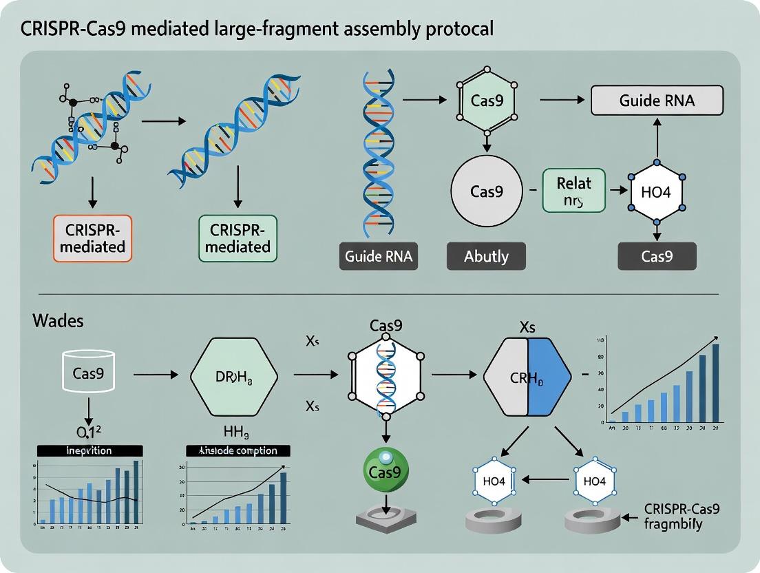
Mastering Large-Fragment DNA Assembly: A Comprehensive CRISPR-Cas9 Protocol for Synthetic Biology and Therapeutic Development
This article provides a detailed, step-by-step protocol for assembling large DNA fragments (10-100+ kb) using CRISPR-Cas9-mediated homology-directed repair.
Mastering Large-Fragment DNA Assembly: A Comprehensive CRISPR-Cas9 Protocol for Synthetic Biology and Therapeutic Development
Abstract
This article provides a detailed, step-by-step protocol for assembling large DNA fragments (10-100+ kb) using CRISPR-Cas9-mediated homology-directed repair. Tailored for researchers and drug development professionals, it covers foundational principles, a robust methodological workflow, critical troubleshooting and optimization strategies, and comprehensive validation approaches. By integrating the latest advancements in precision genome engineering, this guide enables the reliable construction of complex genetic circuits, synthetic pathways, and therapeutic gene cassettes, accelerating research in synthetic biology, gene therapy, and biomanufacturing.
Understanding CRISPR-Cas9 for Large DNA Assembly: Principles, Advantages, and Design Rules
Why Large-Fragment Assembly? Key Applications in Synthetic Biology and Therapeutics.
This document presents Application Notes and Protocols developed within a broader thesis research project focused on optimizing CRISPR-Cas9 mediated large-fragment assembly (LFA). The central hypothesis of the thesis is that CRISPR-Cas9, beyond its gene-editing applications, provides a highly precise and efficient mechanism for the in vivo assembly of large DNA constructs (>10 kb), overcoming key limitations of in vitro methods like Gibson Assembly or Golden Gate. This protocol is designed for researchers in synthetic biology and therapeutic development who require robust, scalable methods for constructing complex genetic systems.
Application Notes
Large-fragment assembly is critical for engineering biological systems that require extensive genetic reprogramming. The table below summarizes key quantitative benchmarks and applications.
Table 1: Applications and Benchmarks of Large-Fragment Assembly
| Application Domain | Typical Fragment Size | Key Challenge Addressed | Therapeutic/Synthetic Biology Example |
|---|---|---|---|
| Biosynthetic Pathway Engineering | 20 - 100+ kb | Reconstituting multi-gene pathways from heterologous sources | Assembly of polyketide synthase (PKS) or non-ribosomal peptide synthetase (NRPS) clusters for novel antibiotic production. |
| Genome-Scale Editing | 10 - 50 kb | Inserting large transgenes or multiple gene cassettes | Knock-in of synthetic cytokine gene circuits into safe-harbor loci (e.g., AAVS1) for CAR-T cell therapy enhancement. |
| Synthetic Chromosome/Vector Construction | 50 - 500+ kb | Building minimal genomes or large episomal vectors | De novo assembly of synthetic yeast chromosomes (Sc 2.0 project) or mammalian artificial chromosomes (MACs) for gene therapy. |
| Viral Vector Engineering | 5 - 15 kb (payload) | Packaging large or multiple transgenes into viral capsids | Assembly of complete "gutless" adenovirus or lentivirus genomes carrying multiple tumor-suppressor genes and reporters. |
| Metabolic Engineering | 10 - 30 kb | Stacking multiple enzyme genes and regulatory elements | Constructing an entire heterologous biofuel production pathway (e.g., isoprenoid pathway) in a microbial chassis. |
Core Protocol: CRISPR-Cas9 MediatedIn VivoAssembly
This detailed protocol describes the assembly of two large linear DNA fragments (Fragment A and Fragment B) into a circular plasmid in vivo using homology-directed repair (HDR) triggered by double-strand breaks (DSBs) created by CRISPR-Cas9.
Materials & Reagent Solutions
Table 2: Research Reagent Solutions for CRISPR-Cas9 LFA
| Reagent / Material | Function / Description | Example Product/Catalog |
|---|---|---|
| CRISPR-Cas9 Expression Plasmid | Expresses S. pyogenes Cas9 and a single-guide RNA (sgRNA). Targets and cleaves the recipient vector backbone. | Addgene #62988 (pX330-U6-Chimeric_BB-CBh-hSpCas9) |
| Linear DNA Fragments (A & B) | Large fragments to be assembled. Must contain >500 bp homology arms to each other and to the cut site on the backbone. | PCR-amplified or enzymatically excised, gel-purified. |
| Recipient/Backbone Vector | Circular plasmid that will be linearized by Cas9, providing the scaffold for fragment assembly. Contains the sgRNA target sequence. | Standard high-copy cloning vector (e.g., pUC19 derivative). |
| Competent Cells | E. coli or yeast strains with high transformation efficiency and robust HDR machinery. | E. coli HST08 Stbl3 (for unstable constructs), S. cerevisiae (for yeast-based assembly). |
| Homology-Directed Repair (HDR) Enhancers | Small molecules that inhibit NHEJ or stimulate HDR pathways in host cells. | RecA protein (for E. coli), RS-1 (for mammalian cells), nuclease inhibitors. |
| Selection & Screening Media | Antibiotics and/or chromogenic/fluorescent reporters to identify correct assemblies. | LB + Ampicillin (100 µg/mL), X-Gal/IPTG for blue-white screening. |
Detailed Experimental Methodology
Day 1: Preparation of DNA Components
- Design sgRNA: Design a 20-nt sgRNA sequence targeting a unique site on the recipient vector backbone, ideally between the homology arm regions. Use design tools (e.g., CRISPick, CHOPCHOP).
- Prepare Fragments: Generate Fragment A and B via long-range PCR or restriction digest. Include 500-800 bp homology overlaps at their junctions and to the Cas9 cut site on the backbone. Purify fragments using gel electrophoresis and a gel extraction kit.
- Prepare Backbone: Clone the sgRNA target sequence into the recipient vector if not present. Verify by sequencing.
- Prepare CRISPR Plasmid: Clone the designed sgRNA sequence into the CRISPR-Cas9 expression plasmid.
Day 2: Co-Transformation and In Vivo Assembly
- Transformation Mix: On ice, combine in a 1.5 mL tube:
- 50 µL of high-efficiency competent cells.
- 50 ng CRISPR-Cas9 expression plasmid.
- 100 ng linearized recipient backbone vector.
- 100 ng of Fragment A.
- 100 ng of Fragment B.
- (Optional) 1 µL of 10 mM RS-1 (for E. coli).
- Heat Shock: Incubate on ice for 30 min, heat shock at 42°C for 45 sec (for E. coli), and return to ice for 2 min.
- Recovery: Add 950 µL of SOC medium and incubate at 37°C for 90 minutes with shaking (220 rpm).
Day 3: Selection and Screening
- Plate Cells: Plate 100-200 µL of the recovery culture on pre-warmed agar plates containing the appropriate antibiotic (selecting for the assembled plasmid).
- Incubate: Incubate plates overnight at 37°C (30°C for unstable constructs).
- Screen Colonies: Pick 10-20 colonies. Screen by colony PCR using primers flanking the assembly junctions. For large assemblies (>15 kb), perform restriction digest analysis.
Day 4+: Validation
- Sequence Verification: Purify plasmid DNA from positive clones and subject to long-read sequencing (e.g., Oxford Nanopore, PacBio) to confirm the integrity of the entire assembled large fragment.
- Functional Validation: Depending on the application, perform functional assays (e.g., enzyme activity, protein expression, pathway output).
Visualized Workflows and Pathways
CRISPR-Cas9 LFA Experimental Workflow
Molecular Mechanism of Cas9-Mediated In Vivo Assembly
This application note details protocols for CRISPR-Cas9-mediated large-fragment assembly, a cornerstone methodology for advanced genome engineering. Moving beyond simple gene knockout, these techniques leverage the precision of Cas9-induced double-strand breaks (DSBs) to direct the integration of multi-kilobase DNA sequences via homologous recombination (HR). This work supports a broader thesis on optimizing fidelity and efficiency in complex genomic edits for therapeutic and synthetic biology applications.
Table 1: Comparison of CRISPR-Cas9 Mediated Assembly Methods
| Method | Key Feature | Typical Insert Size | Efficiency Range* | Primary Repair Pathway |
|---|---|---|---|---|
| HR with ssODN Donors | Short homology arms (30-60 nt) | < 200 bp | 0.1% - 10% | Homology-Directed Repair (HDR) |
| dsDNA with Long Homology Arms | Plasmid or PCR fragment donors | 200 bp - 10+ kbp | 0.01% - 5% | Homology-Directed Repair (HDR) |
| CRISPR-Cas9 Assisted HDR (CA-HDR) | Concurrent Cas9 cleavage of donor & target | 1 - 5 kbp | 5% - 30% | Homology-Directed Repair (HDR) |
| Non-Homologous End Joining (NHEJ)-Mediated Ligation | Microhomology-independent | 1 - 3 kbp | 1% - 20% | Non-Homologous End Joining (NHEJ) |
*Efficiency is highly cell-type and locus dependent. *Refers to relative increase over standard HDR in difficult-to-edit cells.*
Table 2: Optimized Reagent Concentrations for Mammalian Cell Transfection (HEK293T)
| Reagent | Final Concentration (nM) | Purpose | Notes |
|---|---|---|---|
| SpCas9 mRNA or Protein | 50-100 nM | Creates targeted DSB | Protein gives faster kinetics, lower off-target. |
| sgRNA | 50-100 nM | Guides Cas9 to target locus | Chemically modified for stability. |
| dsDNA Donor Template | 50-200 nM | Provides repair template | Linearized, homology arms 500-800 bp. |
| NHEJ Inhibitor (e.g., SCR7) | 5-10 µM | Suppresses NHEJ, enriches HDR | Add 1-2 hours before transfection. |
| HDR Enhancer (e.g., RS-1) | 5-10 µM | Stimulates Rad51, promotes HR | Titrate carefully; can be cytotoxic. |
Detailed Protocols
Protocol 1: CA-HDR for Large Fragment Integration in Mammalian Cells
Objective: Integrate a 3-kb expression cassette into a defined genomic locus. Workflow:
- Design & Preparation:
- Design sgRNAs targeting the genomic locus and a linearized donor plasmid (outside homology arms).
- Prepare donor DNA with 500-800 bp homology arms flanking the insert. Linearize via PCR or restriction digest.
- Synthesize high-quality sgRNA and SpCas9 protein.
- Cell Preparation & Transfection (HEK293T):
- Seed 2e5 cells/well in a 24-well plate 24h prior.
- Solution A: Mix 50nL SpCas9 protein (50nM final) + 50ng sgRNA. Incubate 10min at 25°C for RNP complexing.
- Solution B: Mix 200ng linear donor DNA + 1µL HDR enhancer (RS-1, 7.5µM final).
- Combine Solutions A & B with 2µL lipofectamine-based transfection reagent in 50µL Opti-MEM. Incubate 15min.
- Add complex dropwise to cells with fresh medium.
- Post-Transfection & Analysis:
- Add NHEJ inhibitor (SCR7, 5µM final) at 2h post-transfection.
- At 48-72h, harvest cells for genomic DNA extraction.
- Validate integration via junctional PCR (primers inside insert + outside homology arm) and Sanger sequencing.
Protocol 2: NHEJ-Mediated dsDNA Fragment Assembly in Vitro
Objective: Ligate multiple DNA fragments in a one-pot reaction using Cas9. Workflow:
- Fragment Generation:
- Design overlapping sgRNAs such that Cas9 digestion of each PCR-amplified fragment creates complementary, 5-10 bp overhangs.
- Generate fragments via PCR with primers adding the sgRNA target sites at termini.
- One-Pot Digestion/Ligation:
- Assemble reaction: 100ng each DNA fragment, 50nM SpCas9 protein, 50nM each sgRNA, 1X T4 DNA Ligase Buffer, 2000U T4 DNA Ligase, in 50µL.
- Incubate: 37°C for 60min (digestion), then 25°C for 60min (ligation), then 80°C for 10min (inactivation).
- Product Recovery:
- Purify DNA using a spin column.
- Transform 5µL into competent E. coli. Screen colonies by colony PCR and restriction digest for correct assembly.
Visualizations
Title: CRISPR-Cas9 Mediated DSB Repair Pathways for Gene Editing
Title: CA-HDR Protocol Workflow for Large Fragment Integration
The Scientist's Toolkit: Research Reagent Solutions
Table 3: Essential Reagents for CRISPR-Cas9 Precision Assembly
| Reagent / Solution | Function & Role in Protocol | Example Product / Note |
|---|---|---|
| High-Fidelity Cas9 Nuclease | Creates clean, specific DSBs. Protein form allows rapid RNP delivery. | Alt-R S.p. Cas9 Nuclease V3 (IDT), TruCut Cas9 Protein (Thermo). |
| Chemically Modified sgRNA | Increases stability and reduces immune response in cells. Essential for high efficiency. | Alt-R CRISPR-Cas9 sgRNA (IDT), Synthego sgRNA EZ Kit. |
| Long-Fragment DNA Donor Template | Provides homology-directed repair template. Must be high-purity and linear. | PCR-amplified using Q5 High-Fidelity DNA Polymerase (NEB). |
| HDR Enhancer (Small Molecule) | Temporarily inhibits NHEJ or stimulates Rad51 to shift repair balance toward HDR. | RS-1 (Rad51 stimulator), SCR7 pyrazine (NHEJ inhibitor). |
| Electroporation/Lipofection Reagent | For efficient co-delivery of large RNP complexes and donor DNA into cells. | Neon Transfection System (Thermo), Lipofectamine CRISPRMAX (Thermo). |
| NHEJ Reporter Cell Line | Enables rapid, quantitative assessment of HDR vs. NHEJ activity for protocol optimization. | U2OS DR-GFP (HDR) / EJ5-GFP (NHEJ) reporter lines. |
| Next-Gen Sequencing Analysis Service | For unbiased, genome-wide assessment of on-target efficiency and off-target effects. | Illumina-based amplicon sequencing with tools like CRISPResso2. |
Within the context of CRISPR-Cas9 mediated large-fragment assembly protocol research, Homology-Directed Repair (HDR) is the primary cellular engine for achieving seamless, scarless integration of DNA fragments. This application note details the protocols and considerations for leveraging HDR in advanced genome engineering workflows, moving beyond simple knockouts to sophisticated knock-ins and multi-kilobase assemblies.
Table 1: Comparison of HDR Efficiency Factors
| Factor | Typical Range/Value | Impact on HDR Efficiency |
|---|---|---|
| Homology Arm Length | 30-1000 bp (linear) / 800-2000 bp (ssODN) | Longer arms (>800 bp) significantly increase efficiency for large fragments. |
| Donor DNA Form | dsDNA (plasmid, PCR), ssODN | ssODNs optimal for <200 bp; linear dsDNA donors superior for large fragments. |
| Cell Cycle Phase | S/G2 Phase | HDR is 5-10x more efficient in S/G2 vs. G1 phase. |
| NHEJ Inhibition (e.g., SCR7, NU7024) | 5-20 µM | Can enhance HDR:NHEJ ratio by 2-5 fold. |
| Cas9 Delivery Method | RNP, Plasmid, mRNA | RNP delivery often yields highest HDR efficiency with reduced toxicity. |
| Template Concentration | 10-200 nM (ssODN), 1-50 µg (plasmid) | High concentration critical, but can be cytotoxic. |
Table 2: Common HDR Donor Templates
| Template Type | Optimal Insert Size | Key Advantages | Key Limitations |
|---|---|---|---|
| Single-Stranded Oligodeoxynucleotides (ssODNs) | < 200 bp | High efficiency, low toxicity, easy synthesis. | Limited cargo capacity. |
| PCR-amplified Linear dsDNA | 200 bp - 5 kb | Flexible, no bacterial cloning, good efficiency. | Prone to degradation, may require purification. |
| Plasmid DNA | > 1 kb | Stable, high yield, can include selection markers. | Low efficiency, risk of random integration. |
| Viral Vectors (AAV) | < 4.7 kb | Very high transduction efficiency. | Complex production, size constraint. |
| rAAV-based Donors | < 5 kb | High HDR rates in dividing & non-dividing cells. | Production complexity, immunogenicity concerns. |
Experimental Protocols
Protocol 1: HDR-Mediated Large-Fragment Knock-in Using Linear dsDNA Donor
This protocol is designed for inserting fragments from 0.5 to 5 kb into a mammalian genome using Cas9 RNP and a PCR-generated donor.
Materials:
- Cas9 protein and sgRNA (or synthetic crRNA/tracrRNA)
- Target-specific sgRNA (designed with minimal off-targets)
- Donor DNA template (PCR-amplified with ≥500 bp homology arms)
- Electroporation buffer or transfection reagent (e.g., Neon Buffer, Lipofectamine CRISPRMax)
- Cultured mammalian cells (e.g., HEK293T, iPSCs, RPE1)
- NHEJ inhibitor (optional, e.g., 5 µM SCR7)
Procedure:
- Donor Template Preparation:
- Design primers to amplify your insert flanked by homology arms (800-1000 bp each). Include a PAM-disrupting mutation in the homology arm to prevent re-cleavage.
- Perform high-fidelity PCR. Purify the product using a silica-column or gel extraction kit to remove primer dimers and template DNA. Elute in nuclease-free water or TE buffer. Quantify via spectrophotometry.
RNP Complex Assembly:
- For one reaction, complex 30 pmol of Cas9 protein with 36 pmol of sgRNA (or equimolar crRNA:tracrRNA duplex) in nuclease-free duplex buffer.
- Incubate at room temperature for 10-20 minutes to form active RNP.
Cell Preparation and Transfection/Electroporation:
- Harvest cells in log growth phase. For electroporation (e.g., Neon System), resuspend 1e5 - 1e6 cells in Buffer R with RNP complex and 1-2 µg of purified donor DNA. Electroporate using appropriate settings (e.g., 1400V, 20ms, 1 pulse for HEK293T).
- For lipid-based transfection, follow manufacturer's guidelines for CRISPR RNP delivery, adding donor DNA simultaneously.
Post-Transfection Culture:
- Seed transfected cells into pre-warmed culture media. If using, add NHEJ inhibitor 1-2 hours post-transfection.
- Culture cells for 48-72 hours to allow for repair and expression.
Analysis:
- Harvest genomic DNA. Screen using junction PCR (primers outside homology arm and within insert) followed by Sanger sequencing.
- For clonal isolation, single-cell sort or dilute into 96-well plates 3-5 days post-transfection. Expand and validate clones via PCR and sequencing.
Protocol 2: Enhancing HDR Efficiency via Cell Cycle Synchronization
HDR is most active in S and G2 phases. Synchronizing cells can boost knock-in rates.
Procedure:
- Synchronization (Double Thymidine Block):
- Treat cells with 2 mM thymidine for 18 hours.
- Wash cells 3x with PBS and release into fresh medium for 9 hours.
- Treat again with 2 mM thymidine for 17 hours.
Release and Transfection:
- Wash cells thoroughly and release into complete medium. Cells will now be largely synchronized at the G1/S border.
- Perform the RNP + donor transfection/electroporation (as in Protocol 1) 3-5 hours post-release, when most cells are progressing through S phase.
Continue Culture and Analysis as in Protocol 1, Step 4 & 5.
Visualization: HDR Pathway and Workflow
Diagram 1: HDR Molecular Pathway & NHEJ Competition.
Diagram 2: HDR-Mediated Large-Fragment Knock-in Workflow.
The Scientist's Toolkit: Research Reagent Solutions
Table 3: Essential Reagents for HDR Experiments
| Reagent / Solution | Function / Purpose | Example / Notes |
|---|---|---|
| High-Fidelity DNA Polymerase | Amplifies donor template with ultra-low error rates. | Q5 (NEB), KAPA HiFi, PrimeSTAR GXL. Critical for long homology arm integrity. |
| Cas9 Nuclease (WT) | Generates the target double-strand break (DSB). | Recombinant Cas9 protein (IDT, Thermo). RNP format offers fast action and reduced off-targets. |
| Synthetic sgRNA or crRNA:tracrRNA | Guides Cas9 to the specific genomic locus. | Chemically modified (e.g., Alt-R CRISPR-Cas9 sgRNA) for enhanced stability and reduced immunogenicity. |
| NHEJ Inhibitors | Temporarily suppresses the error-prone NHEJ pathway to favor HDR. | SCR7, NU7024, NU7441. Use with caution due to potential cytotoxicity. |
| Cell Synchronization Agents | Enriches cell population in S/G2 phase where HDR is active. | Thymidine, Aphidicolin, RO-3306 (CDK1 inhibitor). |
| Electroporation System & Buffer | Enables high-efficiency co-delivery of RNP and donor DNA, especially in hard-to-transfect cells. | Neon (Thermo), Nucleofector (Lonza) systems. Buffer R, SE Cell Line Kit. |
| Lipid-based Transfection Reagent (CRISPR-optimized) | Alternative non-viral delivery method for RNP and donor. | Lipofectamine CRISPRMax, RNAiMAX. |
| Single-Cell Cloning Supplement | Enhances survival of single cells after sorting/dilution to isolate clones. | CloneR (Stemcell), RevitaCell (Thermo). |
| HDR Donor Constructs | Provides template for repair. Can be supplied as ultramer ssODNs or linearized plasmid. | GeneArt Precision gRNA Synthesis Kit, custom dsDNA fragments (IDT, Twist). |
This application note is framed within the context of a broader thesis on CRISPR-Cas9 mediated large-fragment assembly protocol research. It provides a comparative analysis of key DNA assembly methodologies—CRISPR-Cas9, Gibson Assembly, Golden Gate Assembly, and Yeast Assembly—with a focus on their principles, quantitative performance metrics, and detailed protocols for implementation in research and drug development.
The following table summarizes the core characteristics and performance data of the four assembly technologies.
Table 1: Comparative Summary of DNA Assembly Technologies
| Feature | CRISPR-Cas9 Assembly | Gibson Assembly | Golden Gate Assembly | Yeast Assembly (TAR) |
|---|---|---|---|---|
| Core Principle | Homology-directed repair (HDR) triggered by Cas9-induced double-strand breaks. | In vitro one-pot isothermal assembly using 5' exonuclease, polymerase, and ligase. | In vitro, type IIS restriction enzyme-based, scarless assembly of multiple fragments. | In vivo homologous recombination in Saccharomyces cerevisiae. |
| Typical Assembly Size | Up to 10-100 kb (limited by delivery). | 2-6 fragments, up to ~20 kb. | 4-10+ fragments in a single reaction, modular. | Very large constructs (100 kb - 2 Mb). |
| Assembly Time (Hands-on) | High (requires cloning of guide RNAs, often requires selection). | Low (~2 hours in vitro reaction). | Low (1-2 hour digestion/ligation). | High (requires yeast transformation and culture, days). |
| Throughput | Low to medium. | High (standardized fragments). | Very High (modular, hierarchical). | Low (for large, complex assemblies). |
| Scarlessness | Scarless if using HDR with perfect repair. | Can be scarless if designed appropriately. | Scarless by design using type IIS sites. | Scarless via homologous recombination. |
| Fidelity | Medium (can have HDR errors, NHEJ). | High (commercial master mix). | Very High (digestion is irreversible). | Medium (yeast can rearrange DNA). |
| Primary Application | Genome editing, targeted insertion of large fragments. | Cloning, pathway assembly, mutagenesis. | Modular cloning (MoClo), combinatorial libraries, synthetic biology. | Assembly of whole pathways, chromosomes, or entire genomes. |
| Typical Cost per Reaction | High (Cas9, guides, repair templates). | Medium. | Low to Medium. | Low (yeast culture media). |
Detailed Experimental Protocols
CRISPR-Cas9 Mediated Large-Fragment Assembly Protocol
This protocol is central to the thesis research, detailing the replacement of a genomic locus with a large donor DNA fragment.
A. Materials (Research Reagent Solutions):
- sgRNA Expression Plasmid: Encodes target-specific guide RNA (e.g., pX330 derivative).
- Donor DNA Template: Linear or circular DNA containing the insert flanked by >800 bp homology arms.
- Cas9 Source: Expressed from the sgRNA plasmid or as purified protein.
- Cells: Adherent or suspension mammalian cell line with good HDR efficiency (e.g., HEK293T).
- Transfection Reagent: PEI or commercial lipid-based transfection kit.
- Selection Antibiotics/Puromycin: If a selection marker is included in the donor.
- Lysis Buffer & PCR Reagents: For genotyping.
- Surveyor or T7E1 Nuclease: For initial cleavage efficiency check.
B. Protocol Steps:
- Design & Cloning:
- Design two sgRNAs targeting the 5' and 3' boundaries of the genomic region to be replaced.
- Clone sgRNA sequences into the expression vector.
- Prepare the donor construct with homology arms. Purify as high-quality linear fragment or supercoiled plasmid.
Cell Transfection:
- Seed cells in a 6-well plate to reach 70-80% confluency at transfection.
- For each well, mix 1 µg of sgRNA/Cas9 plasmid and 1-2 µg of donor DNA in 250 µL of serum-free medium.
- Add 6 µL of PEI reagent (1 mg/mL), vortex, and incubate 15 min at RT.
- Add the mixture dropwise to cells. Change media after 6-8 hours.
Selection & Screening:
- 48-72 hours post-transfection, begin antibiotic selection if applicable. Maintain selection for 5-7 days.
- For puromycin selection, use 1-3 µg/mL (concentration must be pre-determined for the cell line).
Genotype Analysis:
- Harvest a portion of cells 72 hrs post-transfection (pre-selection) to check cutting efficiency via T7E1 assay.
- After selection, isolate genomic DNA from pooled populations or single-cell clones.
- Perform PCR using primers outside the homology arms and inside the inserted donor. Confirm correct integration by Sanger sequencing of PCR products.
- For large insertions (>1 kb), use long-range PCR or Southern blot for validation.
Gibson Assembly Protocol
A. Materials:
- Gibson Assembly Master Mix (Commercial): Contains T5 exonuclease, Phusion polymerase, and Taq DNA ligase.
- DNA Fragments: PCR-amplified or digested with 15-40 bp overlapping ends.
- Competent E. coli: High-efficiency DH5α or similar.
B. Protocol:
- Fragment Preparation: Generate inserts and vector with designed overlaps. Gel-purify fragments.
- Assembly Reaction: In a 10-20 µL total volume, mix vector and insert(s) at a molar ratio of 1:3 (vector:insert). Use 0.02-0.5 pmols of vector DNA. Add an equal volume of 2X Gibson Master Mix. Incubate at 50°C for 15-60 minutes.
- Transformation: Transform 2-5 µL of the reaction into 50 µL of competent cells. Plate on selective media and incubate overnight.
Golden Gate Assembly Protocol
A. Materials:
- Type IIS Restriction Enzyme (e.g., BsaI-HFv2, Esp3I): Cleaves outside its recognition site.
- T4 DNA Ligase: With suitable buffer.
- DNA Parts ("Modules"): Flanked by defined, non-palindromic 4-bp overhangs.
B. Protocol:
- Reaction Setup: In a single tube, combine 50-100 ng of destination vector, equimolar amounts of each insert part, 10 U of BsaI-HFv2, 400 U of T4 DNA Ligase, and 1X T4 Ligase Buffer.
- Thermocycling: Run the following program: (37°C for 2-5 min + 16°C for 5 min) x 25-30 cycles, then 50°C for 5 min, 80°C for 10 min.
- Transformation: Transform 2 µL directly into competent E. coli.
Yeast Assembly (Transformation-Associated Recombination - TAR) Protocol
A. Materials:
- Yeast Strain: S. cerevisiae with high recombination efficiency (e.g., VL6-48N).
- Linearized Vector Backbone: Containing yeast origin, marker, and terminal homology to fragments.
- Co-transformed DNA Fragments: With 40-60 bp overlaps.
- LiAc Transformation Mix: 1X TE, 1X LiAc, PEG 3350, single-stranded carrier DNA.
B. Protocol:
- Yeast Culture: Grow yeast to mid-log phase (OD600 ~0.5-0.8).
- Transformation: Mix 100-200 ng of linearized vector and equimolar amounts of each overlapping fragment with 50 µL of competent yeast cells and 500 µL of LiAc/PEG mix. Heat shock at 42°C for 20-40 minutes.
- Plating & Screening: Plate on synthetic drop-out media lacking the appropriate nutrient to select for the marker. Incubate at 30°C for 2-3 days. Screen colonies by yeast colony PCR.
Visualized Workflows and Pathways
Title: CRISPR-Cas9 Large-Fragment Assembly Workflow
Title: Logical Comparison of Assembly Method Principles
The Scientist's Toolkit: Key Research Reagent Solutions
Table 2: Essential Reagents for Featured Assembly Experiments
| Reagent/Material | Function in Experiment | Example/Note |
|---|---|---|
| Cas9 Nuclease (WT) | Creates targeted double-strand break (DSB) to initiate homology-directed repair (HDR). | Can be delivered as plasmid, mRNA, or ribonucleoprotein (RNP) complex. RNP offers rapid action and reduced off-targets. |
| sgRNA Expression Construct | Guides Cas9 to the specific genomic locus for cleavage. | Requires careful design to minimize off-target effects. Can be chemically synthesized as crRNA:tracrRNA duplex. |
| Homology-directed Repair (HDR) Donor Template | Provides the template for precise insertion of the large fragment. Can be single-stranded oligo (ssODN) or double-stranded (dsDNA). | For large fragments (>1 kb), dsDNA with long homology arms (>800 bp) is critical. Often includes a selectable marker. |
| Gibson Assembly Master Mix | All-in-one enzymatic mix for seamless, in vitro assembly of multiple overlapping fragments. | Commercial mixes (e.g., from NEB) offer high efficiency and reproducibility for 2-6 fragment assemblies. |
| Type IIS Restriction Enzyme (e.g., BsaI) | Enzyme core to Golden Gate Assembly. Cleaves outside its recognition site to generate unique, non-palindromic overhangs. | High-fidelity (HF) versions minimize star activity, enabling more complex, multi-fragment assemblies. |
| T4 DNA Ligase | Joins DNA fragments with compatible cohesive ends. Used in Golden Gate and standard cloning. | Requires ATP. Used in the same buffer with Type IIS enzymes during Golden Gate cycling. |
| Competent S. cerevisiae Cells | The host for Yeast Assembly, providing highly efficient endogenous homologous recombination machinery. | Specific strains like VL6-48N are auxotrophic for multiple markers, allowing selection for assembled constructs. |
| Polyethyleneimine (PEI) Max | A cost-effective cationic polymer for transient co-transfection of plasmid DNA into mammalian cells. | Optimal ratio of PEI:DNA must be determined for each cell line to balance efficiency and toxicity. |
| Puromycin Dihydrochloride | A commonly used selection antibiotic for mammalian cells. Kills non-transfected cells within 1-3 days. | Effective concentration (typically 1-5 µg/mL) must be titrated for each cell line prior to the experiment. |
| Surveyor or T7 Endonuclease I | Mismatch-specific nucleases used to detect and quantify Cas9-induced indel mutations at the target site. | Provides an initial, rapid assessment of genome editing efficiency prior to HDR screening. |
Within the broader thesis on CRISPR-Cas9 mediated large-fragment assembly protocols, the success of homology-directed repair (HDR) is critically dependent on rational pre-design. This application note details the quantitative relationships and protocols governing three fundamental parameters: the size of the donor DNA fragment, the length of homology arms (HAs), and the GC content of these homologous regions. Optimization of these factors is essential for achieving high-efficiency, precise assembly of large genomic constructs, a cornerstone technology for advanced therapeutic development.
Table 1: Optimal Ranges for Pre-Design Parameters in CRISPR-Cas9 HDR
| Parameter | Recommended Range | Typical Optimal Value | Key Rationale & Impact |
|---|---|---|---|
| Donor Fragment Size | < 5 kb for ssODNs; >5 kb for dsDNA donors (e.g., plasmids) | ssODN: 100-200 bp; dsDNA: 1-3 kb | Larger dsDNA donors are necessary for large insertions but show lower HDR efficiency. Electroporation efficiency drops significantly >5 kb. |
| Homology Arm Length | 30 - 1000 bp per arm | Plasmid donor: 500-800 bp; ssODN: 30-90 bp | Longer arms increase HDR efficiency for large fragments by stabilizing recombination. Shorter arms are sufficient for point mutations. |
| GC Content | 40% - 60% | ~50% | GC < 40% may impede stable annealing; GC > 60% can cause secondary structures, inhibiting recombination machinery. |
| Optimal Total Homology | 600 - 1600 bp (for dsDNA donors) | ~1000 bp | Provides a balance between recombination efficiency and practical donor synthesis/cloning constraints. |
Table 2: Impact of Parameter Deviation on HDR Outcomes
| Parameter | Deviation | Potential Experimental Consequence |
|---|---|---|
| Homology Arm Length | Too Short (< 200 bp for large fragments) | Drastic reduction in HDR efficiency (<1%); increased dominance of error-prone NHEJ. |
| Homology Arm Length | Excessively Long (> 1500 bp) | Diminishing returns on efficiency; increased difficulty in donor template preparation with no significant HDR gain. |
| GC Content | Too Low (< 30%) | Reduced thermal stability of donor-target heteroduplex, leading to poor recombination. |
| GC Content | Too High (> 70%) | Formation of stable secondary structures (e.g., hairpins) in donor DNA, blocking Rad51/RecA filament invasion. |
| Fragment Size | Very Large (> 5 kb) | Challenging delivery into cells; significantly reduced HDR efficiency even with long homology arms. |
Detailed Protocols
Protocol 1:In SilicoDesign and Analysis of Homology Arms
Objective: To design optimal homology arms for a given genomic locus and donor fragment.
Materials:
- Genomic sequence of target locus (from databases like ENSEMBL or UCSC Genome Browser).
- Sequence of the donor insert.
- Bioinformatics tools: Primer3, NEB Tm Calculator, IDT OligoAnalyzer, or SnapGene.
Procedure:
- Locus Identification: Extract a 2-3 kb genomic sequence flanking the intended Cas9 cut site.
- Arm Definition:
- For plasmid-based donors, select 500-800 bp sequences immediately 5’ and 3’ of the cut site.
- For ssODN donors, select 30-90 bp sequences.
- GC Content Analysis:
- Calculate the GC percentage for each arm using any sequence analysis tool.
- If GC content is outside 40-60%, consider slightly extending or trimming the arm to adjust the value. Avoid regions of extreme AT- or GC-richness.
- Secondary Structure Check:
- Input the full donor sequence (including HAs and insert) into a tool like OligoAnalyzer.
- Analyze for hairpins and self-dimers. A ΔG value more negative than -9 kcal/mol for secondary structures may be problematic.
- Repeat & Unique Sequence Check: Use BLAST against the host genome to ensure homology arms are unique and lack repetitive elements.
Protocol 2: Empirical Testing of HA Length via a Modular Donor System
Objective: Experimentally determine the minimal effective HA length for a specific large-fragment insertion.
Materials:
- Backbone plasmid with your gene of interest (GOI) but lacking HAs.
- PCR primers to amplify 200 bp, 500 bp, and 1000 bp homology arms from the target cell line's genomic DNA.
- Gibson Assembly or Golden Gate Assembly reagents.
- Cells (e.g., HEK293T, iPSCs), Cas9 RNP, and electroporation/nucleofection system.
Procedure:
- Modular Donor Construction:
- Amplify the three pairs of HAs (200, 500, 1000 bp) from genomic DNA.
- Perform separate assembly reactions to clone the same GOI with each HA pair into the backbone plasmid. Verify plasmids by sequencing.
- Co-Delivery and Editing:
- For each donor plasmid (200, 500, 1000 bp HAs), complex with a fixed amount of Cas9 RNP targeting the intended locus.
- Electroporate/nucleofect each complex into separate cell aliquots.
- Include a "Cas9-only" negative control.
- Analysis:
- Harvest cells 72 hours post-editing.
- Extract genomic DNA and perform PCR across the 5’ and 3’ junctions.
- Quantify HDR efficiency via next-generation sequencing (NGS) of amplicons or digital droplet PCR (ddPCR).
- Plot HDR efficiency (%) versus HA length to identify the point of diminishing returns.
The Scientist's Toolkit
Table 3: Essential Research Reagent Solutions for HDR Optimization
| Reagent / Material | Function & Rationale |
|---|---|
| High-Fidelity DNA Polymerase (e.g., Q5, Phusion) | Accurately amplifies long homology arms and donor fragments to prevent mutations. |
| Gibson Assembly Master Mix | Enables seamless, one-pot assembly of multiple DNA fragments (e.g., GOI + variable HAs) into a vector. |
| Cas9 Nuclease (WT) and sgRNA | Generates the target double-strand break (DSB) to initiate repair. Chemical modification of sgRNA enhances stability. |
| Recombinant Rad51 Protein | Can be added in vitro to stabilize ssDNA overhangs on donor templates, potentially boosting HDR rates for difficult loci. |
| HDR Enhancers (e.g., RS-1, SCR7) | Small molecules that inhibit NHEJ (SCR7) or stimulate Rad51 activity (RS-1), used during/post-electroporation to shift repair toward HDR. |
| Electrocompetent Cells (e.g., NEB Stable) | For high-efficiency transformation of large, complex donor plasmids during cloning stages. |
| Nucleofector System & Kit (e.g., Lonza 4D-Nucleofector) | Enables efficient co-delivery of bulky RNP and large donor DNA plasmids into difficult cell types (primary cells, stem cells). |
| ddPCR HDR Detection Kit | Provides absolute, sensitive quantification of precise knock-in events without the need for NGS. |
Visualizations
Diagram Title: Workflow for Optimizing HDR Pre-Design Parameters
Diagram Title: Interdependence of Key HDR Design Parameters
Step-by-Step Protocol: From sgRNA Design to Transformed Colonies
This protocol constitutes Stage 1 of a comprehensive thesis on CRISPR-Cas9 mediated large-fragment assembly. The efficiency of assembling multi-kilobase DNA constructs hinges on precise in silico design. This stage focuses on the computational selection of optimal sgRNA targets and the design of homology-directed repair (HDR) templates, forming the blueprint for subsequent molecular cloning and cellular engineering experiments.
Application Notes & Protocols
sgRNA Selection and Design Protocol
Objective: To identify and rank high-efficiency, specific sgRNAs for creating double-strand breaks (DSBs) at predefined genomic loci to facilitate large-fragment insertion.
Detailed Methodology:
- Define Target Loci: Identify the precise genomic coordinates (GRCh38/hg38) for the DSB. For large-fragment insertion, two sgRNAs are typically required: one for the 5' and one for the 3' end of the insertion site.
- Retrieve Sequence: Use the UCSC Genome Browser or ENSEMBL to extract a 500bp sequence flanking each target site.
- sgRNA Candidate Generation: Input sequences into prediction tools (see Table 1). The core 20-nt protospacer sequence must be immediately 5' of a Protospacer Adjacent Motif (PAM). For Streptococcus pyogenes Cas9 (SpCas9), the PAM is 5'-NGG-3'.
- Filter for Specificity:
- Perform a BLAST search against the relevant genome to ensure minimal off-target potential.
- Utilize tools to calculate off-target scores, discarding sgRNAs with significant hits (≤3 mismatches).
- Filter for Efficiency: Score remaining candidates using predictive algorithms. Select the top 2-3 candidates per target site for empirical validation.
- Final Selection: Choose the final sgRNA pair considering both high on-target efficiency and minimal off-target risk. Ensure the DSBs will produce compatible ends for the intended assembly.
Key Data Table: Comparison of sgRNA Design Tools Table 1: Features of prominent sgRNA design platforms.
| Tool Name | Key Algorithm/Scoring Method | Output Metrics | Best For | URL (as of 2024) |
|---|---|---|---|---|
| CRISPRscan | Model based on zebrafish data; considers nucleosome position | Efficiency score (0-100) | High on-target efficiency in vivo | crisprscan.org |
| CHOPCHOP | Multiple models (Doench ’16, Moreno-Mateos ’17), specificity check | Efficiency & specificity scores, off-target list | Balanced design & ease of use | chopchop.cbu.uib.no |
| CRISPick (Broad) | Rule Set 2 (Doench et al., 2016) | On-target & off-target scores | Standardized workflows & reproducibility | design.synthego.com |
| CRISPRdirect | Bowtie alignment for specificity | Specificity score, off-target list | Rapid specificity screening | crispr.dbcls.jp |
Homology Arm Construction and Vector Design Protocol
Objective: To design the HDR donor plasmid containing the large fragment of interest flanked by homology arms (HAs) complementary to the genomic target site.
Detailed Methodology:
- Determine Arm Length: Based on recent literature, optimal HA length balances recombination efficiency and cloning difficulty. For large fragments (>5 kb), longer arms are beneficial (see Table 2).
- Extract Arm Sequences: Using the genomic coordinates, extract the sequences for the left and right homology arms. The DSB site should be located within the sequence between the two arms.
- Sequence Optimization (Optional but Recommended):
- Avoid Repetitive Elements: Screen arms for simple repeats or common transposable elements using RepeatMasker.
- Prevent Re-cutting: Introduce silent mutations (synonymous codon changes) within the protospacer sequence in the donor template to prevent Cas9 from cleaving the newly integrated DNA.
- Vector Backbone Selection: Choose a high-copy plasmid backbone (e.g., pUC19-derived) with an appropriate bacterial selection marker (Ampicillin or Kanamycin resistance).
- In Silico Assembly:
- Use sequence editing software (e.g., SnapGene, Geneious) to assemble the final plasmid map in this order: 5' Homology Arm - Large Insert Fragment - 3' Homology Arm, all cloned into the prepared vector backbone.
- Include a selectable or screenable marker (e.g., Puromycin resistance, GFP) within the insert or between the arms for rapid enrichment of edited cells.
- Verification: Simulate diagnostic restriction digests and Sanger sequencing primer binding sites to ensure the final construct can be verified post-cloning.
Key Data Table: Homology Arm Length Guidelines Table 2: Recommended homology arm lengths based on experimental goals.
| Application Context | Recommended HA Length (each arm) | Rationale & Evidence |
|---|---|---|
| Standard Gene Insertion (<5 kb) | 800 - 1000 bp | Robust efficiency across cell lines; balances cloning ease and HDR rate. |
| Large Fragment Assembly (>5 kb) | 1000 - 2000 bp | Longer arms increase HDR efficiency for complex inserts by providing more sequence context for homologous recombination. |
| ssODN Donor Templates | 50 - 120 nt (total) | Short, single-stranded donors are effective for point mutations or small tags. |
| Primary or Hard-to-Transfect Cells | ≥1500 bp | Maximizes HDR efficiency in cell types with low recombination activity. |
The Scientist's Toolkit: Research Reagent Solutions
Table 3: Essential materials and tools for in silico design and vector preparation.
| Item | Category | Function & Rationale |
|---|---|---|
| SnapGene Software | Bioinformatics Tool | Visual plasmid design, restriction cloning simulation, and primer design. Critical for error-free in silico assembly. |
| Benchling Molecular Biology Suite | Bioinformatics Platform | Cloud-based design, shared team workflows, and direct integration with genomic databases. |
| NEB Builder HiFi DNA Assembly Master Mix | Cloning Reagent | High-fidelity enzyme mix for seamless assembly of multiple DNA fragments (e.g., HAs, insert, backbone) in a single reaction. |
| Gibson Assembly Master Mix | Cloning Reagent | Alternative one-step, isothermal assembly method for joining multiple overlapping DNA fragments. |
| Q5 High-Fidelity DNA Polymerase | PCR Reagent | PCR amplification of homology arms and insert fragments with ultra-low error rate to prevent mutations in HDR templates. |
| Genome-Compatible Plasmid Backbone | Vector | e.g., pUC19-based vectors; provides high-copy replication in E. coli for ample yield during plasmid preparation. |
Visualized Workflows
Diagram 1: sgRNA selection and filtering workflow.
Diagram 2: HDR donor plasmid construction workflow.
This protocol describes the generation of high-fidelity DNA fragments via PCR amplification, purification, and quality assessment, a critical stage within a broader research framework for CRISPR-Cas9 mediated large-fragment assembly. The assembly of large genetic constructs requires precisely defined, high-quality DNA fragments with overlapping homology or specific end sequences compatible with Cas9-assisted ligation. The integrity and purity of these initial building blocks directly determine the efficiency and success of subsequent assembly steps. This application note provides a standardized workflow and troubleshooting guide for researchers and drug development professionals.
Key Research Reagent Solutions
| Reagent / Material | Function & Rationale |
|---|---|
| High-Fidelity DNA Polymerase | Enzyme with proofreading activity (3'→5' exonuclease) to minimize PCR-induced errors, essential for maintaining sequence integrity in assembled constructs. |
| dNTP Mix | Deoxynucleotide triphosphate solution providing the building blocks for DNA synthesis. Must be of high purity and balanced concentration. |
| Template DNA | Plasmid, genomic DNA, or synthetic oligonucleotide serving as the source for the target fragment amplification. |
| Primers (Forward & Reverse) | Oligonucleotides designed with sequence-specific binding regions and necessary 5' extensions (e.g., homology arms, overhangs) for downstream assembly. |
| PCR Purification Kit | Silica-membrane based spin column system for rapid removal of enzymes, primers, dNTPs, and salts from amplification reactions. |
| Gel Extraction Kit | Kit for isolating DNA fragments from agarose gels, used to purify the specific product from non-specific amplifications or primer-dimer. |
| Quantitative Fluorometer & dsDNA Assay Kit | Instrument and dye-based assay (e.g., Qubit with HS dsDNA reagents) for accurate, specific quantification of double-stranded DNA concentration. |
| Bioanalyzer or TapeStation | Microfluidics-based capillary electrophoresis systems for precise sizing and quality assessment of DNA fragments (e.g., sizing, detecting degradation). |
Experimental Protocol: PCR Amplification
Primer Design Guidelines
- Specific Binding Region: 18-25 bp, Tm ~60°C.
- 5' Extensions: Add required sequences for assembly (e.g., 15-30 bp homology arms for Gibson Assembly, 4-bp overhangs for Golden Gate, or guide RNA targets for Cas9-mediated assembly).
- Final Primer Length: Typically 35-55 nucleotides.
- Purification: Use PAGE or HPLC purification for primers >40 bp.
PCR Reaction Setup
Perform reactions in a nuclease-free PCR tube or plate.
| Component | Volume (µL) - 50 µL Rxn | Final Concentration |
|---|---|---|
| Nuclease-Free Water | To 50 µL | - |
| 5X High-Fidelity Buffer | 10 µL | 1X |
| dNTP Mix (10 mM each) | 1 µL | 200 µM each |
| Forward Primer (10 µM) | 2.5 µL | 0.5 µM |
| Reverse Primer (10 µM) | 2.5 µL | 0.5 µM |
| Template DNA | Variable (e.g., 1-10 ng plasmid) | - |
| High-Fidelity DNA Polymerase | 0.5-1 µL | - |
| Total Volume | 50 µL | - |
Thermal Cycling Conditions
- Initial Denaturation: 98°C for 30 seconds.
- Cycling (30-35 cycles):
- Denaturation: 98°C for 10 seconds.
- Annealing: 60-72°C (based on primer Tm) for 15-30 seconds.
- Extension: 72°C for 15-30 seconds/kb of product length.
- Final Extension: 72°C for 2-5 minutes.
- Hold: 4°C.
Experimental Protocol: PCR Product Purification
Method A: Purification of a Single, Specific Band
- Analyze the entire PCR reaction on an agarose gel (0.8-1.2%).
- Excise the band of correct size under low-wavelength UV light, minimizing gel volume.
- Purify DNA using a Gel Extraction Kit following manufacturer's protocol. Elute in nuclease-free water or low-EDTA TE buffer.
Method B: Purification of a Clean PCR Product (No non-specific bands)
- Use a PCR Purification Kit directly on the reaction mixture.
- Perform all wash steps thoroughly to remove residual primers.
- Elute in 20-30 µL of nuclease-free water or provided elution buffer.
Experimental Protocol: Quality Assessment
Quantitative Analysis
- Use a fluorometric dsDNA assay (e.g., Qubit) for accurate concentration measurement. Record yield (ng/µL and total ng).
Qualitative Analysis
- Option 1: Run 1 µL of purified product on a high-resolution agarose gel (1-2%) alongside a DNA ladder.
- Option 2 (Recommended): Use a fragment analyzer system (e.g., Agilent Bioanalyzer, TapeStation, or Fragment Analyzer).
- Provides a digital electropherogram, precise sizing, and a DNA Integrity Number (DIN) or similar score.
- Detects contamination from primer-dimer, degradation, or RNA.
Data Presentation & Troubleshooting
Table 1: Expected Outcomes and Quality Metrics for Purified DNA Fragments
| Parameter | Acceptable Range | Assessment Method |
|---|---|---|
| Concentration | ≥ 10 ng/µL | Fluorometric assay |
| Total Yield | ≥ 500 ng | Fluorometric assay |
| Purity (A260/A280) | 1.8 - 2.0 | Spectrophotometry (note: less reliable for low concentration) |
| Size Accuracy | Within 5% of expected size | Agarose Gel / Fragment Analyzer |
| Degradation / Integrity | Single, sharp peak/band; DIN > 7.0 | Fragment Analyzer / Gel |
Table 2: Common PCR Amplification Issues and Solutions
| Problem | Potential Cause | Recommended Solution |
|---|---|---|
| No Amplification | Poor primer design, low template quality/quantity, incorrect Tm | Re-design primers, check template, run gradient PCR for optimal Tm. |
| Non-specific Bands | Primer-dimer, low annealing temperature, excess Mg2+ | Increase annealing temperature, use touchdown PCR, optimize Mg2+, switch to hot-start polymerase. |
| Low Yield | Too many cycles (polymerase exhaustion), short extension time | Reduce cycles, increase extension time, add DMSO (3-5%) for GC-rich templates. |
| Smeared Bands | Degraded template, excess template, contamination | Use fresh reagents, reduce template amount, perform purification in clean area. |
Title: PCR Fragment Generation & Quality Control Workflow
Title: Protocol Stage 2 in CRISPR-Cas9 Large-Fragment Assembly Thesis
This protocol, within the context of a thesis on CRISPR-Cas9 mediated large-fragment assembly, details the critical stage of co-delivering three key components: Cas9 endonuclease, sequence-specific single-guide RNA (sgRNA), and a donor DNA fragment. Efficient, simultaneous delivery is paramount for achieving high rates of homology-directed repair (HDR) and successful genomic integration of large DNA constructs. This application note compares current methodologies, provides quantitative data on efficiency and toxicity, and outlines optimized, detailed protocols for mammalian cell systems.
Comparative Analysis of Co-delivery Methods
Table 1: Quantitative Comparison of Primary Co-delivery Strategies
| Method | Typical Max. Donor Size (kb) | Avg. HDR Efficiency (%) (Reported Range) | Cytotoxicity (Relative) | Key Advantages | Key Limitations | Optimal Cell Type(s) |
|---|---|---|---|---|---|---|
| Lipid Nanoparticles (LNPs) | 10+ | 15-40 (5-60) | Low-Medium | High cargo capacity, low immunogenicity, clinically relevant. | Complex formulation, potential batch variability. | HEK293, HeLa, Primary cells. |
| Electroporation (Nucleofection) | 10+ | 10-30 (1-50) | Medium-High | High efficiency, works in hard-to-transfect cells. | High cell death, requires specialized equipment. | Immune cells (T-cells), iPSCs, cell lines. |
| Polyethylenimine (PEI) | 5-10 | 5-20 (1-30) | Medium | Simple, inexpensive, good for nucleic acids. | High toxicity at high doses, lower efficiency for large donors. | HEK293, adherent cell lines. |
| Viral Vectors (AAV) | ~4.7 | 1-10 (0.5-20) | Low | Extremely high transduction, stable expression. | Strict cargo size limit, immunogenicity concerns. | Neurons, in vivo models, primary cells. |
| Microinjection | 100+ | 20-60 (10-80) | Low (per cell) | Precise, no cargo size limit, direct to nucleus. | Low throughput, technically demanding. | Zygotes, oocytes. |
Table 2: Key Reagent Formats for Component Delivery
| Component | Common Formats for Delivery | Notes on Co-delivery Optimization |
|---|---|---|
| Cas9 | Plasmid DNA, mRNA, Ribonucleoprotein (RNP) complex | RNP offers fast action, reduced off-targets, and no risk of genomic integration of Cas9 sequence. |
| sgRNA | Plasmid DNA (U6 promoter), in vitro transcribed (IVT) RNA, synthetic crRNA+tracrRNA, pre-complexed RNP | Synthetic sgRNA or RNP complex reduces transcriptional load and accelerates editing. |
| Donor DNA | Plasmid (circular), PCR fragment, ssODN (for <200 bp), dsDNA with homology arms (linear) | Linear dsDNA with 500-1000 bp homology arms is optimal for large fragment HDR. Avoid plasmid backbone integration. |
Detailed Protocols
Protocol 3.1: Co-delivery via Lipid Nanoparticles (Formulated RNP + Donor)
This protocol uses pre-assembled Cas9 RNP and a linear dsDNA donor co-encapsulated in LNPs for high-efficiency, low-toxicity delivery.
Materials:
- Purified Cas9 protein
- Synthetic sgRNA (or crRNA+tracrRNA)
- Linear dsDNA donor fragment (gel-purified)
- Commercial LNP formulation kit (e.g., GenVoy-ILM, Lipofectamine CRISPRMAX Cas9 Transfection Reagent)
- Opti-MEM I Reduced Serum Medium
- Target cells (e.g., HEK293T) at 70-80% confluency
Procedure:
- RNP Complex Assembly: In a sterile tube, combine 5 µg of Cas9 protein and 2 µg of sgRNA in nuclease-free buffer. Incubate at room temperature for 10-20 minutes to form the RNP complex.
- Donor Preparation: Dilute 1-2 µg of linear dsDNA donor fragment in Opti-MEM to a total volume of 25 µL.
- LNP Formation: a. Dilute the recommended volume of lipid reagent in 25 µL of Opti-MEM in Tube A. b. Add the assembled RNP complex to the donor DNA in Tube B. Mix gently. c. Combine Tube A and Tube B. Mix by gentle pipetting. Do not vortex. d. Incubate the mixture at room temperature for 10-15 minutes to allow LNP formation.
- Cell Transfection: While complexes form, aspirate media from cells and replace with fresh, pre-warmed complete media. Add the 50 µL LNP mixture dropwise to the cells. Gently swirl the plate.
- Incubation and Analysis: Incubate cells at 37°C, 5% CO2. Analyze editing efficiency via flow cytometry, PCR, or sequencing 48-72 hours post-transfection.
Protocol 3.2: Co-delivery via Electroporation (Nucleofection)
This protocol is optimized for hard-to-transfect cells such as primary T cells or induced pluripotent stem cells (iPSCs).
Materials:
- Cas9 mRNA or RNP complex
- Synthetic sgRNA
- Linear dsDNA donor
- Nucleofector Device and appropriate Cell Line/ Primary Cell Nucleofector Kit
- Supplemented Nucleofection Solution
Procedure:
- Cell Preparation: Harvest and count cells. Centrifuge to pellet. For 1 reaction, resuspend 0.5-1 x 10^6 cells in 20 µL of pre-warmed Nucleofection Solution from the kit.
- Cargo Preparation: In a separate tube, combine Cas9 mRNA (2-5 µg) or RNP complex (2 µg Cas9 + 1 µg sgRNA), 1-2 µg of donor DNA, and optionally 0.5 µg of a fluorescent reporter plasmid to assess efficiency.
- Nucleofection: Add the cargo mixture to the cell suspension. Mix gently and transfer to a certified cuvette. Select the appropriate pre-optimized program on the Nucleofector device (e.g., for HEK293: CM-130; for T cells: EO-115).
- Recovery: Immediately after pulsing, add 500 µL of pre-warmed culture medium to the cuvette. Gently transfer the cell suspension to a pre-coated culture plate containing warm medium.
- Incubation and Analysis: Place plate in incubator. Medium can be changed 12-24 hours post-nucleofection. Analyze cells after 72-96 hours.
The Scientist's Toolkit: Key Research Reagent Solutions
Table 3: Essential Materials for Co-delivery Experiments
| Item | Function | Example/Supplier | Notes |
|---|---|---|---|
| High-Purity Cas9 Protein | Endonuclease component of the RNP complex. Essential for clean, efficient cleavage. | IDT Alt-R S.p. Cas9 Nuclease V3, Thermo Fisher TrueCut Cas9 Protein | Nuclease-free, carrier protein-free versions reduce cytotoxicity. |
| Chemically Modified Synthetic sgRNA | Guides Cas9 to the target genomic locus. Chemical modifications increase stability and reduce immunogenicity. | IDT Alt-R CRISPR-Cas9 sgRNA, Synthego sgRNA EZ Kit | 2'-O-methyl and phosphorothioate modifications are standard. |
| Homology-Directed Repair (HDR) Donor Template | Provides the correct template for repair after Cas9 cleavage. Can be ssODN, dsDNA fragment, or plasmid. | IDT Ultramer DNA Fragment, Gibson Assembly for plasmid donors | For large fragments (>1kb), use gel-purified linear dsDNA with long homology arms. |
| Transfection/Lipid Reagent | Forms complexes with nucleic acids/RNPs to facilitate cellular uptake. | Thermo Fisher Lipofectamine CRISPRMAX, Mirus Bio TransIT-X2 | CRISPRMAX is optimized for RNP delivery. |
| Electroporation/Nucleofection System | Applies electrical pulses to create transient pores in cell membranes for cargo entry. | Lonza 4D-Nucleofector X Unit, Bio-Rad Gene Pulser Xcell | Gold standard for hard-to-transfect and primary cells. |
| HDR Enhancers | Small molecules that transiently inhibit the NHEJ pathway or promote HDR. | SCR7, RS-1, NU7026, L755507 | Add at time of transfection. Toxicity and efficacy are cell-type specific; titrate carefully. |
Visualizations
Title: CRISPR Component Co-delivery Workflow
Title: DNA Repair Pathway Decision After Co-delivery
Within the broader thesis on CRISPR-Cas9 mediated large-fragment assembly, Stage 4 is critical for isolating and validating successful assemblies. Following transfection of the engineered constructs and CRISPR-Cas9 components, this stage involves the cultivation of transfected cells, application of selective pressure to enrich for correct assemblies, and implementation of screening strategies to identify clones harboring the intended large-fragment insertion or replacement.
Post-Transfection Culturing Protocol
Immediate Post-Transfection Handling
- Time Point: 24-48 hours post-transfection.
- Protocol:
- Recovery Period: Allow cells to recover in complete growth medium without selection for 24-48 hours to permit expression of resistance markers and genome editing.
- Cell Passaging: Gently passage cells at a lower density (e.g., 1:3 to 1:6 split) to prevent over-confluence.
- Medium Change: Replace medium 24 hours post-transfection to remove transfection reagents and cellular debris.
Initiation of Selection
- Time Point: 48 hours post-transfection.
- Protocol:
- Antibiotic Selection: Begin culturing cells in complete growth medium containing the appropriate selection antibiotic (e.g., Puromycin, G418, Hygromycin B). The concentration must be pre-determined via a kill curve.
- Density Maintenance: Maintain cells at sub-confluent densities (typically 50-70%) during the selection period, which can last 7-14 days.
- Medium Refreshment: Change selection medium every 2-3 days, observing for the death of non-transfected/unstable cells and the emergence of resistant foci.
Table 1: Common Selection Agents and Typical Working Concentrations for Mammalian Cells
| Selection Agent | Target / Mechanism | Typical Working Concentration Range | Time to Foci Appearance |
|---|---|---|---|
| Puromycin | Protein synthesis inhibitor (ribosome) | 1.0 - 5.0 µg/mL | 3-7 days |
| G418 (Geneticin) | Protein synthesis inhibitor (ribosome) | 200 - 800 µg/mL | 7-14 days |
| Hygromycin B | Protein synthesis inhibitor (ribosome) | 50 - 300 µg/mL | 7-14 days |
| Blasticidin | Protein synthesis inhibitor (peptide bond) | 2.0 - 10 µg/mL | 5-10 days |
Screening for Correct Assemblies
Following successful selection, a multi-tiered screening approach is necessary to identify clones with the correctly assembled large fragment.
Primary Screening: PCR-Based Genotyping
This rapid, high-throughput method screens for the presence of the insert and correct junction sequences.
- Protocol:
- Lysis: Harvest a portion of each resistant clone (~10^4 cells) into 20-50 µL of direct PCR lysis buffer with Proteinase K. Incubate at 56°C for 60 min, then 95°C for 10 min.
- Primer Design: Design three PCR reactions per clone:
- Insert Check: Forward primer in endogenous locus upstream of 5' homology arm, reverse primer within the inserted fragment.
- 5' Junction Check: Forward primer upstream of 5' homology arm, reverse primer just downstream of the Cas9-induced cut site within the insert.
- 3' Junction Check: Forward primer just upstream of the Cas9-induced cut site at the insert's 3' end, reverse primer downstream of 3' homology arm in the endogenous locus.
- PCR Amplification: Use a high-fidelity polymerase. Cycle conditions: 98°C for 30s; 35 cycles of (98°C for 10s, 65°C for 30s, 72°C for 1 min/kb); 72°C for 5 min.
- Analysis: Resolve products via agarose gel electrophoresis. Clones positive for all three reactions proceed to secondary screening.
Table 2: Primary Screening PCR Results Interpretation
| Clone Result (Insert / 5' Junction / 3' Junction) | Interpretation | Action |
|---|---|---|
| + / + / + | Positive for correct assembly. | Proceed to secondary screening. |
| + / - / + | Potential rearrangement at 5' junction. | Lower priority. Sequence if necessary. |
| + / + / - | Potential rearrangement at 3' junction. | Lower priority. Sequence if necessary. |
| - / - / - | No insert. False positive selection. | Discard. |
Secondary Screening: Southern Blot Analysis
Confirms correct integration, copy number, and absence of random integrations.
- Protocol (Key Steps):
- Genomic DNA Extraction: Isolate high-molecular-weight gDNA from candidate clones using a phenol-chloroform or column-based method.
- Restriction Digest: Digest 10-15 µg of gDNA with two different restriction enzymes that: a) cut once inside and once outside the insert to determine a diagnostic fragment length, and b) cut only outside the modified locus to assess copy number.
- Gel Electrophoresis & Transfer: Run on a 0.8% agarose gel, depurinate, denature, neutralize, and transfer via capillary action to a nylon membrane.
- Probe Labeling & Hybridization: Prepare a digoxigenin (DIG)-labeled probe targeting a region internal to the insert or specific to a junction. Hybridize overnight at 42°C in appropriate buffer.
- Detection: Perform stringency washes, incubate with anti-DIG-AP antibody, and develop using a chemiluminescent substrate. Compare band sizes to expected diagnostic fragments.
Tertiary Screening: Long-Range Sequencing
Definitively validates the sequence integrity of the entire assembled locus.
- Protocol: Utilize long-read sequencing platforms (e.g., Oxford Nanopore, PacBio). Design PCR primers >1 kb outside the modified locus to amplify the entire region. Prepare sequencing library from the amplicon and run on the platform. Align reads to the reference sequence to confirm perfect assembly.
The Scientist's Toolkit: Key Research Reagent Solutions
Table 3: Essential Materials for Post-Transfection Culturing and Screening
| Item | Function | Example Product/Type |
|---|---|---|
| Selection Antibiotics | Eliminates cells that did not stably integrate the resistance marker, enriching for transfected population. | Puromycin dihydrochloride, G418 sulfate. |
| Direct PCR Lysis Buffer | Enables rapid cell lysis and direct genotyping PCR without lengthy DNA purification, crucial for high-throughput primary screening. | QuickExtract DNA Extraction Solution, homemade buffer with Proteinase K and Triton X-100. |
| High-Fidelity DNA Polymerase | Provides accurate amplification of junction regions and large fragments for screening PCR with low error rates. | Phusion HF, Q5 High-Fidelity. |
| DIG DNA Labeling and Detection Kit | Enables non-radioactive, highly specific probe generation and detection for Southern blot confirmation of integration. | Roche DIG-High Prime DNA Labeling and Detection Starter Kit II. |
| Long-Range PCR Kit | Amplifies the entire modified genomic locus (potentially >10 kb) for tertiary validation via sequencing. | KAPA HiFi HotStart ReadyMix, PrimeSTAR GXL. |
| Nanopore Sequencing Kit | Allows for direct, real-time sequencing of long amplicons to validate the entire assembly in a single read. | Oxford Nanopore Ligation Sequencing Kit (SQK-LSK114). |
Title: Post-Transfection Culturing and Screening Workflow
Title: Multi-Tiered Screening Strategy Pyramid
Application Notes
The assembly of large, multi-component therapeutic gene expression cassettes represents a critical step in advanced cell and gene therapy development. Within the broader thesis on CRISPR-Cas9 mediated large-fragment assembly, this stage demonstrates the application of precise genome editing tools for the targeted integration of complex, functionally optimized genetic payloads. The protocol enables the replacement of a disease-associated genomic locus with a therapeutic cassette containing a promoter, transgene, polyadenylation signal, and regulatory elements. This methodology overcomes limitations associated with viral vector capacity and random integration, offering a path for precise, safe, and durable therapeutic gene expression for monogenic disorders, such as hemophilia A or severe combined immunodeficiency (SCID). The key quantitative metrics for evaluation include integration efficiency, cassette integrity, and functional protein output.
Table 1: Key Performance Metrics for CRISPR-Cas9 Mediated Cassette Integration
| Metric | Typical Range (HEK293T Cells) | Target for Therapy | Measurement Method |
|---|---|---|---|
| HDR Efficiency | 10-35% | >20% | NGS of junction sites |
| Cassette Integrity | 60-90% | >95% | Long-range PCR + sequencing |
| Indel Frequency | 5-25% | <10% | T7E1 assay or NGS |
| Expression Level | 40-120% of endogenous reference | >70% of physiological need | ELISA / Western Blot |
| Clonal Purity | 50-80% | >99% | Single-cell cloning & screening |
Detailed Experimental Protocol
Objective: To assemble and integrate a therapeutic gene expression cassette containing a EF1α promoter, a FVIII cDNA (for hemophilia A model), a WPRE element, and a synthetic polyA signal into a defined "safe harbor" locus (e.g., AAVS1) in human HEK293T cells using CRISPR-Cas9 mediated homology-directed repair (HDR).
Materials:
- HEK293T cell line
- pSpCas9(BB)-2A-Puro (AAVS1 gRNA plasmid) (Addgene #62988)
- Donor DNA template: High-quality, linearized dsDNA fragment or AAVS1-targeting donor plasmid containing homology arms (~800 bp each) flanking the therapeutic cassette.
- Transfection reagent (e.g., Lipofectamine 3000)
- Puromycin
- Genomic DNA extraction kit
- PCR reagents for screening (including primers external to homology arms and internal to the cassette)
- Nuclease (e.g., T7 Endonuclease I)
- ELISA kit for human Factor VIII
Procedure:
Day 1: Cell Seeding
- Culture HEK293T cells in DMEM + 10% FBS at 37°C, 5% CO₂.
- One day prior to transfection, seed 2.5 x 10⁵ cells per well in a 6-well plate to achieve ~80% confluence at transfection.
Day 2: Transfection
- Prepare two separate mixes: A. Plasmid Mix: Combine 1.5 µg Cas9/gRNA plasmid and 1.0 µg donor DNA template in 125 µL Opti-MEM. B. Reagent Mix: Dilute 7.5 µL Lipofectamine 3000 reagent in 125 µL Opti-MEM. Incubate for 5 minutes.
- Combine Mix A and Mix B. Incubate for 15-20 minutes at room temperature.
- Add the 250 µL transfection complex dropwise to the cells. Gently swirl the plate.
- Incubate cells at 37°C.
Day 3: Selection
- 24 hours post-transfection, replace medium with fresh medium containing 1-2 µg/mL puromycin.
- Continue selection for 48-72 hours to eliminate non-transfected cells.
Day 5-6: Expansion and Screening
- Allow surviving cells to recover and expand in standard medium for 3-5 days.
- Harvest a portion of the population for genomic DNA extraction.
- Perform diagnostic PCR:
- Use one primer outside the 5' homology arm and one primer inside the inserted cassette.
- Use one primer inside the cassette and one primer outside the 3' homology arm.
- Use primers for the unmodified allele as a control.
- Analyze PCR products by gel electrophoresis. Correct integration yields specific bands of predicted size.
- For indel analysis at potential off-target sites, perform T7E1 assay on PCR products from known off-target loci.
Day 7+: Clonal Isolation and Validation
- Following population screening, dilute cells to ~0.5 cells/well in a 96-well plate for clonal isolation.
- Expand individual clones for 2-3 weeks.
- Screen clones using junction PCR as above. Confirm cassette integrity via Sanger sequencing of the PCR products.
- For positive clones, quantify therapeutic protein (e.g., Factor VIII) secretion via ELISA from conditioned medium collected over 24-48 hours.
- Validate genomic insertion site and copy number by Southern blot or ddPCR.
Visualizations
Therapeutic Cassette Assembly & Integration Workflow
Structure of the Donor DNA Therapeutic Cassette
The Scientist's Toolkit
Table 2: Key Research Reagent Solutions
| Reagent / Material | Supplier Examples | Function in the Protocol |
|---|---|---|
| High-Fidelity DNA Assembly Mix | NEB HiFi DNA Assembly, Gibson Assembly Master Mix | Seamless assembly of multiple DNA fragments (promoter, gene, etc.) into the donor vector. |
| Cas9 Nuclease (WT) + sgRNA | Integrated DNA Technologies (IDT), Synthego | Forms the RNP complex to create a precise double-strand break at the genomic target site. |
| Chemically Synthesized dsDNA Donor | Twist Bioscience, IDT gBlocks | Provides the homology-flanked therapeutic cassette; avoids cloning, ideal for large, complex sequences. |
| HDR Enhancers (e.g., Rad51 stimulator) | Merck (RS-1), Selleckchem (L755507) | Small molecule additives to transiently increase HDR efficiency over error-prone NHEJ. |
| Genomic DNA Cleanup Kit | Qiagen DNeasy, Promega Wizard | Provides high-quality, PCR-ready genomic DNA from transfected cell populations for screening. |
| Long-Range PCR Kit | Takara LA Taq, KAPA HiFi HotStart | Amplifies the full integrated cassette (up to 10+ kb) from genomic DNA to verify integrity. |
| T7 Endonuclease I | NEB | Detects indels at on- and off-target sites by cleaving mismatches in heteroduplex DNA. |
| Recombinant Protein Standard | R&D Systems, Abcam | Provides a quantified standard for ELISA to measure therapeutic protein output from edited clones. |
Solving Common Pitfalls: A Troubleshooting Guide for Efficiency and Fidelity
This application note, framed within a thesis on CRISPR-Cas9 mediated large-fragment assembly protocol research, addresses critical bottlenecks in genome engineering workflows. Low assembly efficiency for large DNA fragments is a multi-factorial challenge, primarily hinging on three interdependent variables: single-guide RNA (sgRNA) on-target efficacy, the Homology-Directed Repair (HDR) rate, and the optimization of delivery methods. We present a systematic diagnostic framework, supported by current data and detailed protocols, to identify and rectify inefficiencies.
Table 1: Key Factors Impacting CRISPR-Cas9 Mediated Large-Fragment Assembly Efficiency
| Factor | Sub-factor | High-Efficiency Range / Ideal Characteristic | Typical Low-Efficiency Indicator | Key Measurement Method |
|---|---|---|---|---|
| sgRNA Efficacy | On-target Activity (Predicted) | >60 (Cutting Frequency Determination, CFD) Score | <40 CFD Score | In silico prediction (e.g., CFD, Doench '16 Rule Set 2) |
| On-target Activity (Empirical) | >40% Indel Rate (T7E1/Sanger) | <20% Indel Rate | T7 Endonuclease I assay, Next-Generation Sequencing (NGS) | |
| Specificity (Off-targets) | 0-1 predicted high-risk sites | ≥3 predicted high-risk sites with high CFD scores | Whole-genome sequencing (WG-S), GUIDE-seq | |
| HDR Rate | Donor Template Design | Homology Arm Length: 800-1000 bp | Homology Arm Length: <200 bp | PCR amplification, Sequencing |
| Donor Delivery & Form | Linear dsDNA, ssODN co-delivered with RNP | Supercoiled plasmid, delivered separately from RNP | Gel electrophoresis, Qubit fluorometry | |
| Cell Cycle Synchronization | >50% cells in S/G2 phase | Un-synchronized population | Flow cytometry (FUCCI, EdU staining) | |
| Delivery Optimization | Delivery Method (Common) | Electroporation (Nucleofection) for primary/immune cells | Lipofection for hard-to-transfect cells | Fluorescence microscopy (GFP reporter), Flow cytometry |
| Cas9 Format | RNP (pre-complexed sgRNA + Cas9 protein) | Plasmid DNA expressing Cas9/sgRNA | SDS-PAGE, Bradford assay | |
| Cell Health Post-Delivery | Viability >70% at 24h | Viability <50% at 24h | Trypan blue exclusion, ATP-based assays |
Table 2: Troubleshooting Matrix for Low Assembly Efficiency
| Observed Symptom | Primary Suspect | Secondary Suspect | Diagnostic Experiment |
|---|---|---|---|
| High Indels, No HDR | Low HDR rate | Donor template delivery failure | qPCR for donor template presence in sorted cells |
| Low Indels, No HDR | sgRNA efficacy | Cas9 activity / Delivery failure | T7E1 assay on bulk population 48h post-delivery |
| High Cell Death | Delivery cytotoxicity | Cas9/sgRNA dosage too high | Titrate RNP complex; optimize electroporation parameters |
| Inconsistent Clonal Results | Off-target effects | Low HDR rate / Monoallelic modification | NGS of target locus and top predicted off-target sites from multiple clones |
Experimental Protocols
Protocol 3.1: Empirical sgRNA Efficacy Validation (T7E1 Assay)
Purpose: Quantify indel formation rate at target locus to confirm sgRNA cutting activity in vitro or in vivo. Materials: Genomic DNA extraction kit, PCR reagents, T7 Endonuclease I (NEB, M0302S), Agarose gel electrophoresis system. Procedure:
- Sample Collection: Harvest cells 48-72 hours post-CRISPR delivery. Extract genomic DNA.
- PCR Amplification: Design primers ~500bp flanking the target site. Perform PCR to generate amplicons. Purify PCR products.
- Heteroduplex Formation: Denature and reanneal PCR products (95°C for 10 min, ramp down to 25°C at -0.1°C/sec).
- T7E1 Digestion: Digest reannealed products with T7E1 enzyme (37°C, 60 min).
- Analysis: Run digested products on agarose gel (2-3%). Cleavage bands indicate presence of indels.
- Quantification: Calculate indel frequency using formula: % Indels = 100 * (1 - sqrt(1 - (b+c)/(a+b+c))), where a is integrated intensity of undigested band, and b & c are cleavage bands.
Protocol 3.2: HDR Rate Quantification via Droplet Digital PCR (ddPCR)
Purpose: Precisely measure the percentage of alleles that have undergone correct HDR-mediated integration. Materials: ddPCR Supermix for Probes (No dUTP) (Bio-Rad), ddPCR system (Bio-Rad QX200), Target-specific FAM probe (HDR allele), HEX probe (reference locus). Procedure:
- Assay Design: Design a FAM-labeled probe spanning the novel junction created by successful HDR. Design a HEX-labeled probe for a reference sequence on the same amplicon or a control locus.
- Genomic DNA Preparation: Extract genomic DNA 5-7 days post-editing. Restrict DNA with a frequent cutter (e.g., EcoRI) to reduce viscosity.
- ddPCR Setup: Prepare 20µL reaction with ddPCR Supermix, primers, probes, and ~50ng digested genomic DNA. Generate droplets.
- PCR Amplification: Run thermal cycling: 95°C for 10 min; 40 cycles of 94°C for 30s and 60°C for 60s; 98°C for 10 min.
- Reading & Analysis: Read droplets on QX200 Droplet Reader. Set thresholds based on negative controls. HDR rate = (FAM+ droplets / HEX+ droplets) * 100 * (ploidy correction factor).
Protocol 3.3: RNP Electroporation Optimization for Primary T Cells
Purpose: Maximize delivery efficiency and cell viability for difficult-to-transfect cell types. Materials: Human primary T cells, P3 Primary Cell 4D-Nucleofector X Kit (Lonza), Cas9 protein (e.g., Alt-R S.p. HiFi), chemically synthesized sgRNA (Alt-R), dsDNA HDR donor template, 4D-Nucleofector System. Procedure:
- RNP Complex Formation: Complex Alt-R Cas9 protein (60pmol) and sgRNA (60pmol) in duplex buffer. Incubate at room temperature for 10 minutes.
- Cell Preparation: Isolate and activate T cells. 48 hours post-activation, count and centrifuge 1e6 cells per condition.
- Electroporation Mixture: Resuspend cell pellet in 20µL P3 Primary Cell Solution. Add pre-formed RNP complex and 2µg of linear dsDNA HDR donor (in minimal volume). Mix gently.
- Electroporation: Transfer mixture to a 16-well Nucleocuvette Strip. Run program EH-115 on the 4D-Nucleofector.
- Recovery: Immediately add 80µL pre-warmed complete media. Transfer cells to a 96-well plate. Add 100µL more media after 30 minutes.
- Assessment: Monitor viability at 24h (Trypan blue). Assay editing efficiency at 72-96h (flow cytometry or genomic analysis).
Visualization Diagrams
Diagram Title: Diagnostic Workflow for Low Assembly Efficiency
Diagram Title: HDR Pathway & Key Influencing Factors
The Scientist's Toolkit: Research Reagent Solutions
| Reagent / Material | Primary Function | Key Considerations for Large-Fragment Assembly |
|---|---|---|
| Alt-R S.p. HiFi Cas9 Nuclease V3 (IDT) | High-fidelity Cas9 protein for RNP formation. | Reduces off-target effects, crucial for maintaining clone integrity during long in vitro culture post-assembly. |
| Alt-R CRISPR-Cas9 sgRNA (IDT) | Chemically synthesized, two-part sgRNA (crRNA + tracrRNA). | High purity and consistency; can be chemically modified (e.g., phosphorothioates) to enhance stability. |
| Linear dsDNA Donor Fragment | HDR template with long homology arms. | Generate via PCR (with modified bases) or enzymatic assembly. Purify extensively (column + gel) to remove template DNA. |
| Neon Transfection System / 4D-Nucleofector (Thermo/Lonza) | Electroporation devices for RNP/donor delivery. | Essential for hard-to-transfect cells. Optimization of program, tip type, and cell number is critical for viability. |
| NHEJ Inhibitors (e.g., SCR7, NU7026) | Small molecules to temporarily inhibit the NHEJ pathway. | Can boost HDR rates 2-5 fold. Must titrate and assess cytotoxicity for each cell type. |
| Cell Cycle Synchronization Reagents (e.g., Aphidicolin, Nocodazole) | Chemicals to enrich cells in S/G2 phase. | Increases proportion of cells competent for HDR. Recovery time post-synchronization is a critical variable. |
| Droplet Digital PCR (ddPCR) Assay Kits (Bio-Rad) | For absolute quantification of HDR and reference alleles. | Provides precise, digital counting of successful integration events without reliance on standard curves. |
| T7 Endonuclease I (NEB) | Enzyme for detecting indel mutations via mismatch cleavage. | Fast, cost-effective initial screen for sgRNA cutting activity. Less sensitive than NGS for low-frequency events. |
Minimizing Off-Target Effects and Vector Re-Circularization
Within the development of a robust, high-fidelity CRISPR-Cas9 mediated large-fragment assembly protocol, two primary technical hurdles persist: off-target DNA cleavage and the re-circularization of linearized vector backbones. Off-target effects compromise genomic integrity and experimental validity, while vector re-circularization drastically reduces assembly efficiency by increasing background colonies. This application note details current, validated strategies to mitigate these issues, directly supporting the core thesis of advancing reliable large-fragment genome engineering.
Quantifying and Minimizing CRISPR-Cas9 Off-Target Effects
Current Data on Off-Target Reduction Strategies
The efficacy of various Cas9 variants and design strategies is quantified in recent studies. Data is summarized in Table 1.
Table 1: Comparison of Strategies for Minimizing CRISPR-Cas9 Off-Target Effects
| Strategy / Cas9 Variant | Mechanism of Fidelity Enhancement | Reported Reduction in Off-Target Activity vs. Wild-Type SpCas9 | Key Trade-off or Note | Primary Citation (Year) |
|---|---|---|---|---|
| High-Fidelity Cas9 (SpCas9-HF1) | Weakened non-specific DNA interactions. | ~85% reduction across validated sites. | Some reduction in on-target efficiency. | Nature (2016) |
| HypaCas9 | Enhanced proofreading via allosteric regulation. | ~78% reduction with maintained on-target activity. | Improved specificity in cells. | Nature (2017) |
| eSpCas9(1.1) | Reduced positive charge in non-target strand groove. | >90% reduction for some problematic sites. | Performance varies by guide sequence. | Nature (2016) |
| Modified gRNA (Truncated, 17-18nt) | Shorter complementarity reduces tolerance to mismatches. | 5,000-fold reduction in some cases. | Requires careful design; can lower on-target rate. | Nature Biotechnology (2015) |
| Chemical Modifications (2'-O-Methyl-3'-phosphonoacetate) | Increases binding specificity and nuclease resistance. | Up to 90% reduction in off-target editing. | Cost increase; used primarily for therapeutic R&D. | Nature Biotechnology (2020) |
| Computational Guide Design (CHOPCHOP, CRISPOR) | In silico prediction and avoidance of off-target sites. | Significant reduction in predicted off-target loci. | Dependent on genome assembly quality. | NAR (2019) |
| Circular mRNA for Cas9 Delivery | Transient expression reduces Cas9 exposure window. | ~2-3 fold reduction compared to plasmid delivery. | Optimized for therapeutic applications. | Cell (2023) |
Detailed Protocol: Off-Target Assessment via GUIDE-seq
This protocol is critical for empirically defining off-target profiles in your specific experimental system during protocol development.
Materials:
- Cells of interest
- RNP complexes (High-Fidelity Cas9 protein + target-specific sgRNA)
- GUIDE-seq Oligonucleotide: Double-stranded, blunt-ended, 34-bp dsODN with phosphorothioate modifications.
- Transfection reagent (e.g., Lipofectamine CRISPRMAX)
- Lysis buffer, PCR reagents, NGS library prep kit
- Primers for amplification of integrated GUIDE-seq tag
Procedure:
- Co-Delivery: Co-transfect cells with the Cas9 RNP complex and the GUIDE-seq dsODN (e.g., 100 pmol RNP + 100 pmol dsODN per well in a 24-well plate).
- Incubation: Culture cells for 48-72 hours to allow for editing and tag integration at double-strand break sites.
- Genomic DNA Extraction: Harvest cells and extract genomic DNA using a silica-column based method.
- Tag-Specific PCR: Perform a primary PCR using one primer specific to the integrated GUIDE-seq tag and a second primer targeting a known genomic locus (to assess on-target integration control). Then, perform a nested, tag-specific PCR to enrich for off-target sites.
- Next-Generation Sequencing (NGS): Prepare an indexed sequencing library from the purified nested PCR product. Sequence on an Illumina MiSeq or HiSeq platform.
- Bioinformatic Analysis: Process reads using the open-source GUIDE-seq analysis software to map all double-strand break sites (on- and off-target) in the genome.
Preventing Vector Re-Circularization in Large-Fragment Assembly
Comparative Analysis of Backbone Deactivation Methods
To enable efficient cloning of large inserts, the linearized vector must be irreversibly prevented from self-ligating. Table 2 compares common methods.
Table 2: Methods for Preventing Vector Re-Circularization
| Method | Principle | Efficiency (Background Reduction) | Recommended for Large Fragments? | Key Consideration |
|---|---|---|---|---|
| Dephosphorylation (CIP/SAP) | Removes 5' phosphate groups, preventing ligase activity. | ~10-50 fold. | Conditional. Can hinder insert ligation if not controlled. | Critical: Must be heat-inactivated post-treatment. |
| Dual Asymmetric Digestion | Uses two restriction enzymes creating incompatible ends. | >100 fold. | Yes, highly effective. | Requires careful enzyme selection to avoid star activity. |
| PCR-Based Linearization | Amplifies vector with primers containing desired terminal. | ~50-100 fold (no template DNA). | Yes. Eliminates background from original plasmid. | Polymerase fidelity is critical; requires purification. |
| Cas9-Mediated Linearization | CRISPR-Cas9 cuts at a specific site within the vector backbone. | >100 fold (with proper control). | Optimal. Seamlessly integrates with Cas9 assembly workflow. | Requires careful sgRNA design to avoid cutting insert. |
| Gel Purification | Physical separation of linearized vector from any uncut circular plasmid. | >50 fold. | Essential supplementary step. | Recovery of large linear vectors can be inefficient. |
Detailed Protocol: Cas9-Mediated Vector Linearization and Purification
This protocol integrates directly with a CRISPR-Cas9 assembly pipeline, promoting high efficiency.
Materials:
- Plasmid vector (e.g., ~8-12 kb BAC vector).
- Linearization sgRNA: Designed to target a unique site in the vector's antibiotic resistance gene or multiple cloning site.
- High-Fidelity Cas9 Nuclease (or SpyFi Cas9).
- T7 Endonuclease I or Surveyor Nuclease (for optional digestion check).
- Agarose Gel DNA Extraction Kit (for large fragments >5 kb).
- PCR Purification Kit.
Procedure:
- Digest Preparation: Set up a 50 µL digestion reaction:
- 2 µg Plasmid Vector
- 5 µL 10x Cas9 Reaction Buffer
- 3 µL (150 ng) High-Fidelity Cas9 protein
- 2 µL (2 µg) target-specific sgRNA (chemically synthesized)
- Nuclease-free water to 50 µL. Incubate at 37°C for 2 hours.
- Digestion Verification (Optional): Run 100 ng of digested and uncut vector on a 0.8% agarose gel. A shift from supercoiled to linear form should be visible. For more sensitive detection, use a T7E1 mismatch assay on a PCR product spanning the cut site.
- Enzyme Inactivation: Add 1 µL of Proteinase K (20 mg/mL) to the reaction and incubate at 56°C for 15 minutes.
- Purification (Critical):
- Perform a standard PCR purification to remove salts and proteins. Elute in 30 µL elution buffer.
- Run the entire eluate on a low-melt, low-EEO 0.8% agarose gel at low voltage (4-6 V/cm) to cleanly separate linearized vector from any residual uncut plasmid.
- Excise the linear band under blue-light illumination.
- Use a gel extraction kit optimized for large fragments. Elute in 15-20 µL of warm elution buffer or nuclease-free water.
- Quantification: Accurately measure DNA concentration using a fluorometric assay (e.g., Qubit). The linearized vector is now ready for large-fragment assembly ligation.
The Scientist's Toolkit: Research Reagent Solutions
| Reagent / Material | Supplier Examples | Primary Function in This Context |
|---|---|---|
| High-Fidelity Cas9 (SpyFi, HiFi Cas9) | IDT, Thermo Fisher, MilliporeSigma | Engineered nuclease variant for significantly reduced off-target DNA cleavage. |
| Chemically Modified Synthetic sgRNA (Alt-R) | Integrated DNA Technologies (IDT) | Enhanced stability and specificity; can be modified (e.g., 2'-O-methyl) to reduce immune responses in cells. |
| GUIDE-seq dsODN | Custom synthesis (IDT, Eurofins) | Tag for genome-wide, unbiased identification of double-strand breaks. Phosphorothioate bonds prevent degradation. |
| T7 Endonuclease I / Surveyor Nuclease | NEB, IDT | Detects small indels at predicted cleavage sites via mismatch cleavage assay. |
| Rapid T4 DNA Ligase | Thermo Fisher, NEB | Efficient ligation of large DNA fragments with short incubation times, minimizing vector re-ligation artifacts. |
| Gel Extraction Kit (for Large Fragments >10 kb) | Qiagen, Macherey-Nagel | Optimized buffers and columns for high-yield recovery of large, linear DNA fragments from agarose gels. |
| Fluorometric DNA Quantification Kit (Qubit) | Thermo Fisher | Accurate, selective quantification of double-stranded DNA, unaffected by RNA or nucleotides, critical for ligation stoichiometry. |
| Next-Generation Sequencing Kit (MiSeq) | Illumina | For deep sequencing of GUIDE-seq or other off-target assessment libraries. |
This application note details specialized protocols for overcoming persistent obstacles in CRISPR-Cas9 mediated large-fragment assembly, a core methodology within our broader thesis research. The assembly of synthetic genes, biosynthetic pathways, or therapeutic constructs is frequently hindered by sequences that are intrinsically difficult to clone, propagate, or edit. These include high GC-content regions, which form stable secondary structures; repetitive sequences, which promote recombination; and genes toxic to host bacterial strains, which exert selective pressure against maintenance of the desired construct. This document provides targeted strategies to optimize assembly and stability for such challenging sequences, ensuring the robustness of large-fragment assembly workflows for advanced research and drug development.
Quantitative Challenges and Strategic Solutions
The table below summarizes the primary challenges and the quantitative impact of standard versus optimized protocols.
Table 1: Challenges and Performance Metrics for Problematic Sequences
| Sequence Type | Challenge in Standard Cloning | Key Intervention | Typical Success Rate (Standard) | Success Rate (Optimized Protocol) |
|---|---|---|---|---|
| High GC (>70%) | Polymerase stalling, incomplete synthesis, secondary structure in ssDNA. | Use of high-GC polymerases & additives; elevated elongation temps. | ~20-40% | ~70-85% |
| Direct Repeats (>200 bp) | RecA-mediated homologous recombination in E. coli, leading to deletions. | Use of recombination-deficient strains (recA, recBCD mutants). | <10% (full-length) | >90% (full-length) |
| Toxic Genes (e.g., antimicrobial peptides, membrane disruptors) | Host cell death; severe growth inhibition; plasmid instability. | Tight repression (e.g., araBAD, tet promoters); use of low-copy vectors. | Near 0% in standard strains | ~60-80% in specialized systems |
| Long Homology Arms (>1 kb) for Assembly | Increased off-target recombination; complex plasmid topology. | Truncation to 300-600 bp; use of ssDNA oligos with phosphorothioate bonds. | Variable, high background | High-fidelity, clean assembly |
Detailed Experimental Protocols
Protocol 3.1: Assembly and Cloning of High-GC Fragments
Objective: To successfully amplify and clone DNA fragments with GC content exceeding 70%.
Materials:
- Template DNA: Containing target high-GC region.
- Polymerase: KAPA HiFi HotStart (Roche) or Q5 High-GC Enhancer (NEB).
- PCR Enhancers: Betaine (1-1.5 M final), DMSO (3-5% v/v), or GC-Rich Solution (Roche).
- Cloning System: Gibson Assembly or Golden Gate Assembly reagents.
- Competent Cells: NEB Stable or similar, for transformation.
Method:
- PCR Setup:
- Prepare a 50 µL reaction with 1x High-GC Polymerase buffer.
- Add betaine to 1 M final concentration and DMSO to 3%.
- Use a two-step cycling protocol:
- Denaturation: 98°C for 20 sec.
- Annealing/Extension: 72°C for 30 sec/kb (15-20 cycles).
- Final extension at 72°C for 2 min/kb.
Purification: Clean PCR product using a paramagnetic bead-based system (e.g., SPRIselect) with a 1:1 ratio to remove primers and inhibitors.
Assembly:
- For Gibson Assembly, use 50-100 ng of linearized vector and a 2:1 molar ratio of insert. Incubate at 50°C for 15-60 min.
- For Golden Gate, use BsaI-HFv2 or similar enzyme and incubate in a thermocycler (37°C for 5 min, 20 cycles of 37°C/16°C for 2 min each, then 50°C for 5 min, 80°C for 5 min).
Transformation: Transform 2 µL of assembly reaction into NEB Stable competent cells, recover in SOC at 30°C for 1.5-2 hours before plating on selective media. Lower growth temperature reduces toxicity and recombination.
Protocol 3.2: Stabilizing Repetitive and Toxic Sequences
Objective: To maintain plasmid integrity for sequences with long repeats or genes toxic to E. coli.
Materials:
- Specialized E. coli Strains: Stbl2 (recA1), Stbl3 (recA13), or NEB Stable for repeats. For toxic genes, use Tuner(DE3) with tightly regulated T7/lac promoter or CopyCutter EPI400 (low-copy induction).
- Low-Copy Vectors: pSC101 origin-based vectors (copy number ~5).
- Repressible Promoters: pBAD (arabinose-inducible), pTet (tetracycline-inducible).
Method:
- Cloning Strategy:
- Clone the problematic fragment last in the assembly workflow.
- Use low-copy-number vectors from the outset for toxic genes.
- For repeats, design assembly to avoid placing repeats on the same orientation of replication where possible.
Transformation and Growth:
- Transform directly into the specialized strain (e.g., Stbl2 for repeats) – do not use standard DH5α or TOP10.
- Plate transformations and incubate at 30-32°C (not 37°C) for 18-24 hours. Lower temperature further reduces recombination rates.
Verification:
- Screen colonies by colony PCR using primers that flank the repeat region or toxic gene.
- For toxic genes, maintain cultures in media with the appropriate repressor (e.g., 0.2% glucose for pBAD, tetracycline for pTet) until induction is required.
- Perform diagnostic restriction digest with enzymes that cut within and outside the repeat to confirm size.
Visualization of Workflows and Strategies
High GC Fragment Assembly Workflow
Strategies for Toxic Genes and Repeats
The Scientist's Toolkit: Research Reagent Solutions
Table 2: Essential Reagents for Challenging Sequence Cloning
| Reagent / Material | Supplier Examples | Function & Brief Explanation |
|---|---|---|
| KAPA HiFi HotStart / Q5 High-GC | Roche, NEB | DNA polymerases engineered for high processivity and accuracy through GC-rich templates and secondary structures. |
| Betaine Solution | Sigma-Aldrich, Thermo Fisher | PCR additive that equalizes strand melting temperatures, preventing secondary structure formation in high-GC regions. |
| NEB Stable Competent E. coli | New England Biolabs | recA- endA- strain with additional mutations to enhance plasmid stability for repeats and toxic genes. |
| Stbl2/Stbl3 Competent Cells | Thermo Fisher | Specialized recA1 or recA13 strains designed to suppress recombination of long direct repeats. |
| pSC101 Origin Vectors | Addgene, laboratory constructed | Low-copy-number origin (~5 copies/cell) to reduce metabolic burden and toxicity of expressed genes. |
| pBAD/TOPO or pTet Vectors | Thermo Fisher, Takara | Vectors with tightly regulated, inducible promoters (arabinose or tetracycline) to repress toxic gene expression until induction. |
| Gibson Assembly Master Mix | NEB, SGI-DNA | Isothermal, single-reaction assembly method ideal for joining multiple fragments, including those with difficult ends. |
| Golden Gate Assembly Kit (BsaI-HFv2) | NEB | Type IIS restriction enzyme-based assembly allowing seamless, scarless, and directional multi-fragment cloning. |
| Magnetic Bead Purification Kits | Beckman Coulter, Thermo Fisher | SPRIselect beads for high-efficiency size-selective clean-up of PCR products and assembly reactions. |
Within the broader research for a CRISPR-Cas9-mediated large-fragment assembly protocol, achieving high-efficiency Homology-Directed Repair (HDR) is a critical bottleneck. Non-homologous end joining (NHEJ) dominates in mammalian cells, especially outside the S/G2 phases. This application note details the use of small molecule HDR enhancers and cell cycle synchronization strategies to tilt the DNA repair balance toward HDR, thereby increasing the yield of precise genomic integrations essential for assembling large DNA constructs.
Small Molecule Inhibitors: Mechanisms and Quantitative Data
Small molecule compounds can modulate DNA repair pathways by targeting key enzymes. The following table summarizes the properties and effects of prominent HDR-boosting compounds.
Table 1: Small Molecule Inhibitors for Enhancing HDR Efficiency
| Compound | Primary Target | Proposed Mechanism of Action | Typical Working Concentration | Effect on HDR (Fold Increase)* | Key Considerations |
|---|---|---|---|---|---|
| SCR7 | DNA Ligase IV | Inhibits the final step of NHEJ by blocking DNA Ligase IV, thereby reducing competing repair. | 1-10 µM | 2-5x | Specificity debated; may have off-target effects. Stability can be an issue. |
| RS-1 | RAD51 | Stimulates RAD51 nucleoprotein filament stability and activity, directly promoting homologous recombination. | 5-10 µM | 3-7x | Can be cytotoxic at higher doses. Efficacy varies by cell type. |
| NU7441 | DNA-PKcs | Potent inhibitor of DNA-PKcs, a critical kinase in NHEJ signaling. | 0.5-1 µM | 2-4x | Highly potent NHEJ inhibitor. May increase genomic instability with prolonged use. |
| Brefeldin A | Unclear | May impair endocytosis/vesicle trafficking, indirectly affecting repair pathway choice. | 0.1-1 µM | 1.5-3x | Less characterized for HDR. Broader cellular effects. |
| L755507 | β3-AR / RAD51? | Reported as a RAD51 stimulator, though target specificity requires validation. | 7.5 µM | 2-4x | Requires further independent validation. |
*Fold increase is variable and depends heavily on cell type, target locus, and delivery method. Data compiled from recent literature.
Cell Cycle Synchronization for HDR
HDR is primarily active in the S and G2 phases when sister chromatids are available as repair templates. Synchronizing cells at these phases can dramatically improve HDR outcomes.
Table 2: Cell Cycle Synchronization Strategies for HDR Enhancement
| Method | Target Phase | Common Agent/Protocol | Mechanism | Impact on HDR Efficiency | Drawbacks |
|---|---|---|---|---|---|
| Chemical Inhibition | S/G2 | Aphidicolin, Thymidine (double block) | Reversible inhibition of DNA polymerase, arresting cells at G1/S boundary. Release enriches S phase. | Can increase HDR 2-6x post-release. | Can be stressful; synchronization degrades over time. |
| Chemical Inhibition | G2/M | RO-3306 (CDK1 inhibitor) | Inhibits CDK1, arresting cells at G2 phase. | Directly enriches HDR-competent G2 cells. | Optimal timing is cell line-dependent. |
| Serum Starvation | G0/G1 | Low serum (e.g., 0.1% FBS) for 48-72h | Induces quiescence (G0). Re-feeding with serum creates a wave of synced cells. | Moderate increase as cells progress into S/G2. | Incomplete synchronization; not suitable for all cell types. |
| Mitotic Shake-off | M | Physical detachment of rounded mitotic cells. | Collects naturally dividing cells. Post-mitosis, cells enter G1 and progress synchronously. | High purity sync. Can be combined with chemical agents. | Low yield; only for adherent cells. |
Integrated Experimental Protocols
Protocol 4.1: Combined HDR Enhancement for CRISPR Large-Fragment Assembly
This protocol integrates cell cycle synchronization and small molecule treatment in a HEK293T cell model for assembling a ~5 kb donor fragment.
Materials:
- HEK293T cells
- Cas9 RNP (sgRNA targeting genomic locus)
- HDR donor template (dsDNA or ssDNA with long homology arms >500 bp)
- Transfection reagent (e.g., Lipofectamine CRISPRMAX)
- RO-3306 (5 mM stock in DMSO)
- RS-1 (5 mM stock in DMSO)
- Opti-MEM, complete growth medium (DMEM + 10% FBS)
Procedure:
- Cell Cycle Synchronization (G2 Arrest):
- Plate HEK293T cells to reach ~40% confluency in 24h.
- Treat cells with 9 µM RO-3306 in complete medium for 18-20 hours.
- Validation Point: Analyze a sample by flow cytometry (PI staining) to confirm >70% G2/M arrest.
CRISPR Delivery and Small Molecule Treatment:
- Prepare Cas9 RNP complex with sgRNA according to manufacturer's instructions.
- Mix RNP complex with HDR donor DNA.
- Critical Step: Add RS-1 to the complex mixture at a final planned concentration of 7.5 µM.
- Transfert the cells using your preferred method (e.g., lipofection) according to optimized conditions.
Post-Transfection Incubation:
- Post-transfection, replace medium with fresh complete medium containing 7.5 µM RS-1.
- Incubate cells for 12-16 hours to allow repair in the presence of the HDR enhancer.
Release and Recovery:
- Remove medium containing RS-1. Wash cells gently with PBS.
- Add fresh complete medium without inhibitors.
- Allow cells to recover and express the integrated fragment for 48-72 hours before analysis.
Analysis:
- Assess integration efficiency via long-range PCR across junctions, digital PCR (dPCR), or next-generation sequencing (NGS).
Protocol 4.2: Flow Cytometry Analysis for Cell Cycle Profile (PI Staining)
Materials: PBS, 70% ethanol, RNase A, Propidium Iodide (PI) solution, flow cytometer. Procedure:
- Harvest cells (trypsinization), pellet, and wash with PBS.
- Fix cells by resuspending in ice-cold 70% ethanol dropwise. Incubate at -20°C for ≥2 hours.
- Pellet cells, wash with PBS.
- Resuspend pellet in 500 µL PI/RNase staining solution (e.g., from commercial kit). Incubate at 37°C for 30 min in the dark.
- Analyze on a flow cytometer using a 488 nm laser. Collect at least 10,000 events. Use FL2 or FL3 channel for PI detection.
- Analyze data with ModFit LT or similar software to determine percentage of cells in G1, S, and G2/M phases.
Visualizations
Diagram 1: HDR Boosting via Pathway Inhibition & Cell Cycle Sync
Diagram 2: Integrated HDR Enhancement Workflow
The Scientist's Toolkit: Research Reagent Solutions
Table 3: Essential Materials for HDR Enhancement Experiments
| Reagent/Category | Example Product (Supplier) | Function in HDR Enhancement | Key Notes |
|---|---|---|---|
| NHEJ Inhibitors | SCR7 (Tocris), NU7441 (Selleckchem) | Suppresses the competing NHEJ pathway, increasing chance for HDR. | Verify solubility and stability. Include DMSO-only controls. |
| HDR Stimulators | RS-1 (Sigma-Aldrich), L755507 (MedChemExpress) | Enhances RAD51 activity and homologous recombination machinery. | Titrate for each cell line to balance efficacy and cytotoxicity. |
| Cell Cycle Inhibitors | RO-3306 (CDK1i, Sigma), Aphidicolin (Sigma) | Synchronizes cell population in HDR-permissive phases (S/G2). | Timing is critical. Flow validation is highly recommended. |
| CRISPR Delivery | Lipofectamine CRISPRMAX (Thermo Fisher), Neon Transfection System | Efficient co-delivery of Cas9 RNP and large HDR donor templates. | Optimize for large DNA fragment size. |
| HDR Donor Template | ssDNA (IDT), dsDNA with long homology arms (gBlocks, GeneArt) | Provides repair template for precise integration. | Purity and design (homology arm length >500 bp) are key. |
| Cell Cycle Analysis Kit | PI/RNase Staining Solution (BD Cycletest) | Validates synchronization efficiency pre-transfection. | Essential for protocol optimization and reproducibility. |
| Analysis Reagents | LongAmp Taq PCR Kit (NEB), ddPCR Supermix (Bio-Rad) | Detects and quantifies precise HDR-mediated integration events. | Use assays specific for the knock-in junction. |
Within the broader thesis on CRISPR-Cas9 mediated large-fragment assembly, scaling the core protocol is critical for applications in synthetic biology and multiplexed genetic engineering. This note details adaptations for high-throughput (HT) and multi-fragment (>3 fragments) assembly, addressing key bottlenecks in efficiency, throughput, and data management.
Table 1: Performance Metrics of Scaled Assembly Protocols
| Protocol Variant | Max Fragments | Avg. Assembly Efficiency (%) | Avg. Throughput (Reactions/Day) | Key Limiting Factor |
|---|---|---|---|---|
| Standard (96-well) | 3-4 | 75 ± 12 | 96 | Manual Colony Picking |
| Automated Liquid Handling | 5-8 | 68 ± 15 | 960 | Cas9/gRNA Delivery Efficiency |
| MEGA (Multiplexed Enhanced Genome Assembly) | 12 | 45 ± 10 | 288 | Homology Arm Design Complexity |
| In Vitro Recombination + Electroporation | 10+ | 30 ± 8 (E. coli) | 576 | Transformation Efficiency |
Table 2: Reagent Cost and Time Analysis per 1000 Assemblies
| Component | Standard Protocol Cost ($) | HT-Optimized Protocol Cost ($) | Time Saved (Hours) |
|---|---|---|---|
| Cas9/gRNA RNP | 850 | 620 (bulk, in-house prep) | 12 |
| Gibson/HiFi Master Mix | 1200 | 900 (large-volume purchase) | 4 |
| Microtiter Plates & Consumables | 300 | 150 (bulk, automated-compatible) | 6 |
| Total | ~2350 | ~1670 | ~22 |
Detailed Experimental Protocols
Protocol 1: High-Throughput Screening of gRNA Pairs for Multi-Fragment Assembly
Objective: Rapidly identify functional gRNA pairs that minimize off-target cleavage and maximize on-target fusion efficiency for >5 fragment assemblies.
- Design: Use bioinformatics tools (e.g., CHOPCHOP, CRISPy) to design 3 candidate gRNA pairs per intended fusion junction. Include 25-35bp homology arms.
- Pooled Oligo Synthesis: Synthesize all gRNA scaffolds and homology-arm-flanked fragment templates as pooled oligonucleotide libraries.
- In Vitro Transcription (IVT): Perform high-yield IVT for pooled gRNA templates using T7 RNA polymerase in a 96-well plate format.
- Microscale Assembly Reaction:
- In a 384-well plate, combine per well: 50ng of each DNA fragment, 10pmol of each pooled gRNA, 20pmol Cas9 protein, 15µL 2X HiFi DNA Assembly Master Mix.
- Incubate: 37°C for 15min (CRISPR cleavage), then 50°C for 60min (homology-directed assembly).
- E. coli Transformation: Use automated liquid handler to transfer 2µL of each reaction to 10µL of electrocompetent E. coli in a 96-well electroporation array. Recover in 300µL SOC per well for 1 hour.
- Screening: Plate 50µL onto selective agar in OmniTrays. Use a colony picker to inoculate 1.2mL deep-well culture blocks for plasmid extraction. Sequence pools via NGS.
Protocol 2: MEGA for Parallel Multi-Fragment Assembly
Objective: Assemble up to 12 fragments encoding distinct genetic modules (e.g., promoter, ORF, tags, terminators) in a single reaction.
- Fragment Preparation: Generate fragments via PCR using primers with 40bp homology arms. Purify using a HT SPRI bead-based system on an automated platform.
- CRISPR-Cas9 Digest and Assembly Setup:
- Premix in order: 100ng of each fragment, 2µL of 10X Cas9 buffer, 15pmol of each junction-specific gRNA (n-1 for n fragments).
- Add 20pmol of Cas9 nuclease, bring to 18µL with nuclease-free water. Incubate at 37°C for 30 min.
- Add 20µL of 2X NEBuilder HiFi DNA Assembly Master Mix directly to the digest. Do not purify.
- Incubation: Run in a thermocycler: 50°C for 75 minutes, then hold at 4°C.
- Transformation and Validation: Electroporate 2µL into 25µL of Endura ElectroCompetent cells. Plate on large (150mm) selective plates. Screen 24 colonies by analytical digest and fragment PCR.
Visualization: Workflows and Logical Relationships
Diagram 1: HT and MEGA Workflow Comparison (Max 760px)
Diagram 2: Multi Fragment Assembly Mechanism (Max 760px)
The Scientist's Toolkit: Research Reagent Solutions
Table 3: Essential Materials for Scaled CRISPR Assembly
| Item | Function in Scaled Protocol | Example Product/Supplier |
|---|---|---|
| Automated Liquid Handler | Enables precise, reproducible pipetting in 96-/384-well formats, critical for library assembly and reagent distribution. | Beckman Coulter Biomek i7, Hamilton STARlet. |
| Electrocompetent E. coli (HT Format) | High-efficiency cells supplied in 96-well plates for direct transformation of assembly reactions. | Lucigen Endura ElectroCompetent Cells (96-well array). |
| Nextera XT DNA Library Prep Kit | Facilitates rapid, HT preparation of amplicon libraries from colony PCRs for NGS validation of assemblies. | Illumina. |
| 2X NEBuilder HiFi DNA Assembly Master Mix (Large Volume) | Robust, high-fidelity assembly enzyme mix for simultaneous multiple fragment joining; bulk purchase reduces cost. | New England Biolabs (1mL+ volumes). |
| SPRIselect Beads (Beckman) | For high-throughput, automated PCR cleanup and size selection on liquid handlers. | Beckman Coulter. |
| 384-Well PCR Plates, Low Profile | Optimal for small reaction volumes, ensuring efficient heat transfer during thermocycling. | ThermoFisher Scientific, MicroAmp. |
| Colony Picking Robot | Automates the transfer of individual colonies to deep-well culture blocks, eliminating a major throughput bottleneck. | Singer Instruments PIXL, BioMicroLab LabBuddy. |
Ensuring Success: Validation Methods and Benchmarking Performance
Application Notes
Within a research thesis focused on developing a robust CRISPR-Cas9 mediated large-fragment assembly protocol, primary validation of correctly assembled constructs is a critical, rate-limiting step. Following in vivo or in vitro assembly of large DNA fragments (e.g., multi-gene pathways, synthetic chromosomes), rapid and reliable screening methods are required before proceeding to functional assays. Diagnostic PCR and RFLP analysis serve as complementary, first-tier analytical techniques to verify assembly fidelity, presence of key junctions, and absence of major indels or gross rearrangement.
Diagnostic PCR provides a quick, sensitive method to screen for the presence or absence of specific assembly junctions and inserted fragments. It is the primary tool for initial colony or clone screening. However, it cannot distinguish between sequences of the same length. RFLP analysis adds a layer of sequence confirmation by exploiting the presence or absence of specific restriction endonuclease sites introduced during the assembly design phase. A digestion pattern shift confirms the correct integration and sequence context at the target locus, providing higher confidence than PCR alone. Together, these methods filter out incorrectly assembled constructs, ensuring only putative positives advance to secondary validation (e.g., Sanger sequencing, long-read sequencing).
Protocols
Protocol 1: Diagnostic PCR for Assembly Junction Verification
Objective: To amplify specific regions spanning engineered junctions between assembled fragments to confirm correct linear order and orientation.
Materials:
- Template DNA: Colony lysate or purified plasmid/cellular DNA from putative assembled clones.
- Junction-specific primer pairs (Forward primer in upstream fragment, Reverse in downstream fragment).
- High-fidelity DNA polymerase master mix (e.g., Q5, Phusion).
- Thermocycler.
- Agarose gel electrophoresis system.
Method:
- Primer Design: Design primers (18-25 bp) with Tm ~60°C. The 3' ends should be unique to their respective fragments, spanning the designed junction. Include a positive control (original assembly vector/backbone) and negative control (non-assembled host DNA).
- Template Preparation (Colony PCR): Touch a transformed colony with a sterile pipette tip and resuspend in 20 µL of sterile water or lysis buffer. Heat at 95°C for 5-10 minutes, then centrifuge briefly. Use 1 µL of supernatant as template.
- PCR Reaction Setup (25 µL):
- 12.5 µL 2X High-Fidelity Master Mix
- 1.0 µL Forward Primer (10 µM)
- 1.0 µL Reverse Primer (10 µM)
- 1.0 µL Template DNA
- 9.5 µL Nuclease-free Water
- Thermocycling Conditions:
- Initial Denaturation: 98°C for 30 sec.
- 30 Cycles: [98°C for 10 sec, Tm (primer-specific) for 20 sec, 72°C for (15-30 sec/kb)].
- Final Extension: 72°C for 2 min.
- Analysis: Run 5-10 µL of the PCR product on a 1-2% agarose gel. A band of the expected size indicates the presence of the correct junction.
Protocol 2: RFLP Analysis for Sequence Confirmation
Objective: To verify the sequence integrity of the assembled region by analyzing the restriction digestion fragment length pattern.
Materials:
- Purified plasmid DNA from PCR-positive clones.
- Appropriate restriction endonucleases (selected during construct design).
- Compatible restriction enzyme buffer.
- Incubator or heat block.
- DNA molecular weight marker.
- Agarose gel electrophoresis system.
Method:
- Restriction Site Selection (Design Phase): During assembly design, incorporate unique restriction sites at strategic positions (e.g., at fragment boundaries, within newly assembled cassettes) that will yield a diagnostic pattern.
- Digestion Reaction Setup (20 µL):
- 200-500 ng Purified Plasmid DNA
- 1.0 µL Restriction Enzyme A (10 U/µL)
- 1.0 µL Restriction Enzyme B (10 U/µL) [if double digest]
- 2.0 µL 10X Compatible Buffer
- Nuclease-free Water to 20 µL
- Incubation: Incubate at the recommended temperature (typically 37°C) for 1-2 hours.
- Analysis: Load the entire digest alongside an appropriate DNA ladder on a 0.8-1.2% agarose gel. Compare the fragment pattern to the expected in silico digestion of the correctly assembled construct.
Data Presentation
Table 1: Expected Diagnostic PCR Amplicons for a Model 3-Fragment Assembly
| Junction Tested | Primer Pair (F → R) | Expected Amplicon Size (bp) | Purpose |
|---|---|---|---|
| Fragment A - B | FAterminus → RBorigin | 750 | Verifies fusion of Fragment A to B |
| Fragment B - C | FBterminus → RCorigin | 1200 | Verifies fusion of Fragment B to C |
| Vector - Fragment A | FVectorups → RAorigin | 500 | Verifies correct integration into backbone |
| Fragment C - Vector | FCterminus → RVectordwn | 650 | Verifies circular closure of assembly |
Table 2: Expected RFLP Diagnostic Fragments for Validation of the Assembled Construct
| Restriction Enzymes | Expected Fragments from Correct Assembly (bp) | Expected Fragments from Empty Vector (bp) | Diagnostic Fragment(s) for Insert |
|---|---|---|---|
| EcoRI + XbaI | 3500, 2200, 800 | 4200, 800 | 3500, 2200 |
| SpeI (Single Digest) | 5200, 1800 | 5000 | 5200 (or 1800 shift) |
Visualizations
Title: Diagnostic PCR Screening Workflow for Clone Validation
Title: RFLP Analysis Logic for Sequence Confirmation
The Scientist's Toolkit
Table 3: Essential Research Reagent Solutions for Primary Validation
| Item | Function in Validation | Key Considerations |
|---|---|---|
| High-Fidelity DNA Polymerase (e.g., Q5) | Amplifies junction regions with minimal error rates for reliable screening. | Essential for accuracy; standard Taq may introduce errors. |
| Junction-Specific Oligonucleotide Primers | Bind uniquely to designed fragment boundaries to amplify only correct assemblies. | Specificity of 3' end is critical; must be HPLC-purified. |
| Restriction Endonucleases (e.g., EcoRI-HF) | Cut at specific sequences to generate diagnostic fragment length patterns. | Use high-fidelity (HF) variants to reduce star activity. |
| Rapid DNA Lysis Buffer (Colony PCR) | Quickly releases template DNA from bacterial cells for PCR screening. | Contains detergents and/or lysozyme; avoids lengthy plasmid preps. |
| DNA Molecular Weight Marker (Ladder) | Provides size reference for both PCR amplicons and RFLP fragments. | Choose a ladder with high resolution in the expected size range. |
| Gel Loading Dye (with Tracking Dyes) | Adds density to samples for gel loading and visualizes electrophoresis progress. | Often contains SDS to denature proteins that may affect migration. |
| Thermostable DNA Ladder | A stable DNA size standard that can be added directly to PCR reactions before cycling. | Allows direct size estimation of PCR products post-thermocycling. |
Within CRISPR-Cas9 mediated large-fragment assembly protocol research, verifying the precise assembly of DNA constructs is paramount. Sanger sequencing of junctions and long-range sequencing strategies provide complementary, definitive confirmation of sequence fidelity, structural integrity, and the absence of unwanted rearrangements. These methods are critical for applications in synthetic biology, gene therapy vector construction, and engineered cell line development.
Application Notes
Sanger Sequencing of Assembly Junctions
Sanger sequencing remains the gold standard for validating specific loci with high accuracy (>99.99%). In large-fragment assembly, it is used to confirm that homology-directed repair (HDR) events occurred correctly at each junction between assembled fragments. Key limitations include a read length cap of ~800-1000 bp, necessitating strategic primer design to cover every novel junction created by the assembly process. For a typical 10 kb construct assembled from four fragments, a minimum of three junctional sequences must be verified.
Long-Range Sequencing Strategies
Technologies like Oxford Nanopore Technologies (ONT) and PacBio HiFi enable single-molecule, long-read sequencing, crucial for confirming the overall structure and continuity of large assemblies. They can detect large insertions/deletions, inversions, and translocations missed by short-read technologies.
Table 1: Comparison of Confirmation Sequencing Methods
| Method | Read Length | Accuracy | Primary Use in Assembly Confirmation | Approximate Cost per Sample* | Throughput Time |
|---|---|---|---|---|---|
| Sanger Sequencing | ~800-1000 bp | Very High (>99.99%) | Junction verification, SNP validation | $5-$15 per reaction | 1-2 days |
| ONT (MinION) | 10 kb - >1 Mb | Moderate-High (Q20-Q30 with duplex) | Structural integrity, repeat resolution | $500-$1000 per flow cell | 1-3 days |
| PacBio HiFi | 10-25 kb | Very High (>Q30) | Full-length haploid sequencing, variant detection | $1000-$2000 per SMRT cell | 2-4 days |
| Illumina MiSeq | 2x300 bp | Very High (>Q30) | Deep variant detection, not ideal for structure | $500-$750 per run | 1-2 days |
*Cost estimates are for reagent and consumable costs only.
Detailed Protocols
Protocol 1: Sanger Sequencing of Assembly Junctions
Objective: To design primers and perform PCR amplification and sequencing of all novel junctions in a CRISPR-Cas9 assembled DNA construct.
Materials:
- Purified DNA construct (plasmid or genomic DNA from cloned cells)
- Junction-specific primers (see design rules below)
- PCR master mix (high-fidelity polymerase)
- PCR purification kit
- Sanger sequencing service or capillary sequencer
Procedure:
- Primer Design: For each novel junction formed between assembled fragments, design two sequencing primers.
- Position one primer 150-300 bp upstream of the junction, sequencing across the junction.
- Position a second primer 150-300 bp downstream of the junction, sequencing back across the junction (for bidirectional confirmation).
- Ensure primer Tm ~60°C, length 18-25 bp, and avoid secondary structures.
- PCR Amplification: Amplify a 400-800 bp region encompassing each junction using high-fidelity PCR. This verifies the junction is present and amplifiable.
- PCR Product Purification: Clean PCR products using a spin-column-based purification kit to remove primers and dNTPs.
- Sequencing Reaction Setup: Submit purified PCR products for sequencing with the corresponding designed primers. Use a concentration of 5-10 ng/100 bp of PCR product per reaction.
- Data Analysis: Align sequencing chromatograms to the expected reference sequence using software (e.g., SnapGene, Geneious, Benchling). Confirm exact homology at junctions, absence of indels, and correct sequence of integrated fragments.
Protocol 2: Validation via Oxford Nanopore Long-Range Sequencing
Objective: To prepare and sequence a full-length, assembled construct for structural validation.
Materials:
- High Molecular Weight (HMW) genomic DNA (gDNA) or purified plasmid DNA
- ONT Ligation Sequencing Kit (SQK-LSK114)
- Magnetic bead-based cleanup beads (e.g., AMPure XP)
- Qubit fluorometer and genomic DNA assay kit
- Nanopore flow cell (e.g., R10.4.1)
- MinION or GridION sequencer
Procedure:
- DNA Extraction & QC: Isolate HMW gDNA from engineered cells using a gentle protocol (e.g., Nanobind HMW kit) or purify assembled plasmid. Assess integrity via pulsed-field gel electrophoresis and quantify via Qubit. Aim for >10 µg of DNA with average fragment size >20 kb.
- Library Preparation: Follow the ONT ligation sequencing kit protocol.
- DNA Repair & End-Prep: Repair DNA damage and prepare blunt, phosphorylated ends.
- Adapter Ligation: Ligate sequencing adapters to the DNA ends.
- Purification: Use magnetic beads to clean up and size-select library fragments (>3 kb).
- Priming & Loading: Prime the selected flow cell with sequencing buffer. Load the prepared library onto the flow cell.
- Sequencing: Run sequencing on the device for up to 72 hours, monitoring reads in real-time via MinKNOW software.
- Basecalling & Analysis: Use Dorado or MinKNOW for basecalling. Align reads to the expected reference assembly using minimap2. Visualize alignments with IGV or Genome Spy to check for structural continuity, misassemblies, and large-scale errors.
Visualization of Workflows
Title: Strategic Paths for Definitive Assembly Confirmation
Title: Sanger Sequencing Strategy Across a Novel Junction
The Scientist's Toolkit: Research Reagent Solutions
Table 2: Essential Materials for Assembly Confirmation
| Item | Function in Confirmation | Example Product(s) |
|---|---|---|
| High-Fidelity PCR Mix | Amplifies junction regions for Sanger sequencing with minimal error. | NEB Q5, Kapa HiFi, Platinum SuperFi II. |
| Sanger Sequencing Service/Kit | Provides capillary electrophoresis for base-level accuracy. | Eurofins Genomics, Genewiz, Thermo Fisher BigDye Terminator v3.1. |
| HMW DNA Extraction Kit | Gently isolates long, intact DNA for long-read sequencing. | Circulomics Nanobind HMW DNA Kit, Qiagen MagAttract HMW. |
| Long-Read Sequencing Kit | Prepares library for ONT or PacBio sequencing. | Oxford Nanopore Ligation Sequencing Kit (SQK-LSK114), PacBio SMRTbell prep kit. |
| Magnetic Bead Cleanup Kit | Size-selects and purifies DNA fragments during library prep. | AMPure XP Beads, SPRIselect. |
| Fluorometric DNA Quant Kit | Accurately quantifies low-concentration DNA for library prep. | Thermo Fisher Qubit dsDNA HS/BR Assay. |
| Alignment & Visualization SW | Aligns reads to reference and visualizes structural fidelity. | minimap2 (alignment), IGV/Genome Spy (visualization), Geneious. |
In CRISPR-Cas9 mediated large-fragment assembly protocols, successful genomic integration is only the first step. Functional validation is critical to confirm that the newly assembled construct is correctly expressed and that its encoded protein or regulatory element exhibits the intended biological activity. This protocol details methods for quantitative expression analysis and functional activity assays, framed within the context of validating complex genetic edits for therapeutic development.
The following table summarizes core validation experiments, their readouts, and typical benchmarks for success.
Table 1: Summary of Functional Validation Assays
| Validation Tier | Assay Name | Key Readout | Typical Success Benchmark | Time Required |
|---|---|---|---|---|
| Expression | qRT-PCR | mRNA expression level (Fold change vs. control) | >10-fold increase over background | 6-8 hours |
| Expression | Western Blot | Protein expression level and size | Clear band at expected molecular weight (± 5 kDa) | 1-2 days |
| Expression | Flow Cytometry (for fluorescent reporters) | % of positive cells, Median Fluorescence Intensity (MFI) | >70% positive cells; MFI increase >50x | 4-6 hours |
| Activity | Luciferase Reporter Assay | Relative Luminescence Units (RLU) | RLU increase >20-fold over empty vector control | 24-48 hours |
| Activity | ELISA for Secreted Factors | Concentration (e.g., pg/mL) | Concentration within expected physiological range | 5-8 hours |
| Activity | Antibiotic/Metabolite Resistance | Survival rate / Colony count | >60% survival rate under selection | 3-7 days |
Detailed Experimental Protocols
Protocol 1: qRT-PCR for mRNA Expression Analysis
Objective: Quantify transcript levels of the inserted gene. Materials: RNA extraction kit, Reverse Transcription kit, gene-specific primers, SYBR Green master mix, qPCR instrument. Procedure:
- Harvest Cells: 72 hours post-transfection/transduction, lyse cells for RNA extraction.
- RNA Isolation: Purify total RNA using a column-based kit. Measure concentration via spectrophotometry.
- cDNA Synthesis: Use 1 µg of total RNA in a 20 µL reverse transcription reaction.
- qPCR Setup: Prepare reactions in triplicate: 10 µL SYBR Green mix, 1 µL each forward/reverse primer (10 µM), 2 µL cDNA, 6 µL nuclease-free water.
- Cycling Conditions: 95°C for 10 min; 40 cycles of 95°C for 15 sec, 60°C for 1 min.
- Analysis: Calculate fold change using the 2^(-ΔΔCt) method, normalizing to a housekeeping gene (e.g., GAPDH).
Protocol 2: Western Blot for Protein Expression
Objective: Confirm protein expression and approximate size. Materials: RIPA lysis buffer, protease inhibitors, BCA assay kit, SDS-PAGE gel, PVDF membrane, primary & secondary antibodies, chemiluminescent substrate. Procedure:
- Protein Extraction: Lyse cells in RIPA buffer + inhibitors. Centrifuge at 12,000 x g for 15 min at 4°C.
- Quantification: Use BCA assay to determine protein concentration.
- Gel Electrophoresis: Load 20-30 µg protein per lane on a 4-20% gradient gel. Run at 120 V for 90 min.
- Transfer: Transfer to PVDF membrane at 100 V for 70 min on ice.
- Blocking & Incubation: Block with 5% non-fat milk for 1 hour. Incubate with primary antibody overnight at 4°C, then HRP-conjugated secondary for 1 hour at RT.
- Detection: Apply chemiluminescent substrate and image.
Protocol 3: Luciferase Reporter Assay for Promoter/Enhancer Activity
Objective: Measure the transcriptional activity of an assembled regulatory element. Materials: Luciferase assay kit, cell lysis buffer, luminometer, white-walled 96-well plates. Procedure:
- Seed & Transfect: Seed HEK293T cells at 50% confluency in 96-well plates. Co-transfect with the assembled construct (regulatory element driving luciferase) and a Renilla normalization plasmid.
- Incubate: Culture for 24-48 hours.
- Lysis & Measurement: Aspirate media, add passive lysis buffer. Rock for 15 min. Transfer lysate to a new white plate.
- Readout: Inject luciferase assay substrate, read firefly luminescence immediately. Then inject Renilla substrate for normalization.
- Analysis: Calculate fold change as Firefly/Renilla luminescence relative to control.
Visualization of Validation Workflow and Pathways
Diagram 1: Tiered Functional Validation Workflow
Diagram 2: Central Dogma to Assay for Validation
The Scientist's Toolkit: Research Reagent Solutions
Table 2: Essential Reagents for Functional Validation
| Reagent/Material | Supplier Examples | Critical Function in Validation |
|---|---|---|
| SYBR Green qPCR Master Mix | Thermo Fisher, Bio-Rad | Enables quantitative, real-time detection of PCR products for mRNA measurement. |
| High-Sensitivity Luminescence Substrate | Promega (ONE-Glo, Nano-Glo), PerkinElmer | Provides stable, bright signal for luciferase reporter assays with low background. |
| PE/Cy5-conjugated Antibodies | BioLegend, BD Biosciences | Allows multiplexed detection of surface markers via flow cytometry for cell sorting/analysis. |
| Chemiluminescent HRP Substrate (ECL) | Cytiva (Amersham), Millipore | Visualizes target proteins on Western blots with high sensitivity and dynamic range. |
| Recombinant Protein Standard | R&D Systems, PeproTech | Serves as a quantitative calibrator in ELISA for absolute concentration determination. |
| Column-Based RNA/Protein Extraction Kits | Qiagen, Zymo Research | Provides rapid, pure, and inhibitor-free nucleic acid or protein preparation from cells. |
| Renilla/Firefly Dual-Luciferase Vectors | Promega, Addgene | Allows normalization of transfection efficiency in promoter/enhancer activity studies. |
| CRISPR-Cas9 Positive Control gRNA & Template | Synthego, IDT | Validates editing efficiency of the assembly protocol before functional assays. |
This application note is framed within a broader thesis research project aimed at developing a robust, standardized protocol for CRISPR-Cas9-mediated large-fragment assembly (LFA) in mammalian genomes. A critical, often overlooked, component of such protocol development is establishing realistic performance benchmarks. The success of LFA is a complex function of the genomic target locus, the design of donor DNA, and the cellular context. This document synthesizes current data to provide expected efficiency benchmarks for various experimental parameters and outlines detailed protocols for their determination, enabling researchers to set appropriate expectations and troubleshoot their LFA experiments effectively.
Data Presentation: Benchmarking Tables
Table 1: Expected Knock-in Efficiency Ranges by Fragment Size and Delivery Method Data synthesized from recent literature on HDR-mediated integration in commonly engineered cell lines (e.g., HEK293T, RPE1, U2OS, iPSCs) using Cas9 RNP electroporation.
| Fragment Size | Donor DNA Type | Primary Delivery Method | Expected Efficiency Range (HEK293T) | Expected Efficiency Range (iPSCs) | Key Limiting Factor |
|---|---|---|---|---|---|
| ≤ 500 bp | ssODN | Electroporation | 20% - 60% | 1% - 10% | HDR competition with NHEJ |
| 1 - 2 kb | dsDNA plasmid (linearized) | Electroporation | 10% - 30% | 0.5% - 5% | Cellular dsDNA toxicity, recombination |
| 2 - 5 kb | dsDNA PCR fragment | Electroporation + HDR enhancers | 5% - 15% | 0.1% - 2% | Donor nuclear import, homology arm potency |
| 5 - 10 kb | dsDNA (e.g., Gibson assembly) | Electroporation + HDR enhancers | 1% - 8% | <0.5% - 1% | Chromatin accessibility, donor complexity |
| > 10 kb | AAV, PAC, BAC | Viral/Transfection + CRISPR | 0.1% - 3%* | <0.1%* | Homology-directed repair (HDR) pathway saturation, cytotoxicity |
Efficiencies for very large fragments (>10 kb) are highly variable and depend heavily on advanced strategies like virus-CRISPR hybrids or microcell-mediated transfer.
Table 2: Relative HDR Efficiency Across Common Mammalian Cell Types Benchmark normalized to HEK293T cells (=1.0) for a 1-kb fragment knock-in via RNP electroporation.
| Cell Type | Relative HDR Efficiency | Primary Constraint | Recommended Mitigation Strategy |
|---|---|---|---|
| HEK293T | 1.0 (Reference) | Low | N/A (High baseline) |
| RPE1 | 0.7 - 0.9 | Cell cycle distribution | Synchronize in S/G2 phase |
| U2OS | 0.5 - 0.8 | p53 activity | Transient p53 inhibition |
| HAP1 | 0.8 - 1.2 | Haploid genome | Careful single-copy design |
| iPSCs (Primed) | 0.05 - 0.2 | Low HDR activity, high apoptosis | Use HDR enhancers (e.g., RS-1), clone-based analysis |
| Primary T Cells | 0.1 - 0.4 | Toxicity, low proliferation | Optimized electroporation buffers, IL-2 recovery |
| Neural Stem Cells | 0.02 - 0.1 | Low division rate | Lentiviral donor delivery |
Experimental Protocols
Protocol 1: Benchmarking LFA Efficiency via Flow Cytometry (for Reporter Integration) This protocol quantifies success rates for inserting a fluorescent reporter gene.
A. Materials & Reagents: See The Scientist's Toolkit. B. Procedure:
- Design & Preparation: Design a donor template with your fragment (e.g., GFP-P2A-puromycin) flanked by 800-1000 bp homology arms specific to your safe-harbor locus (e.g., AAVS1). Synthesize as linear dsDNA.
- RNP Complex Formation: For a 20µl reaction, incubate 5µg of purified Cas9 protein with a 1:3 molar ratio of target-specific sgRNA (e.g., targeting AAVS1) at room temperature for 10 minutes.
- Cell Electroporation: Harvest and count your test cell lines (e.g., HEK293T, iPSCs). Resuspend 2e5 cells per condition in 20µl of optimized electroporation buffer (e.g., P3 Primary Cell Buffer). Add pre-formed RNP and 1µg of linear donor DNA to the cell suspension. Electroporate using a 96-well Nucleofector system with the appropriate cell-specific program (e.g., CM-130 for HEK293T).
- Recovery & Culture: Immediately transfer cells to pre-warmed medium. For iPSCs, include 10µM RS-1 and 1µM Alt-R HDR Enhancer V2 in the medium for 48 hours.
- Analysis: At 72-96 hours post-electroporation, analyze cells by flow cytometry for fluorescence signal (e.g., GFP). Gate on live, single cells. The percentage of GFP+ cells represents the raw integration efficiency.
- Calculation:
% Efficiency = (Number of GFP+ cells / Total live cells) * 100. Normalize to the control (cells treated with RNP only).
Protocol 2: Genomic DNA-PCR-Based Validation for Large, Non-Reporter Fragments This protocol assesses correct integration of large, non-fluorescent fragments.
A. Materials: See The Scientist's Toolkit. B. Procedure:
- Transfection & Pooled Culture: Perform LFA experiment as in Protocol 1, but without a fluorescent reporter. Maintain the transfected cell pool under appropriate selection (e.g., puromycin) for 7-14 days to eliminate non-integrated cells.
- Genomic DNA (gDNA) Extraction: Harvest the pooled, selected population. Extract high-molecular-weight gDNA using a silica-membrane column kit.
- Junction PCR Design: Design three PCR assays:
- 5' Junction: Forward primer upstream of the 5' homology arm (genomic), reverse primer within the inserted fragment.
- 3' Junction: Forward primer within the inserted fragment, reverse primer downstream of the 3' homology arm (genomic).
- Internal Control: Amplify a constitutive genomic locus (e.g., GAPDH).
- PCR & Analysis: Perform long-range (~2-5 kb) PCR on the gDNA using a high-fidelity polymerase. Run products on a 0.8% agarose gel. The presence of correctly sized bands for both junction PCRs, alongside the control, indicates precise integration.
- Quantification (qPCR Alternative): To estimate efficiency, perform digital PCR or quantitative PCR (qPCR) with a probe spanning the insert-genome junction, comparing to the internal control locus in the pooled population.
Visualization Diagrams
Diagram Title: LFA Benchmarking Experimental Workflow
Diagram Title: DSB Repair Pathway Competition in LFA
The Scientist's Toolkit: Essential Research Reagents
| Reagent / Solution | Function & Rationale |
|---|---|
| Alt-R S.p. HiFi Cas9 Nuclease V3 | High-fidelity Cas9 variant. Reduces off-target editing, critical for clean background in benchmark studies. |
| Chemically Modified sgRNA (e.g., Alt-R crRNA:tracrRNA) | Enhanced stability and RNP formation efficiency compared to in vitro transcribed sgRNA. |
| Linear dsDNA Donor (PCR or synthesized) | The repair template. Linear dsDNA with long homology arms (>800 bp) is optimal for large fragments. Avoid plasmid backbones to reduce random integration. |
| Electroporation System & Buffer (e.g., Lonza Nucleofector, Neon) | Enables efficient co-delivery of RNP and large DNA donors into difficult cell types. Cell-type-specific buffers are crucial. |
| HDR Enhancers (e.g., RS-1, Alt-R HDR Enhancer V2) | Small molecules that inhibit NHEJ or promote Rad51 activity, selectively boosting HDR rates, especially in recalcitrant cells. |
| Long-Range PCR Kit (e.g., Q5 Hot Start, KAPA HiFi) | For high-fidelity amplification of long homology arms and validation of correctly integrated large fragments via junction PCR. |
| p53 Inhibitor (e.g., Alt-R p53 HiFi Cas9 Protein) | Transient p53 inhibition can improve cell survival post-electroporation in sensitive cells (e.g., iPSCs), increasing editable cell yield. |
| Fluorescent Reporter Plasmid (e.g., GFP donor) | Essential positive control for optimizing delivery and benchmarking maximum achievable efficiency in a new cell line. |
Within the broader context of developing efficient CRISPR-Cas9-mediated large-fragment DNA assembly protocols, a quantitative comparison with established methodologies is essential. This Application Note provides a detailed analysis of cost, time, and fidelity for our optimized CRISPR-Cas9 protocol against traditional restriction enzyme/ligase-based cloning and commercial gene synthesis/services. The data informs strategic decision-making for construct generation in research and therapeutic development.
Table 1: High-Level Comparison of DNA Assembly Methods
| Parameter | CRISPR-Cas9 Mediated Assembly (Lab Protocol) | Traditional Cloning (Restriction/Ligation) | Commercial Gene Synthesis/Fragment Assembly |
|---|---|---|---|
| Typical Turnaround Time | 5-7 days | 7-14 days | 10-20+ business days |
| Hands-On Time | ~8 hours over 3 days | ~10 hours over 5 days | Minimal |
| Cost per 5-10 kb Construct | $150 - $300 | $200 - $500 | $800 - $2,500+ |
| Fidelity (Error Rate) | Very High (Relies on PCR/source fidelity) | High (Depends on enzyme specificity) | Very High (Guaranteed sequence verification) |
| Maximum Practical Insert Size | > 50 kb (in yeast) | 10-20 kb (plasmid-based) | 10-20 kb (standard; larger custom) |
| Flexibility for Iteration/Editing | High (Inherently re-editable) | Low (Requires new RE sites) | None (Must re-order) |
| Primary Bottleneck | Guide RNA design & efficiency | Availability of unique restriction sites | Vendor scheduling & cost |
Table 2: Detailed Cost Breakdown for CRISPR-Cas9 Protocol (Example: 10 kb assembly in yeast)
| Item | Unit Cost | Quantity per Rxn | Total Cost | Notes |
|---|---|---|---|---|
| PCR Amplification of Fragments | ~$40 | High-fidelity polymerase, dNTPs, primers | ||
| CRISPR-Cas9 Reagents | ~$60 | Cas9 Nuclease, in vitro transcription kit for gRNAs, buffers | ||
| Homology Donor DNA | -- | -- | (Included in PCR) | PCR fragments contain 40-60 bp homology arms |
| Yeast Transformation | ~$20 | PEG/LiOAc, carrier DNA, selective plates | ||
| Yeast Plasmid Rescue | ~$30 | Zymolyase, E. coli transformation reagents | ||
| Validation (Sanger Seq) | $15 per reaction | 4-6 reactions | ~$75 | Critical junctions and key regions |
| Total Estimated Range | $225 - $300 | Excludes capital equipment and labor |
Detailed Experimental Protocols
Protocol 1: CRISPR-Cas9 Mediated Large-Fragment Assembly inSaccharomyces cerevisiae
Principle: Utilizes endogenous yeast homologous recombination to assemble multiple linear DNA fragments, with CRISPR-Cas9 counter-selection to eliminate the empty backbone vector, dramatically increasing assembly efficiency.
Materials: See "Research Reagent Solutions" below. Procedure:
- Fragment Preparation: Amplify all DNA fragments (including the linearized vector backbone) using a high-fidelity DNA polymerase. Each fragment must contain 40-60 bp homology overlaps with its neighboring fragments.
- Guide RNA Design & Preparation: Design two gRNAs targeting the intact, circular backbone plasmid outside the region to be replaced. Synthesize gRNAs via in vitro transcription.
- Yeast Transformation-Assembly: a. Prepare competent yeast strain (e.g., VL6-48N) using the standard LiOAc/PEG method. b. In a transformation mix, combine: 100-200 ng of each homologous fragment, 50-100 ng linearized vector, 1 µg Cas9 protein, 500 ng each gRNA, and 10 µg carrier DNA. c. Add 500 µL of transformation mix to 50 µL competent yeast cells, incubate 30 min at 30°C. d. Heat shock at 42°C for 15 min, pellet cells, and plate on appropriate synthetic dropout agar to select for assembled plasmid.
- Plasmid Rescue: After 3 days growth, pick 3-5 colonies, resuspend in zymolyase buffer to lyse, and transform into competent E. coli for plasmid amplification.
- Validation: Isolate plasmid from E. coli and validate by restriction digest and Sanger sequencing across all assembly junctions.
Protocol 2: Traditional Restriction Enzyme/ Ligation Cloning (Reference Protocol)
Principle: Relies on complementary sticky ends generated by restriction enzymes to directionally insert a fragment into a vector, followed by T4 DNA ligase-mediated joining.
Procedure:
- Vector and Insert Digestion: Digest 1-2 µg of vector and insert DNA with the appropriate pair of restriction enzymes in a compatible buffer. Incubate 1-3 hours at recommended temperatures.
- Gel Purification: Run digested products on an agarose gel. Excise bands corresponding to linearized vector and insert. Purify using a gel extraction kit.
- Ligation: Set up ligation reaction with a 3:1 molar ratio of insert to vector, T4 DNA Ligase, and buffer. Incubate at 16°C for 4-16 hours.
- Transformation: Transform 1-5 µL of the ligation reaction into chemically competent E. coli. Plate on LB agar with appropriate antibiotic.
- Colony Screening: The next day, pick 6-12 colonies for colony PCR or analytical restriction digest to identify positive clones.
- Validation: Propagate a positive clone and validate by Sanger sequencing.
Visualizations
The Scientist's Toolkit: Research Reagent Solutions
| Item | Function in CRISPR-Cas9 Assembly | Example/Note |
|---|---|---|
| High-Fidelity DNA Polymerase | Error-free PCR amplification of assembly fragments with long homology arms. | Q5 (NEB), KAPA HiFi, PrimeSTAR GXL. |
| Cas9 Nuclease (purified) | Forms ribonucleoprotein (RNP) complex with gRNA to cleave empty backbone vector in vivo. | Alt-R S.p. Cas9 Nuclease (IDT), NEB Cas9. |
| In Vitro Transcription Kit | For high-yield, cost-effective synthesis of target-specific guide RNAs. | HiScribe T7 (NEB), MEGAshortscript (Thermo). |
| Yeast Transformation Kit | Provides optimized reagents for efficient co-transformation of DNA/RNP complexes. | Frozen-EZ Yeast Transformation II Kit (Zymo). |
| Zymolyase | Digests yeast cell wall to permit plasmid rescue from yeast colonies into E. coli. | Zymolyase 100T (from Arthrobacter luteus). |
| Agarose Gel DNA Extraction Kit | Critical for purifying linearized vector and fragments in traditional cloning. | QIAquick Gel Extraction (Qiagen). |
| T4 DNA Ligase | Joins compatible sticky ends in traditional restriction/ligation cloning. | Quick T4 DNA Ligase (NEB). |
| Chemically Competent E. coli | For plasmid rescue from yeast and routine cloning propagation. | DH5α, NEB 5-alpha, Stbl3 (for repeats). |
Conclusion
CRISPR-Cas9-mediated large-fragment assembly represents a powerful and versatile paradigm shift in genetic engineering, merging precision cutting with cellular repair mechanisms to seamlessly build complex DNA constructs. This protocol demystifies the process, providing a clear path from design to validated assembly. The key takeaways emphasize rigorous in silico design, optimized HDR conditions, and multi-layered validation as pillars of success. As the field advances, further integration of next-generation Cas variants, base editing for creating seamless junctions, and automation will push the boundaries of assemblable fragment size and complexity. This technology is poised to be a cornerstone for next-generation therapeutic development—from multigene cell therapies to synthetic microbial consortia—enabling researchers to build the genetic blueprints of tomorrow's medicines. Future directions include in vivo assembly strategies and standardization for clinical-grade vector production.