Mastering Gibson Assembly: A Complete Guide for Gene Cluster Assembly in Synthetic Biology and Drug Discovery
This comprehensive guide explores Gibson Assembly as a cornerstone technique for assembling large gene clusters, essential for synthetic biology, natural product discovery, and therapeutic development.
Mastering Gibson Assembly: A Complete Guide for Gene Cluster Assembly in Synthetic Biology and Drug Discovery
Abstract
This comprehensive guide explores Gibson Assembly as a cornerstone technique for assembling large gene clusters, essential for synthetic biology, natural product discovery, and therapeutic development. Designed for researchers and drug development professionals, the article provides foundational knowledge, detailed methodology, troubleshooting solutions, and comparative validation against other assembly methods. Readers will gain practical insights for optimizing multi-fragment assembly workflows to engineer metabolic pathways and produce novel bioactive compounds.
What is Gibson Assembly? Unlocking Seamless DNA Assembly for Complex Genetic Constructs
Within the broader thesis on advancing gene cluster assembly for natural product discovery and drug development, Gibson Assembly stands as a foundational technology. Its efficiency and fidelity are critical for constructing large, complex biosynthetic pathways, enabling the heterologous expression and engineering of novel bioactive compounds. This application note details the core enzymatic principle and provides optimized protocols for robust, high-throughput assembly in a research setting.
Core Biochemical Mechanism
Gibson Assembly is a single-tube, isothermal (50°C) reaction that seamlessly assembles multiple overlapping DNA fragments. Three enzymatic activities act in concert:
- 5' Exonuclease: Selectively chews back 5' ends, generating single-stranded 3' overhangs that allow fragments with complementary overlaps to anneal.
- DNA Polymerase: Fills gaps in the annealed strands once the exonuclease has dissociated. A thermostable polymerase is essential for the 50°C reaction.
- DNA Ligase: Seals the nicks in the annealed and extended backbone, creating a covalently closed, double-stranded molecule.
Table 1: Key Enzymatic Activities in Gibson Assembly Master Mix
| Enzyme | Primary Function in Gibson Assembly | Optimal Temperature | Role in the One-Pot Reaction |
|---|---|---|---|
| 5' Exonuclease | Creates complementary 3' overhangs by controlled resection of 5' ends. | 50°C | Initiates assembly by enabling fragment annealing. |
| DNA Polymerase | Synthesizes DNA to fill gaps between annealed fragments. | 50°C (thermostable) | Replaces excised nucleotides and repairs the backbone. |
| DNA Ligase | Catalyzes phosphodiester bond formation to seal nicks. | 50°C (thermostable) | Finalizes assembly, producing intact double-stranded DNA. |
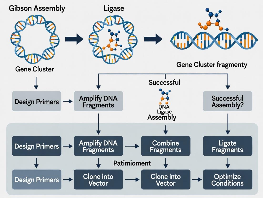
Diagram 1: Gibson Assembly Reaction Workflow
Detailed Protocols
Protocol A: Standard Gibson Assembly for 2-4 Fragments
Objective: Assemble a plasmid from 2-4 linear DNA fragments with 20-40 bp overlaps.
Materials: See "The Scientist's Toolkit" (Section 5.0).
Procedure:
- Fragment Preparation: Generate insert(s) and linearized vector via PCR or restriction digest. Gel-purify all fragments. Determine concentration via spectrophotometry (e.g., Nanodrop).
- Molar Ratio Calculation: Use the following equation to calculate the required mass of each fragment for a 0.03 pmol standard reaction:
- Mass (ng) = (0.03 pmol × fragment length (bp) × 660 g/mol/bp) / 1000
- For the vector backbone, use a 1:2 vector-to-total-insert molar ratio.
- Reaction Setup: Combine in a thin-walled PCR tube:
- 5-100 ng total DNA (sum of all fragments).
- 10 µL of 2X Gibson Assembly Master Mix.
- Nuclease-free water to a final volume of 20 µL.
- Positive Control: 10 ng of provided control linearized vector + control insert.
- Negative Control: Water + Master Mix only.
- Incubation: Place in a thermal cycler at 50°C for 15-60 minutes. For large constructs (>5 kb or >5 fragments), extend time to 60 minutes.
- Transformation: Place tube on ice. Transform 2-5 µL of the assembly reaction into 50 µL of competent E. coli (chemical, >1×10⁸ cfu/µg). Recover in SOC medium for 1 hour at 37°C, then plate on selective agar.
- Validation: Screen colonies by colony PCR and/or restriction digest. Confirm final constructs by Sanger sequencing across all assembly junctions.
Protocol B: High-Throughput Gibson Assembly for Gene Cluster Construction
Objective: Assemble >5 fragments, such as those constituting a biosynthetic gene cluster, in a single reaction.
Procedure:
- Design & Synthesis: Design all gene fragments with 40 bp homology overlaps. Optimize codon usage for the host. Synthesize fragments via pooled oligo synthesis or as gBlocks.
- Normalization: Dilute all purified fragments to 10-20 ng/µL. Use a fluorometric assay (e.g., Qubit) for accurate quantification.
- Optimized Reaction Setup: Combine in a PCR tube:
- Equal molar amounts of each fragment (final 0.005-0.02 pmol each).
- 15 µL of 2X Gibson Assembly Master Mix.
- Nuclease-free water to 30 µL.
- Incubation: 50°C for 60 minutes, followed by a 10-minute hold at 4°C.
- Clean-up (Optional): For large assemblies (>15 kb), clean the reaction using a DNA clean-up kit (elute in 10 µL) to remove enzymes and salts.
- Transformation: Use high-efficiency electrocompetent cells (>1×10⁹ cfu/µg). Electroporate 1-2 µL of the assembly or cleaned product. Add 1 mL SOC, recover with shaking for 2-3 hours before plating on large selective plates.
- Analysis: Screen using long-range PCR or diagnostic digest. Validate the complete cluster via next-generation sequencing (NGS) or pulsed-field gel electrophoresis (PFGE).
Table 2: Protocol Comparison & Optimization Guide
| Parameter | Protocol A (Standard) | Protocol B (High-Throughput) | Optimization Tips |
|---|---|---|---|
| Fragment Number | 2-4 | 5-15+ | Increase overlap length (40-60 bp) for >10 fragments. |
| Fragment Amount | 0.03 pmol total DNA | 0.005-0.02 pmol per fragment | For large clusters, a slight excess of middle fragments can improve yield. |
| Incubation Time | 15-30 min | 60 min | Extend to 90 min for assemblies >50 kb. |
| Competent Cells | Chemical (>1×10⁸ cfu/µg) | Electrocompetent (>1×10⁹ cfu/µg) | Always include a transformation control plasmid. |
| Downstream Analysis | Colony PCR, Sanger | Long-range PCR, NGS, PFGE | Use yeast or bacterial artificial chromosomes (YACs/BACs) for megabase clusters. |
Diagram 2: Gene Cluster Assembly and Validation Workflow
Troubleshooting Guide
Table 3: Common Issues and Solutions
| Problem | Potential Cause | Recommended Solution |
|---|---|---|
| Low Colony Count | Insufficient fragment amount/quality, short incubation, inefficient cells. | Re-quantify fragments fluorometrically. Increase reaction time to 60 min. Use fresh, high-efficiency competent cells. |
| High Background (Empty Vector) | Incomplete vector digestion or PCR linearization. | Treat vector with DpnI (if from PCR) to digest methylated template. Re-purify vector post-digestion. Use alkaline phosphatase treatment with caution. |
| Scrambled Assemblies/ Mutations | Misannealing of repetitive sequences or PCR errors in fragments. | Redesign overlaps to be unique. Use high-fidelity polymerase for fragment generation. Sequence intermediate fragments. |
| Large Cluster Assembly Failure | Complexity limits, secondary structure in overlaps, DNA damage. | Use a hierarchical assembly strategy (assemble sub-clusters first). Increase overlap homology to 60 bp. Ensure DNA is high molecular weight and clean. |
The Scientist's Toolkit
Table 4: Essential Research Reagent Solutions for Gibson Assembly
| Reagent/Material | Function & Role in the Workflow | Example/Notes |
|---|---|---|
| 2X Gibson Assembly Master Mix | Proprietary blend of T5 exonuclease, Phusion polymerase, and Taq DNA ligase in buffer. The core reagent. | Available commercially from NEB, Thermo Fisher, etc. Critical for one-pot isothermal reaction. |
| High-Fidelity DNA Polymerase | Generates error-free PCR fragments for assembly with clean ends. | Phusion U Green, Q5 (NEB), or KAPA HiFi. Essential for fragment preparation. |
| DNA Clean-Up & Gel Extraction Kits | Purifies PCR/digest products and removes enzymes, salts, and primers. | Qiagen, Macherey-Nagel, or Zymo Research kits. Clean fragments are vital for efficiency. |
| Fluorometric DNA Quantification Assay | Accurately measures DNA concentration, unaffected by salts/RNA. | Qubit dsDNA HS/BR Assay (Thermo Fisher). More accurate than absorbance (A260) for assembly. |
| Electrocompetent E. coli | High-efficiency cells for transforming large or complex assemblies. | NEB 10-beta, MegaX DH10B T1R, or homemade cells (>1×10⁹ cfu/µg). |
| Next-Generation Sequencing (NGS) Service | Validates the sequence of large, assembled gene clusters. | Illumina MiSeq for clusters; Nanopore for very long reads. Final quality control step. |
Application Notes
Within the framework of Gibson Assembly for gene cluster assembly research, the synergistic action of exonuclease, polymerase, and DNA ligase is foundational. This one-pot, isothermal method enables the seamless assembly of multiple overlapping DNA fragments into large constructs, such as entire biosynthetic gene clusters for natural product discovery and drug development. The precise coordination of the three enzymatic activities circumvents the need for multiple cloning steps, significantly accelerating the construction of genetic pathways for functional expression and engineering.
The quantitative efficiency of Gibson Assembly is influenced by several key parameters, as summarized below.
Table 1: Key Quantitative Parameters for Gibson Assembly Optimization
| Parameter | Typical Range | Impact on Assembly Efficiency |
|---|---|---|
| Fragment Length | 200 bp - 80 kb | Longer fragments (>10 kb) may require optimization of overlap length and enzyme concentration. |
| Overlap Length | 15-40 bp | 20-40 bp is standard. Shorter overlaps (15-20 bp) can work but may reduce efficiency for complex assemblies. |
| Fragment Molar Ratio | 1:1 for 2 fragments; 0.2:1 for >5 fragments (vector:insert) | A slight molar excess of inserts is critical for multi-fragment assemblies to drive reactions forward. |
| Reaction Incubation Time | 15-60 minutes | 15-30 minutes is often sufficient for simple assemblies; 60 minutes recommended for >5 fragments. |
| Total DNA Amount | 0.02-0.5 pmol of total DNA | Excessive DNA can inhibit the reaction; staying within the linear range of the enzymes is crucial. |
| Assembly Efficiency | 90-100% for 2-3 fragments; 30-80% for >5 fragments | Efficiency decreases with increasing fragment number but remains robust with optimized protocols. |
Experimental Protocols
Protocol 1: Standard Gibson Assembly for Gene Cluster Construction
Objective: To assemble 3-5 linear DNA fragments with 20-40 bp homologous overlaps into a circular plasmid.
Research Reagent Solutions & Materials:
| Item | Function |
|---|---|
| Gibson Assembly Master Mix (2X) | Commercial or homemade mix containing T5 exonuclease, Taq DNA polymerase, and DNA ligase in an optimized buffer. |
| Linearized Vector DNA | Gel-purified plasmid backbone, digested to have ends homologous to the terminal inserts. |
| PCR-Amplified Insert Fragments | Gel- or column-purified DNA fragments with designed homologous ends. |
| Nuclease-Free Water | To adjust reaction volume and prevent enzymatic degradation. |
| Thermocycler or Heating Block | To maintain a constant isothermal reaction temperature of 50°C. |
| Competent E. coli Cells (e.g., DH5α) | For transformation and propagation of the assembled plasmid. |
| SOC Recovery Medium | Nutrient-rich medium for outgrowth of transformed cells. |
| Selection Agar Plates | Antibiotic-containing plates for selecting successful transformants. |
Methodology:
- Design & Preparation: Design all DNA fragments to have 20-40 bp overlapping ends. Generate fragments via PCR or synthesis, and purify using a gel extraction or PCR cleanup kit. Quantify DNA concentration via spectrophotometry.
- Reaction Setup: In a sterile, nuclease-free microtube, combine the following on ice:
- 10 µL of 2X Gibson Assembly Master Mix.
- X µL of linearized vector (final amount 0.02-0.05 pmol).
- Y µL of insert fragment(s) (use a 1.5-2x molar excess of each insert relative to the vector).
- Nuclease-free water to a final volume of 20 µL.
- Gently mix and briefly centrifuge.
- Incubation: Incubate the reaction at 50°C for 15-60 minutes (30 minutes is typical for 3-5 fragments).
- Transformation: Transform 2-5 µL of the assembly reaction into 50 µL of chemically competent E. coli cells following standard heat-shock protocols. Add 950 µL of SOC medium and incubate at 37°C with shaking for 60 minutes.
- Analysis: Plate 50-100 µL of the culture on appropriate antibiotic selection plates. Screen colonies by colony PCR or restriction digest to verify correct assembly.
Protocol 2: Optimization for High-Number Fragment Assembly (>5 fragments)
Objective: To improve the efficiency of assembling 6-15 fragments, such as a large gene cluster, in a single reaction.
Methodology:
- Fragment Preparation: Rigorously purify all fragments by agarose gel electrophoresis and extraction to remove primers, template, and non-specific products. Quantify precisely.
- Molar Ratio Optimization: Use a modified insert:vector ratio. For
ninserts, use a molar ratio of vector:insert1:insert2:...:insertn= 1:0.2:0.2:...:0.2. This prevents incorrect annealing pathways. - Two-Step Assembly (if needed): For very large clusters (>50 kb), consider assembling sub-clusters of 3-4 fragments first, sequence-verifying them, and then performing a final assembly of the sub-clusters.
- Reaction Scaling: Double the reaction volume (40 µL total) and incubation time (60 minutes) to ensure sufficient enzyme activity for all fragments.
- Transformation: Use high-efficiency electrocompetent cells (>10^9 cfu/µg) instead of chemically competent cells. Dialyze or dilute the assembly reaction 5-fold with water before electroporation to reduce salt concentration.
Visualizations
Gibson Assembly Enzymatic Synergy
Gene Cluster Assembly Workflow
Within the broader thesis on Gibson Assembly's transformative role in synthetic biology, this application note focuses on its specific superiority for assembling gene clusters—large, multi-gene DNA constructs essential for studying metabolic pathways, natural product biosynthesis, and therapeutic development. Traditional cloning methods, such as restriction enzyme/ligase cloning and TA cloning, become increasingly inefficient and laborious as construct size and complexity increase. Gibson Assembly (and related isothermal assembly methods) overcomes these limitations through a seamless, one-pot, isothermal reaction that efficiently assembles multiple overlapping DNA fragments.
Advantages Over Traditional Cloning: A Quantitative Comparison
Table 1: Comparison of Key Cloning Methods for Gene Cluster Assembly
| Feature | Gibson Assembly | Restriction Enzyme/Ligase Cloning | TA Cloning | Gateway Cloning |
|---|---|---|---|---|
| Assembly Type | Seamless, scarless | Leaves scars (restriction sites) | Leaves scars (vector sequences) | Leaves scars (att sites) |
| Multi-Fragment Capacity | High (5-10+ fragments in one reaction) | Very Low (typically 1-2 fragments) | Low (typically 1 fragment) | Moderate (via multi-step LR reaction) |
| Hands-On Time | Low (single reaction) | High (multiple enzymatic steps, purification) | Moderate | High (multiple recombination steps) |
| Success Rate for Large Constructs (>10 kb) | High (>80%) | Very Low (<20%) | Not Applicable | Moderate (~50-60%) |
| Cost per Assembly | Moderate | Low (per simple assembly) | Low | High |
| Flexibility in Insert Design | High (any sequence via overlap design) | Low (dependent on restriction sites) | Low (requires compatible ends) | Moderate (requires specific att sites) |
| Typical Timeline | 1-2 days | 3-5 days | 2-3 days | 3-5 days |
Detailed Protocols
Protocol 1: Designing and Assembling a Biosynthetic Gene Cluster (BGC)
Objective: Assemble a 15 kb polyketide synthase (PKS) gene cluster from three ~5 kb fragments into a bacterial expression vector.
Materials:
- DNA Fragments: Gel-purified PCR products or synthesized fragments with 20-40 bp homologous overlaps.
- Vector: Linearized destination vector with 5' phosphorylation.
- Enzyme Master Mix: Commercial Gibson Assembly Master Mix (e.g., from NEB).
- Competent Cells: High-efficiency E. coli (e.g., NEB 10-beta).
- Controls: Vector-only control assembly.
Methodology:
- Fragment Preparation: Amplify the three PKS gene fragments and the linearized vector using a high-fidelity DNA polymerase. Design primers to generate 30-40 bp overlaps between adjacent fragments and the vector ends.
- Purification: Gel-purify all PCR products to remove primers and non-specific amplifications.
- Concentration Measurement: Quantify DNA concentration via fluorometry.
- Assembly Reaction: In a thin-walled PCR tube, combine:
- 0.02-0.5 pmol of each DNA fragment (vector + inserts).
- 15 µL of Gibson Assembly Master Mix.
- Nuclease-free water to a final volume of 20 µL.
- Incubation: Incubate the reaction at 50°C for 15-60 minutes. For complex assemblies (>5 fragments or >20 kb total), extend time to 60 minutes.
- Transformation: Transform 2-5 µL of the assembly reaction into 50 µL of competent E. coli following standard heat-shock protocols. Plate on selective media.
- Screening: Pick 5-10 colonies for colony PCR or analytical restriction digest. Confirm the full assembly by diagnostic PCR across junctions and Sanger sequencing of the entire cluster.
Protocol 2: Rapid Combinatorial Assembly of Pathway Variants
Objective: Generate a library of biosynthetic pathway variants by swapping modular enzymatic domains using Gibson Assembly.
Materials: As in Protocol 1, with fragments designed for modular overlaps.
Methodology:
- Modular Design: Design each enzyme or domain as a separate fragment with standardized overlapping ends (e.g., using the MoClo or Golden Gate-inspired syntax adapted for Gibson overlaps).
- One-Pot Modular Assembly: Mix equimolar amounts of the chosen variant fragments for each module along with the linear vector backbone in a single Gibson Assembly reaction.
- Incubation and Transformation: Perform as in Protocol 1, steps 5-6.
- Library Analysis: Screen multiple colonies to assess library diversity. High-throughput sequencing of the pooled colonies or plasmid minipreps can be used to characterize the variant library.
Visualization of Workflows
Gibson Assembly Mechanism and Workflow
The Scientist's Toolkit: Key Research Reagent Solutions
Table 2: Essential Materials for Gibson Assembly of Gene Clusters
| Item | Function & Rationale | Example Product |
|---|---|---|
| High-Fidelity DNA Polymerase | Generates error-free PCR fragments for assembly; critical for large, complex gene clusters. | NEB Q5, Thermo Fisher Phusion. |
| Commercial Gibson Assembly Mix | Pre-mixed, optimized cocktail of T5 exonuclease, Phusion polymerase, and Taq DNA ligase. Saves time and increases reproducibility. | NEB Gibson Assembly Master Mix, Synthetic Genomics Gibson HiFi Assembly. |
| Gel Extraction Kit | Purifies PCR fragments from agarose gels to remove primers, dimers, and non-specific products that can hinder assembly. | Qiagen QIAquick, Zymoclean Gel DNA Recovery. |
| High-Efficiency Competent Cells | Essential for transforming large, potentially toxic gene cluster constructs. | NEB 10-beta E. coli, Lucigen ElectroTen-Blue. |
| Fragment DNA Synthesesis Service | For large or complex gene clusters where PCR amplification from genomic DNA is not feasible. | IDT gBlocks, Twist Biosynthesis Gene Fragments. |
| Long-Range Sequencing Service | Confirms the fidelity and correct assembly of the entire gene cluster, not just junctions. | PacBio HiFi, Nanopore sequencing. |
As substantiated in this thesis, Gibson Assembly is the method of choice for gene cluster construction due to its seamless, multi-fragment, one-pot reaction scheme. It dramatically reduces the time and complexity associated with assembling large DNA constructs compared to traditional methods, enabling rapid iteration and combinatorial library generation—key capabilities for advancing research in natural product discovery, metabolic engineering, and gene therapy vector development.
Application Notes: The Evolution of Gene Cluster Assembly
The field of synthetic biology has evolved from conceptual frameworks in the early 2000s to a discipline with standardized, robust methodologies. A pivotal thesis in this progression is the establishment of Gibson Assembly as the gold standard for gene cluster assembly, crucial for pathway engineering in natural product drug discovery. The development from isolated, error-prone techniques (e.g., restriction-ligation) to seamless, multi-fragment assembly represents a paradigm shift, enabling the reliable construction of biosynthetic gene clusters (BGCs) exceeding 50 kb.
Key Quantitative Milestones in Assembly Methodology Development:
Table 1: Evolution of DNA Assembly Techniques and Their Impact on Synthetic Biology
| Technique (Year Introduced) | Typical Assembly Efficiency (%) | Max Fragment No. | Max Construct Size (kb) | Key Limitation |
|---|---|---|---|---|
| Restriction/ Ligation (1970s) | 1-10 | 1-2 | 10-20 | Scar sequence dependency, multi-fragment incompatibility |
| BioBrick Assembly (2003) | 30-50 | 2-3 | ~5 | Standardized scars, slow iterative process |
| Golden Gate Assembly (2008) | 80-95 | 10+ | 20+ | Requires specific, absent restriction sites |
| Gibson Assembly (2009) | 70-90 | 15+ | 100+ | High homology region requirement |
| Yeast TAR/Gap Repair (1990s/2010s) | 10-50 | 5-10 | 200+ | Low efficiency in bacteria, yeast-specific |
Recent Data (2023-2024): A meta-analysis of 47 published studies utilizing Gibson Assembly for BGC construction shows an average assembly success rate of 87% for constructs between 20-40 kb when using high-fidelity polymerase and optimized fragment overlap design (40-60 bp). Success rates drop to ~65% for constructs >70 kb, highlighting the frontier for ongoing methodological refinement.
Detailed Protocols
Protocol 2.1: Gibson Assembly for Large Biosynthetic Gene Cluster Assembly
Thesis Context: This protocol is central to the thesis that optimized Gibson Assembly is the most reliable and flexible method for constructing complex natural product pathways for heterologous expression in Streptomyces or E. coli.
Research Reagent Solutions:
Table 2: Essential Toolkit for Gibson Assembly-Based Gene Cluster Construction
| Reagent/Material | Function/Benefit | Example Product/Catalog # |
|---|---|---|
| High-Fidelity DNA Polymerase | Amplifies inserts and vector with ultra-low error rates for large fragments. | NEB Q5 High-Fidelity, Thermo Fisher Phusion Plus |
| Commercial Gibson Assembly Master Mix | Contains T5 exonuclease, Phusion polymerase, and Taq DNA ligase for one-step, isothermal assembly. | NEB HiFi DNA Assembly Master Mix, SGI-DNA Gibson Assembly Master Mix |
| Chemically Competent E. coli | High-efficiency cells for transformation of large, complex assemblies. | NEB 10-beta, NEB Stable, Thermo Fisher One Shot TOP10 |
| RecET/Lambda Red Cloning Strain | Facilitates in vivo recombineering for final assembly or troubleshooting in E. coli. | E. coli GB05-dir (GeneBridges) |
| Gel/PCR DNA Cleanup Kit | Purifies DNA fragments from enzymatic reactions and gels to remove inhibitors. | Zymo Research DNA Clean & Concentrator, Qiagen QIAquick Gel Extraction Kit |
| Sanger & Long-Read Sequencing | Confirms assembly fidelity and corrects sequence errors. | PacBio HiFi, Oxford Nanopore Technologies MinION |
Methodology:
Fragment Design & Preparation:
- Design all fragments (gene cassettes, promoters, resistance markers) with 30-60 bp homologous overlaps to adjacent fragments. Use software (e.g., j5, SnapGene) to avoid repetitive sequences.
- Amplify each fragment via PCR using high-fidelity polymerase. Use genomic DNA, synthesized fragments, or existing plasmids as templates.
- Purify all PCR products using a gel extraction kit. Quantify via fluorometry (Qubit). Aim for equimolar ratios.
Assembly Reaction:
- In a 0.2 mL tube, combine:
- 50-100 ng of linearized vector backbone.
- Insert fragments (equimolar, 2-5:1 insert:vector molar ratio).
- Commercial Gibson Assembly Master Mix to 1/2 total volume.
- Mix gently by pipetting. Incubate at 50°C for 15-60 minutes (15 min for <6 kb, 60 min for >20 kb constructs).
- In a 0.2 mL tube, combine:
Transformation & Screening:
- Transform 2-5 µL of the assembly reaction into 50 µL of high-efficiency competent E. coli via heat shock.
- Plate on selective media. Incubate overnight at 37°C.
- Screen 5-10 colonies by colony PCR using junction-spanning primers.
- Inoculate positive clones for plasmid isolation.
Validation:
- Verify assembly by diagnostic restriction digest.
- Submit the construct for long-read sequencing (PacBio or Nanopore) to confirm the entire sequence of the assembled gene cluster, especially critical for large BGCs.
Protocol 2.2: Hierarchical Assembly of Very Large Clusters (>50 kb)
Thesis Context: For assemblies exceeding practical single-reaction limits, a hierarchical strategy using Gibson Assembly for sub-cluster construction, followed by final integration, demonstrates the method's scalability—a core argument for its gold-standard status.
Methodology:
- Sub-Cluster Assembly: Use Protocol 2.1 to assemble 10-20 kb sub-clusters (e.g., individual operons) into intermediate vectors.
- Intermediate Validation: Fully sequence each intermediate sub-cluster.
- Final Assembly: Design the intermediate vectors with homology to each other and the final destination vector (e.g., a bacterial artificial chromosome). Perform a final Gibson Assembly using the purified intermediate plasmids as large fragments.
- In Vivo Rescue (Alternative): If in vitro assembly fails, co-transform the overlapping intermediate plasmids into a recombinogenic E. coli strain (e.g., GB05-dir) expressing RecET. Select for the final construct, which assembles via homologous recombination in vivo.
Mandatory Visualizations
Diagram Title: Hierarchical Gibson Assembly Workflow for Large BGCs
Diagram Title: Gibson Assembly Molecular Mechanism
Within the broader thesis on Gibson Assembly for gene cluster assembly research, this document details the critical pre-assembly phase. Successful assembly of large, complex genetic constructs—such as biosynthetic gene clusters for natural product discovery in drug development—hinges on meticulous planning of DNA fragment overlaps and rigorous fragment preparation. This protocol outlines the standardized methodologies for these foundational steps.
Key Principles of Overlap Design
Overlaps are the single-stranded homologous regions that facilitate the annealing step in Gibson Assembly. Their design directly dictates assembly efficiency and accuracy.
Quantitative Design Parameters
The following table summarizes optimal parameters for overlap design, synthesized from current literature and experimental validation.
Table 1: Quantitative Parameters for Gibson Assembly Overlap Design
| Parameter | Recommended Value | Rationale & Impact |
|---|---|---|
| Overlap Length | 20-40 bp | <40 bp minimizes mispriming in PCR; >20 bp ensures stable annealing. |
| Melting Temperature (Tm) | 55-65°C | Ensures simultaneous annealing of all fragments during isothermal step. |
| GC Content | 40-60% | Promotes stable hybridization; extremes can cause secondary structures. |
| Terminal Homology | Minimum 15 bp | Absolute minimum for successful recombination; 20+ bp strongly advised. |
| Overlap Uniformity | Tm within 2°C for all fragments | Prevents preferential annealing and ensures synchronous assembly. |
Strategic Considerations for Gene Clusters
- Junction Placement: Avoid overlaps that place recombination junctions within predicted secondary structures or essential protein domains.
- Repeated Sequences: Unique overlaps must be designed for any homologous repetitive elements (e.g., identical promoter sequences) to prevent scrambling.
- Directional Control: Overlaps must be designed to enforce the correct, directional order of fragments in the final assembly.
Fragment Preparation Strategies and Protocols
Fragments can be sourced via PCR amplification from templates or as synthesized dsDNA oligos/blocks. Preparation quality is paramount.
Research Reagent Solutions Toolkit
Table 2: Essential Reagents for Fragment Preparation and QC
| Item | Function & Critical Notes |
|---|---|
| High-Fidelity DNA Polymerase (e.g., Q5, Phusion) | PCR amplification with ultra-low error rates to prevent incorporation of mutations in assembly fragments. |
| DNA Clean-Up & Gel Extraction Kits | For purification of PCR products and isolation of correctly sized fragments from agarose gels. |
| dsDNA Fragmentase or Restriction Enzymes | For generating complex fragment libraries from genomic DNA, as an alternative to synthesis. |
| Fluorometric dsDNA Quantification Assay (e.g., Qubit) | Accurate quantification of fragment concentration for stoichiometric mixing. Critical for multi-fragment assemblies. |
| Capillary Electrophoresis System (e.g., Fragment Analyzer, Bioanalyzer) | Gold-standard for assessing fragment size and purity, detecting primer dimers, and verifying absence of gDNA contamination. |
| In-Fusion or Gibson Assembly Master Mix | Commercial enzyme mixes containing 5’ exonuclease, polymerase, and DNA ligase for the assembly reaction itself. |
Protocol: PCR-Based Fragment Generation with Overhangs
Objective: Amplify a gene fragment with designed 5’ and 3’ overlaps for Gibson Assembly.
Materials:
- Template DNA (plasmid, genomic DNA, cDNA)
- High-fidelity DNA polymerase and corresponding buffer
- dNTP mix (10 mM each)
- Forward and Reverse primers (with 5’ overhangs, see design below)
- Nuclease-free water
- Thermocycler
Methodology:
- Primer Design:
- The 5’ end of each primer consists of the designed overlap sequence (20-40 bp) that is homologous to the adjacent fragment.
- The 3’ end of each primer consists of the gene-specific sequence (18-25 bp, Tm ~60°C) for template annealing.
- Example Primer:
5’-[40 bp Overlap to Fragment A]-[20 bp Gene-Specific Sequence]-3’
PCR Setup (50 µL Reaction):
- Nuclease-free water: to 50 µL
- 2X High-Fidelity Master Mix: 25 µL
- Forward Primer (10 µM): 2.5 µL
- Reverse Primer (10 µM): 2.5 µL
- Template DNA: 1-100 ng (optimize)
- Run in duplicate to ensure sufficient yield.
Thermocycling Conditions:
- Initial Denaturation: 98°C for 30 sec.
- 35 Cycles: Denature at 98°C for 10 sec, Anneal at (Primer Tm +3°C) for 20 sec, Extend at 72°C (30 sec/kb).
- Final Extension: 72°C for 2 min.
- Hold: 4°C.
Post-PCR Purification & QC:
- Pool duplicate reactions.
- Purify using a PCR clean-up kit, eluting in nuclease-free water or low-EDTA TE buffer.
- Quantify using a fluorometric assay.
- Verify size and purity via capillary electrophoresis or agarose gel analysis.
Protocol: Fragment Preparation Workflow from Design to QC
Diagram Title: Fragment Prep Workflow from Design to QC
Pre-Assembly Planning and Stoichiometry
Prior to the assembly reaction, fragments must be mixed in an optimal ratio.
Table 3: Recommended Stoichiometry for Multi-Fragment Gibson Assembly
| Component | Molar Ratio | Calculation Basis | Notes |
|---|---|---|---|
| Linearized Vector Backbone | 1x | Reference amount. | Use 50-100 ng total vector as starting point. |
| Each Insert Fragment | 2x - 3x | Relative to vector molarity. | Equal molarity of all inserts is standard. For difficult assemblies, a 5:1 insert:vector ratio can be tested. |
| Total DNA in Reaction | 0.02-0.5 pmol | For a 20 µL reaction. | Keep total DNA mass <200 ng to avoid inhibition. |
Mixing Protocol:
- Calculate the molar concentration of each purified fragment.
- In a sterile tube, combine fragments according to the ratios in Table 3.
- Adjust the total volume with nuclease-free water so that the fragment mix constitutes 50% of the final assembly reaction volume (e.g., 5 µL of fragment mix for a 10 µL Gibson reaction).
- This pre-mixed fragment pool is now ready to be combined with an equal volume of Gibson Assembly Master Mix.
Step-by-Step Gibson Assembly Protocol: Building Functional Gene Clusters from Fragments
This article, framed within a broader thesis on Gibson Assembly for gene cluster assembly research, details the design principles for overlap sequences. In Gibson Assembly and related methods, overlaps are the single-stranded termini of DNA fragments that facilitate homologous recombination. Optimizing their length, melting temperature (Tm), and sequence composition is critical for the efficient, high-fidelity assembly of large genetic constructs, such as biosynthetic gene clusters for drug discovery.
Quantitative Design Guidelines
The following tables summarize key quantitative parameters for designing optimal overlap sequences.
Table 1: Overlap Length and Tm Recommendations by Assembly Complexity
| Assembly Complexity | Recommended Overlap Length (bp) | Target Tm Range (°C) | Primary Considerations |
|---|---|---|---|
| Standard (2-3 fragments) | 20 - 40 | 48 - 60 | Balancing efficiency and specificity. |
| High-Complexity (>5 fragments) | 30 - 60 | 55 - 65 | Enhanced specificity to prevent misassembly. |
| Large Gene Clusters (>10 kb) | 40 - 80 | 60 - 72 | Increased stability for handling complex repeats. |
| Isothermal (e.g., Gibson) | 15 - 80 (typ. 20-40) | 48 - 65 | Must be compatible with the enzyme's optimal working T (~50°C). |
Table 2: Sequence Composition and Penalty Guidelines
| Parameter | Optimal Condition | Penalty / Avoidance |
|---|---|---|
| GC Content | 40% - 60% | <30% or >70% can destabilize annealing. |
| Terminal Base Pairs | 5' end: G/C; 3' end: A/T | Avoid long A/T stretches at termini. |
| Self-Complementarity | None (hairpins ΔG > -2 kcal/mol) | Strong secondary structures (ΔG < -5 kcal/mol). |
| Cross-Homology | Unique across assembly set | >10 bp of identical sequence in non-overlap regions. |
| Repetitive Sequences | None | Direct or inverted repeats >8 bp within overlap. |
Protocols for Overlap Design and Validation
Protocol 1:In SilicoDesign of Overlap Sequences for Gibson Assembly
Objective: To computationally design and screen optimal overlap sequences for assembling a target multi-fragment gene cluster.
Materials (Research Reagent Solutions Toolkit):
- Software: Geneious, SnapGene, or custom Python/Biopython scripts.
- Tm Calculator: Nearest-neighbor method (e.g., NEB Tm Calculator algorithm).
- Sequence Alignment Tool: BLAST or EMBOSS needle for homology checking.
- Secondary Structure Predictor: mfold or UNAFold.
Methodology:
- Define Fragment Boundaries: Determine the breakpoints for each DNA fragment in the final assembly. Ensure each internal fragment has overlaps with its upstream and downstream partners.
- Generate Candidate Sequences: Extract 20-80 bp sequences from the junction regions. For de novo synthesis, design sequences meeting GC% criteria.
- Calculate Tm: Use the nearest-neighbor method with salt concentrations matching Gibson Assembly Master Mix (~50 mM Na+). Adjust length to bring all fragment overlaps within a 5°C Tm range, ideally 55-60°C.
- Screen for Hazards: a. Perform self-dimer and cross-dimer analysis for all overlaps. b. Check for internal homology (>75% identity) to other genomic regions in the host. c. Verify absence of restrictive enzyme sites if downstream cloning is needed.
- Final Selection: Select the set of overlaps with the most uniform Tm, highest internal sequence uniqueness, and absence of secondary structure.
Protocol 2: Empirical Testing of Overlap Efficiency
Objective: To experimentally validate the assembly efficiency of designed overlaps using a model assembly system.
Materials (Research Reagent Solutions Toolkit):
- Assembly Reagent: Gibson Assembly Master Mix (NEB) or equivalent.
- DNA Fragments: PCR-amplified or synthesized fragments with designed overlaps.
- Competent Cells: High-efficiency E. coli (e.g., NEB 5-alpha, DH5α).
- Selection Media: LB agar plates with appropriate antibiotic.
- Analytical Tools: Colony PCR reagents, gel electrophoresis system, sequencing primers.
Methodology:
- Setup Assembly Reactions: Assemble 3-5 test fragments with varying overlap lengths (e.g., 15, 25, 40 bp) but identical assembly points. Use a standardized fragment molar ratio (typically 1:1 or 2:1 insert:vector).
- Incubation: Incubate reactions at 50°C for 15-60 minutes.
- Transformation: Transform 2-5 µL of each assembly reaction into 50 µL of competent cells. Plate on selective media.
- Efficiency Analysis: a. Count colonies after 16-20 hours of incubation. b. Calculate transformation efficiency (CFU/µg of assembled DNA) for relative comparison. c. Pick 10-20 colonies per condition for colony PCR to verify correct assembly size. d. Send 3-5 positive clones from each condition for Sanger sequencing across all junctions.
- Data Interpretation: The overlap set yielding the highest colony count with >90% sequence verification is considered optimal for that specific assembly context.
Visualizations
Title: Overlap Design and Validation Protocol
Title: Impact of Overlap Melting Temperature
Within a research thesis focused on assembling complex gene clusters via Gibson Assembly, the generation of high-quality DNA fragments is the critical first step. The fidelity, purity, and terminal compatibility of these fragments directly determine the success of downstream seamless assembly. This application note details best practice protocols for the three primary methods of fragment generation—PCR Amplification, Gene Synthesis, and Restriction Digestion—framed within the context of preparing parts for Gibson Assembly.
PCR Amplification of Gene Fragments
PCR is the most common method for amplifying specific fragments from genomic or plasmid DNA. For Gibson Assembly, amplicons must have sufficient overlap (typically 15-40 bp) with adjacent fragments and be free of mutations.
Detailed Protocol: High-Fidelity PCR for Gibson Assembly Fragments
Objective: Amplify a target gene with 20-30 bp overlaps matching adjacent assembly fragments. Reagents:
- Template DNA: 1-10 ng plasmid DNA or 10-100 ng genomic DNA.
- High-Fidelity DNA Polymerase (e.g., Q5, Phusion, KAPA HiFi).
- dNTP Mix: 10 mM each.
- Forward and Reverse Primers: 10 µM each, designed with 5' Gibson overhangs.
- Nuclease-Free Water.
- 5X PCR Buffer (supplied with polymerase).
Procedure:
- Reaction Setup (50 µL):
- Nuclease-Free Water: to 50 µL final volume.
- 5X Buffer: 10 µL.
- dNTPs (10 mM each): 1 µL.
- Forward Primer (10 µM): 2.5 µL.
- Reverse Primer (10 µM): 2.5 µL.
- Template DNA: variable.
- High-Fidelity DNA Polymerase: 0.5-1 unit.
- Thermocycling:
- Initial Denaturation: 98°C for 30 seconds.
- 30-35 Cycles:
- Denature: 98°C for 10 seconds.
- Anneal: Tm + 3°C of the gene-specific portion of primer for 20 seconds.
- Extend: 72°C for 20-30 seconds/kb.
- Final Extension: 72°C for 2 minutes.
- Hold: 4°C.
- Post-PCR Purification: Purify the PCR product using a spin column-based PCR purification kit or gel extraction to remove primers, template, and enzymes. Elute in nuclease-free water or 10 mM Tris-HCl (pH 8.0).
- Quantification: Measure concentration via spectrophotometry (Nanodrop) or fluorometry (Qubit). Verify size and purity by agarose gel electrophoresis.
Best Practices:
- Primer Design: Ensure the 5' overlap region does not form secondary structures or homodimers.
- Template Quality: Use high-quality, minimal-passage template DNA.
- Minimize Cycles: Use the minimum number of cycles necessary to reduce mutation burden.
- Verification: Sequence the final amplified fragment, especially for large (>1 kb) amplicons.
Restriction Digestion for Fragment Preparation
Restriction digestion is ideal for liberating fragments from existing plasmids. For Gibson Assembly, digestion must be complete to prevent parental plasmid carryover.
Detailed Protocol: Preparation of Vector and Insert via Restriction
Objective: Generate a vector backbone and an insert fragment with compatible ends for subsequent Gibson Assembly. Reagents:
- Plasmid DNA: 1-5 µg.
- Appropriate Restriction Enzymes (two for vector, often with a single cut-site each; may be one or two for insert).
- Recommended Reaction Buffer (10X).
- BSA (if required by enzyme).
- Nuclease-Free Water.
Procedure (Double Digestion):
- Reaction Setup (50 µL):
- Plasmid DNA: 1-5 µg (in ≤ 20 µL volume).
- 10X Buffer: 5 µL (use a compatible buffer for both enzymes).
- Enzyme 1: 10-20 units.
- Enzyme 2: 10-20 units.
- Nuclease-Free Water: to 50 µL.
- If needed, add 5 µL of 10X BSA.
- Incubation: Incubate at the recommended temperature(s) for 1-3 hours. For sequential digestion with incompatible buffers, purify DNA after the first digestion.
- Dephosphorylation (for Vector Backbone): To prevent re-ligation, add 1 µL of Antarctic Phosphatase or CIP directly to the reaction after digestion and incubate for an additional 30-60 minutes.
- Purification: Run the entire reaction on an agarose gel. Excise the correct bands and purify using a gel extraction kit. This step is critical to remove enzymes, buffers, and unwanted fragments.
- Quantification: Accurately measure the concentration of the purified fragments.
Best Practices:
- Complete Digestion: Use sufficient enzyme units and incubation time. Verify completeness by analytical gel.
- Buffer Compatibility: Use double-digest buffers or perform sequential digests with purification in between.
- Gel Purification: Essential for removing undigested plasmid and small stuffer fragments.
Gene Synthesis forDe NovoFragment Generation
Gene synthesis is used for de novo generation of optimized sequences, codon-optimized genes, or complex fragments not available from natural sources.
Workflow and Considerations:
- Sequence Design: Design the fragment with optimal codon usage for the host organism. Include Gibson Assembly overlaps (15-40 bp) at the 5' and 3' ends. Avoid internal regions homologous to the overlaps.
- Provider Selection: Select a commercial gene synthesis provider. Key parameters are turnaround time, cost, error rate, and maximum length (often 1.5-3 kb for standard synthesis; longer fragments require assembly).
- Cloning Format: Specify delivery in a standard cloning vector (e.g., pUC57) or as a linear, PCR-ready fragment.
- Validation: Upon receipt, sequence the entire synthesized fragment to confirm accuracy. For linear fragments, amplify with a high-fidelity polymerase before Gibson Assembly.
Table 1: Comparison of Fragment Generation Methods
| Method | Best For | Typical Length | Key Advantage | Primary Consideration for Gibson Assembly |
|---|---|---|---|---|
| PCR Amplification | Amplifying existing sequences | 0.1 - 10 kb | Fast, inexpensive; easy to add overlaps | Fidelity is critical; requires sequencing verification |
| Restriction Digestion | Reusing parts from existing plasmids | 0.1 - 15 kb | Sequence integrity maintained | Must remove all trace of parental plasmid; gel purification essential |
| Gene Synthesis | De novo, optimized, or unnatural sequences | 0.1 - 3 kb (per fragment) | Complete sequence control | Cost for long fragments; requires subcloning for very large pieces |
Table 2: Recommended High-Fidelity Polymerases
| Polymerase | Avg. Error Rate (mutations/bp) | Processivity | Best Suited For |
|---|---|---|---|
| Q5 (NEB) | 2.8 x 10⁻⁷ | High | Standard & complex amplicons, high GC content |
| Phusion (Thermo) | 4.4 x 10⁻⁷ | Very High | Fast PCR, long amplicons (>10 kb) |
| KAPA HiFi (Roche) | 3.5 x 10⁻⁷ | Medium | High yield from limited template, multiplex PCR |
The Scientist's Toolkit: Research Reagent Solutions
Table 3: Essential Reagents for Fragment Generation
| Reagent/Kit | Function | Key Consideration |
|---|---|---|
| High-Fidelity DNA Polymerase (e.g., Q5) | Amplifies target DNA with minimal errors. | Critical for generating mutation-free assembly fragments. |
| PCR Purification Kit | Removes primers, salts, and enzymes from PCR reactions. | Essential for clean-up before Gibson Assembly or digestion. |
| Gel Extraction Kit | Isolates DNA fragments from agarose gels. | Mandatory for purifying restriction fragments from undigested plasmid. |
| Restriction Enzymes (Type IIs, e.g., BsaI) | Cut DNA at specific sequences. | Useful for Golden Gate subcloning before Gibson, or for creating defined ends. |
| DNA Clean & Concentrator Kit | Rapidly desalts and concentrates DNA in small elution volumes. | Useful for adjusting DNA concentration/purity before assembly. |
| Fluorometric dsDNA Quantification Assay | Accurately measures DNA concentration. | More accurate than absorbance for low-concentration or impure samples. |
| Commercial Gene Synthesis Service | Provides de novo DNA fragments. | Specify "cloning-ready" format and sequence-verify upon receipt. |
Experimental Workflow Visualization
Fragment Generation Paths for Gibson Assembly
Fragment Role in Gibson Assembly Thesis
Within a research thesis focused on assembling complex biosynthetic gene clusters via Gibson Assembly, the choice between commercial kits and homemade reagents for master mix preparation is critical. This decision impacts assembly fidelity, throughput, cost, and reproducibility—key factors for successful large-construct cloning for natural product discovery and drug development.
Table 1: Cost and Time Comparison per 50 µL Reaction
| Component | Commercial Kit (e.g., NEB HiFi Gibson Assembly) | Homemade Gibson Assembly Mix |
|---|---|---|
| Reagent Cost | $5.00 - $10.00 | $1.50 - $3.00 |
| Preparation Time | ~5 minutes (thaw & aliquot) | 4-6 hours (enzyme prep & optimization) |
| Shelf-life | 12 months at -20°C | 3-6 months at -20°C (with aliquoting) |
| Hands-on Time | Minimal | High |
Table 2: Performance Metrics in Gene Cluster Assembly
| Metric | Commercial Kit | Homemade Mix | Notes |
|---|---|---|---|
| Transformation Efficiency (CFU/µg) | 1-5 x 10⁴ | 0.5-5 x 10⁴ | Highly dependent on fragment purity & size. |
| Assembly Success Rate (≥4 fragments) | 70-90% | 60-85% | Homemade requires precise pH & ionic optimization. |
| Optimal Fragment Length | 200 bp - 10 kb+ | 200 bp - 10 kb+ | Comparable when optimized. |
| Key Advantage | Consistency, convenience, QC | Cost-saving, customizable enzyme ratios |
Experimental Protocols
Protocol A: Reaction Setup Using a Commercial Kit (Adapted from NEB)
- Thaw Components: Thaw 2X Gibson Assembly Master Mix (containing T5 exonuclease, Phusion polymerase, and Taq ligase in a proprietary buffer) and nuclease-free water on ice.
- Setup Reaction: In a sterile PCR tube, combine:
- Nuclease-free water: to 20 µL final volume.
- 2X Gibson Assembly Master Mix: 10 µL.
- DNA fragments/vector: 10 µL total volume, with a molar ratio of insert:vector typically 2:1 to 3:1. Total DNA amount should be 0.02-0.5 pmols.
- Incubate: Place reaction in a thermocycler at 50°C for 15-60 minutes. For large or complex assemblies (>5 fragments), 60 minutes is recommended.
- Transform: Use 2-5 µL of the assembly reaction to transform 50 µL of competent E. coli cells via heat shock or electroporation.
- Plate & Screen: Plate on selective media and incubate overnight. Screen colonies via colony PCR or analytical restriction digest.
Protocol B: Preparation and Use of Homemade Gibson Assembly Reagents
This protocol is adapted from Gibson et al., 2009 (Nature Methods), with optimizations for gene cluster assembly.
Part I: Stock Solution Preparation
- 5X IsoTherm Buffer: 1M Tris-HCl pH 7.5, 100mM MgCl₂, 100mM DTT, 10mM each dNTP, 5mM NAD⁺, 25% (w/v) PEG-8000. Filter sterilize (0.22 µm), aliquot, and store at -80°C.
- Enzyme Procurement & Dilution:
- T5 Exonuclease (e.g., Thermo Scientific): Dilute to 1 U/µL in supplied storage buffer.
- Phusion DNA Polymerase (e.g., NEB): Use at 0.4 U/µL.
- Taq DNA Ligase (e.g., NEB): Dilute to 40 U/µL in supplied storage buffer.
- Aliquot and store all enzymes at -80°C.
Part II: Master Mix Assembly & Reaction
- Prepare 2X Homemade Master Mix Fresh or from Aliquots: For one reaction, combine in order on ice:
- 5X IsoTherm Buffer: 8 µL
- Nuclease-free water: 2.4 µL
- T5 Exonuclease (1 U/µL): 0.4 µL
- Phusion Polymerase (0.4 U/µL): 2 µL
- Taq DNA Ligase (40 U/µL): 1.2 µL
- Total Volume: 14 µL. Mix by gentle pipetting. Use immediately or flash-freeze in liquid nitrogen for storage at -80°C.
- Setup Reaction: In a PCR tube, combine:
- 2X Homemade Master Mix: 14 µL
- DNA fragments/vector: 6 µL total volume (molar ratio as in Protocol A).
- Incubate and Transform: Follow steps 3-5 from Protocol A. A 60-minute incubation is standard for homemade mixes.
Visualization of Workflow & Decision Logic
Title: Decision Logic for Gibson Assembly Mix Selection
Title: Comparative Workflow: Kit vs. Homemade Gibson Assembly
The Scientist's Toolkit: Research Reagent Solutions
Table 3: Essential Materials for Gibson Assembly-Based Gene Cluster Assembly
| Item | Function in Workflow | Example Product/Supplier |
|---|---|---|
| 2X Gibson Assembly Master Mix (Commercial) | All-in-one optimized enzyme/buffer solution; enables rapid, consistent one-step assembly. | NEB Gibson Assembly HiFi Master Mix, Thermo Fisher GeneArt Gibson Assembly. |
| High-Fidelity DNA Polymerase | For PCR amplification of assembly fragments with minimal errors. Critical for large gene clusters. | Phusion HF Polymerase (NEB), Q5 (NEB). |
| T5 Exonuclease | Creates 3’ overhangs for homologous recombination. Core component of homemade mixes. | Thermo Scientific T5 Exonuclease. |
| Taq DNA Ligase | Seals nicks in the annealed DNA backbone. Thermostable for isothermal reaction. | NEB Taq DNA Ligase. |
| 5X IsoTherm-Style Buffer | Provides optimal pH, ions, cofactors (NAD⁺), and crowding agents (PEG) for the three-enzyme reaction. | Custom formulation per Gibson et al. protocol. |
| Electrocompetent E. coli | High-efficiency cells essential for transforming large, complex gene cluster assemblies. | NEB 10-beta, Lucigen ElectroTen-Blue. |
| Fragment Purification Kit | Cleanup of PCR products and linearized vector to remove enzymes, salts, and primers that inhibit assembly. | Zymo DNA Clean & Concentrator, Qiagen MinElute. |
| Gel Extraction Kit | Isolation of correctly sized DNA fragments from agarose gels for assembly. | Zymo Zymoclean Gel DNA Recovery. |
1. Introduction Within the broader thesis on Gibson Assembly (GA) for gene cluster assembly research, a critical methodological decision is the strategy for assembling large, multi-fragment constructs (>20 kb). This application note details and compares two primary strategies: Sequential Assembly (hierarchical, multi-step) and One-Pot Multi-Fragment Assembly (single-step). The choice of strategy impacts efficiency, fidelity, and throughput for applications in synthetic biology and natural product biosynthetic pathway reconstruction for drug development.
2. Comparative Analysis: Sequential vs. One-Pot Assembly The optimal strategy depends on fragment number, size, homology design, and desired throughput.
Table 1: Strategic Comparison of Assembly Methods
| Parameter | Sequential (Hierarchical) Assembly | One-Pot Multi-Fragment Assembly |
|---|---|---|
| Typical Fragment Number | 4 - 10 per round | 5 - 15+ (theoretical limit is high, practical yield decreases with >10) |
| Maximum Final Construct Size | Virtually unlimited (via iterative rounds) | Limited by transformation efficiency (often 50-150 kb) |
| Key Advantage | Higher per-step accuracy; easier troubleshooting; modular. | Speed; reduced handling; no intermediate cloning/verification. |
| Key Disadvantage | Time-consuming; requires multiple intermediate vectors. | Lower overall yield with many fragments; complex design. |
| Error Propagation Risk | Lower (errors isolated to rounds). | Higher (single error fails entire assembly). |
| Best For | Very large clusters (>100 kb), modular library construction. | Rapid assembly of well-characterized clusters (<100 kb). |
Table 2: Quantitative Performance Data from Recent Studies
| Study (Context) | Strategy | Fragment # & Size | Assembly Efficiency | Key Finding |
|---|---|---|---|---|
| Wang et al., 2023 (Polyketide) | Sequential (3 rounds) | 6 frags, 45 kb total | >80% correct intermediates | 100% correct final construct (3/3 clones). |
| Li & Ellington, 2024 (Optimized GA) | One-Pot | 8 frags, 22 kb | ~60% (6/10 clones correct) | Efficiency dropped to <10% with 12 fragments. |
| This Thesis (PKS-NRPS Cluster) | Both Tested | 9 frags, 32 kb total | Sequential: 90%. One-Pot: 40%. | Sequential proved more reliable for this specific complex cluster. |
3. Detailed Protocols
Protocol 1: Sequential Gibson Assembly for Large Clusters Objective: Assemble a 50 kb gene cluster from 9 fragments via 3 hierarchical rounds. Materials: NEBuilder HiFi DNA Assembly Master Mix, chemically competent E. coli (NEB 10-beta), appropriate antibiotic plates, QIAprep Spin Miniprep Kit, PCR reagents, T4 DNA Ligase. Procedure:
- Fragment Preparation: Generate 9 fragments via PCR or synthesis with 20-40 bp homology overlaps. Gel-purify all fragments.
- Round 1 Assembly (3 sub-clusters): Set up 3 separate GA reactions, each combining 3 fragments + linearized vector (e.g., pUC19). Use a 2:1 molar ratio of insert:vector fragment. Incubate at 50°C for 15-60 minutes.
- Transformation & Screening: Transform 2 µL of each assembly into 50 µL competent cells. Plate. Screen 3-5 colonies per assembly by colony PCR and restriction digest. Sequence-verify one correct clone for each sub-cluster (SC1, SC2, SC3).
- Round 2 Assembly (2 sub-clusters): Linearize a medium-copy vector (e.g., pRSFDuet-1). PCR-amplify SC1, SC2, and SC3 from their plasmids, adding new homology arms for the next assembly. Perform two GA reactions: (SC1 + SC2) and (SC3 + vector). Verify as in Step 3 to generate constructs SC1-2 and SC3-V.
- Final Round Assembly: PCR-amplify the large SC1-2 insert. Digest SC3-V to linearize. Perform final GA to combine SC1-2 with SC3-V. Transform into high-efficiency electrocompetent cells (≥ 1 x 10⁹ cfu/µg). Screen via analytical PacBio or long-range PCR.
Protocol 2: One-Pot Multi-Fragment Gibson Assembly Objective: Assemble a 25 kb construct from 8 fragments in a single reaction. Materials: Gibson Assembly Master Mix (or equivalent homemade mix), electrocompetent E. coli (e.g., NEB Stable), electroporator, SOC medium. Procedure:
- Homology Design: Design all fragments with unique, non-overlapping 20-40 bp homology regions. Use tools like j5 or Geneious for automated design.
- Fragment Preparation: Generate fragments with high-fidelity PCR. Treat with DpnI to remove template DNA. Purify using a spin column; quantify precisely via fluorometry.
- Assembly Reaction: Set up reaction on ice. Use equimolar ratios of all fragments and linearized vector (recommended 0.02-0.05 pmol each). For 8 fragments + vector, a total DNA amount of 0.2-0.4 pmol is typical. Add 2x GA Master Mix to equal volume. Incubate at 50°C for 60 minutes.
- Transformation: Desalt the reaction using a spin column or drop dialysis. Transform 1-2 µL into 50 µL electrocompetent cells via electroporation (1.8 kV). Recover in 1 mL SOC at 37°C for 60-90 minutes.
- High-Throughput Screening: Plate on selective agar. Pick 20-30 colonies. Screen via pooled colony PCR or restriction fragment length polymorphism (RFLP) analysis. Sequence-validate positive clones by long-read sequencing.
4. Visualization of Strategies
Diagram 1: Sequential Assembly Workflow
Diagram 2: One-Pot Assembly & Screening
5. The Scientist's Toolkit: Essential Research Reagent Solutions
Table 3: Key Reagents for Advanced Gibson Assembly
| Reagent/Material | Function & Importance | Example Product |
|---|---|---|
| High-Fidelity DNA Polymerase | PCR amplification of fragments with minimal errors, essential for large clusters. | Q5 High-Fidelity DNA Polymerase (NEB), KAPA HiFi. |
| Gibson Assembly Master Mix | Contains T5 exonuclease, DNA polymerase, and DNA ligase for seamless assembly. | NEBuilder HiFi DNA Assembly Mix, Gibson Assembly Master Mix. |
| Electrocompetent E. coli | Essential for transforming large, complex plasmid assemblies (>20 kb). | NEB 10-beta Electrocompetent, MegaX DH10B T1R. |
| Long-Range Sequencing Service | Verification of large assembly fidelity and sequence integrity. | PacBio HiFi, Oxford Nanopore (ONT). |
| Homology Design Software | Automates design of optimal, non-overlapping homology arms for multi-fragment assemblies. | j5 (public), Geneious Prime, SnapGene. |
| Gel/PCR Clean-Up Kit | High-recovery purification of fragments and assembly reactions. | Monarch DNA Gel Extraction Kit, Zymo Clean & Concentrator. |
Within the framework of a thesis focused on Gibson Assembly for the reconstruction of complex biosynthetic gene clusters (BGCs), the steps following in vitro assembly are critical. Successful in vitro assembly of a BGC via Gibson Assembly is merely the first step; the functional product must be delivered into a suitable host organism (transformation) and correct clones must be accurately identified (screening and verification). This document provides detailed application notes and protocols for these downstream processes, which are pivotal for validating assembly success and initiating heterologous expression studies in drug discovery pipelines.
Host Selection Criteria
The choice of host organism is dictated by the source of the BGC, its genetic complexity, and the desired end product. For drug development, the primary hosts are model prokaryotes and engineered fungal strains.
Table 1: Quantitative Comparison of Common Host Organisms for BGC Expression
| Host Organism | Typical Transformation Efficiency (CFU/µg DNA) | Key Advantages | Key Limitations | Ideal for BGCs from |
|---|---|---|---|---|
| Escherichia coli (CLP0, EPI300) | 1 x 10⁷ – 1 x 10⁹ | High efficiency, rapid growth, extensive genetic tools, good for DNA propagation. | Lack of native post-translational modifications, potential toxicity of expressed pathways. | Actinobacteria, other bacteria (for cloning & maintenance). |
| Pseudomonas putida (KT2440) | 1 x 10⁵ – 1 x 10⁷ | Robust metabolism, high tolerance to toxic compounds, versatile secretion. | Lower transformation efficiency than E. coli, more limited toolbox. | Pseudomonas spp., complex metabolites requiring tolerance. |
| Streptomyces coelicolor | 1 x 10³ – 1 x 10⁵ | Native host for many BGCs, possesses necessary precursors, regulators, and secretion machinery. | Very slow growth, complex morphology, low transformation efficiency. | Actinomycetes (for native-like expression). |
| Aspergillus nidulans | 1 x 10² – 1 x 10⁴ | Eukaryotic protein processing & modification, strong promoters, high secretion capacity. | Complex genetics, longer cultivation times, lower efficiency. | Fungi, eukaryotic pathways requiring processing. |
| Saccharomyces cerevisiae (CEN.PK2) | 1 x 10⁴ – 1 x 10⁶ | Efficient homologous recombination, eukaryotic biology, well-characterized. | May lack specific prokaryotic precursors, plasmid instability for large clusters. | Hybrid assemblies, eukaryotic pathways, refactoring studies. |
Detailed Experimental Protocols
Protocol 3.1: Preparation of ElectrocompetentE. coli(for Large Plasmid/BGC Transformation)
Objective: Generate high-efficiency competent cells suitable for transforming large, Gibson-assembled constructs (>50 kb). Materials: E. coli strain EPI300, LB broth, sterile ddH₂O, 10% glycerol (ice-cold), electroporation cuvettes (1 mm gap), electroporator.
- Inoculate 5 mL LB with a single colony and grow overnight at 37°C, 250 rpm.
- Dilute 1:100 into 100 mL fresh LB in a 500 mL flask. Grow at 37°C, 250 rpm to an OD600 of 0.5-0.6.
- Chill culture on ice for 30 min. Centrifuge at 4,000 x g for 10 min at 4°C.
- Gently resuspend pellet in 50 mL of ice-cold, sterile ddH₂O. Centrifuge as before.
- Resuspend in 25 mL of 10% ice-cold glycerol. Centrifuge.
- Resuspend in a final volume of ~1 mL of 10% glycerol. Aliquot 50 µL into pre-chilled tubes, flash-freeze in liquid nitrogen, and store at -80°C.
Protocol 3.2: Electroporation of Large BGC Constructs intoE. coli
Objective: Introduce Gibson Assembly reaction product into a suitable propagation host. Materials: Gibson Assembly product (desalted), electrocompetent EPI300 cells, SOC recovery medium, selective agar plates.
- Thaw a 50 µL aliquot of competent cells on ice.
- Mix 1 µL of the Gibson Assembly product (or 5-10 µL if dialyzed/desalted) with the cells. Do not mix by pipetting.
- Transfer mixture to a pre-chilled 1 mm electroporation cuvette. Avoid bubbles.
- Electroporate using appropriate parameters (e.g., 1.8 kV, 200 Ω, 25 µF for E. coli).
- Immediately add 1 mL of pre-warmed SOC medium. Transfer to a 1.5 mL tube.
- Recover at 37°C for 60-90 min with shaking (250 rpm).
- Plate 100-200 µL on selective agar plates. Incubate at 37°C for 16-24 hours.
Protocol 3.3: Primary Colony PCR Screening
Objective: Rapidly screen transformant colonies for the presence of key BGC junctions or markers. Materials: Colony PCR master mix, insert-specific verification primers, agarose gel electrophoresis system.
- Prepare a standard PCR master mix, using primers designed to span a key Gibson Assembly junction within the assembled cluster (e.g., from a central module to a vector backbone).
- Using a sterile pipette tip, pick a portion of a transformant colony. Smear onto a fresh selective plate in a numbered grid for later recovery.
- Dip the same tip into the PCR mix and swirl.
- Run PCR with a standard cycling protocol (e.g., 95°C for 2 min; 30 cycles of 95°C/30s, 60°C/30s, 72°C/1 min/kb; 72°C for 5 min).
- Analyze 5 µL of the PCR product by agarose gel electrophoresis. Clones showing the expected amplicon size are considered positive primary hits.
Protocol 3.4: Verification by Restriction Fragment Length Polymorphism (RFLP) Analysis
Objective: Provide a higher-order confirmation of clone integrity beyond PCR. Materials: Plasmid DNA from primary positive clones, 2-3 restriction enzymes with predicted unique cut sites in the assembled BGC, agarose gel system.
- Isolate plasmid DNA from overnight cultures of -5 primary PCR-positive clones using a midi-prep kit suitable for large plasmids.
- Digest 500 ng of each plasmid DNA with the selected enzyme(s) in a 20 µL reaction for 2 hours.
- Run the entire digest on a 0.8% agarose gel at 4-6 V/cm for 2-3 hours, alongside a high-molecular-weight ladder.
- Compare the resulting banding pattern to the in silico digested map of the expected final assembly. A correct clone will show a perfect match.
Protocol 3.5: Final Verification by Long-Read Sequencing (Oxford Nanopore)
Objective: Definitive, end-to-end validation of the assembled BGC sequence. Materials: Purified plasmid DNA (≥1 µg in 50 µL), Native Barcoding Expansion kit (EXP-NBD196), Ligation Sequencing Kit (SQK-LSK110), MinION Mk1C.
- Shearing & Repair: Shear 3 µg of plasmid DNA to ~20 kb fragments using a g-TUBE (Covaris). Perform end-repair and dA-tailing per the Ligation Sequencing Kit protocol.
- Barcode Ligation: Ligate native barcodes to each sample. Pool barcoded samples.
- Adapter Ligation: Ligate sequencing adapters to the pooled, barcoded library.
- Sequencing: Load the library onto a primed R9.4.1 flow cell and run on the MinION Mk1C for 24-48 hours.
- Analysis: Basecall with Guppy, demultiplex with Dorado, and map reads to the expected reference sequence using minimap2. Consensus sequence accuracy >Q30 confirms correct assembly.
The Scientist's Toolkit: Research Reagent Solutions
Table 2: Essential Materials for Transformation and Screening of Assembled BGCs
| Item / Reagent Solution | Function & Application Notes |
|---|---|
| EPI300 E. coli Electrocompetent Cells | Specialized host for large, unstable, or toxic DNA inserts. Contains a pir gene for replication of R6Kγ-origin vectors common in Gibson assemblies. |
| CopyControl Induction Solution | Used with EPI300 to induce high-copy replication from the fosmid backbone for increased DNA yield during midi-prep, after initial low-copy propagation. |
| Midi/Maxi Prep Kit for Large Plasmids | Uses modified alkaline lysis and optimized filtration/column binding to isolate high-purity, high-molecular-weight plasmid DNA (>50 kb) suitable for sequencing and re-transformation. |
| Hi-Fi Assembly Master Mix | An optimized Gibson Assembly enzyme mix for seamless, high-efficiency joining of multiple DNA fragments, forming the basis of the BGC construct. |
| Native Barcoding Kit (Oxford Nanopore) | Enables multiplexing of multiple plasmid samples on a single MinION flow cell, making long-read verification cost-effective. |
| Q5 High-Fidelity DNA Polymerase | Used for generating both assembly fragments and high-fidelity verification PCR amplicons with minimal error rates. |
| PacBio SMRTbell Prep Kit | Alternative to Nanopore for generating highly accurate circular consensus sequences (CCS) of the entire BGC in a single read. |
Visualizations
Title: BGC Transformation and Verification Workflow
Title: Host Selection Decision Logic
Application Notes
The heterologous expression of entire biosynthetic gene clusters (BGCs) in tractable host organisms like Streptomyces coelicolor or Saccharomyces cerevisiae is a cornerstone of modern natural product discovery and engineering. Gibson Assembly, with its ability to seamlessly assemble multiple DNA fragments in a single, isothermal reaction, has become a pivotal tool for this purpose. Within the broader thesis on Gibson Assembly for gene cluster research, its application enables the reconstruction of complex pathways—often 30-100 kb in size—from synthesized or PCR-amplified parts, facilitating the production of novel antibiotics and therapeutic compounds in optimized microbial chassis.
Quantitative Performance of Gibson Assembly in BGC Construction
Recent applications demonstrate the efficiency and scalability of the method for pathway assembly.
Table 1: Representative Case Studies of BGC Assembly via Gibson Assembly
| Therapeutic Compound (Class) | BGC Size (kb) | Number of Fragments | Assembly Strain | Final Titer (mg/L) | Key Reference (Year) |
|---|---|---|---|---|---|
| Erythromycin (Polyketide) | 32 | 8 | S. coelicolor M1152 | 45.2 | [Yuzawa et al., 2018] |
| Penicillin (β-lactam) | 22 | 6 | Aspergillus nidulans | 210 | [Pohl et al., 2016] |
| Taxadiene (Terpenoid) | 15 | 5 | S. cerevisiae | 1,250 | [Ajikumar et al., 2010] |
| Daptomycin (Lipopeptide) | 68 | 12 | Streptomyces lividans | 60.8 | [Flinspach et al., 2020] |
| Novel Glycopeptide | 41 | 9 | Pseudomonas putida | 32.5 | [Li et al., 2023] |
Protocols
Protocol 1: Gibson Assembly of a Modular Polyketide Synthase (PKS) Gene Cluster
This protocol details the assembly of a Type I PKS gene cluster from individually synthesized modules.
Reagents & Equipment:
- Gibson Assembly Master Mix (commercial or prepared in-house)
- T5 exonuclease, Phusion DNA polymerase, Taq DNA ligase (for in-house mix)
- Synthesized DNA fragments (2-5 kb each) with 20-40 bp homologous overlaps
- pCAP03 integrative Streptomyces vector (linearized)
- Chemically competent E. coli GB05-dir (for assembly and propagation)
- SOC outgrowth medium
- LB agar plates with appropriate antibiotic (apramycin, 50 µg/mL)
- PCR purification and gel extraction kits
- Electroporator and 2 mm gap cuvettes
Procedure:
- Fragment Preparation: Dilute all synthesized DNA fragments and linearized vector to 100 ng/µL. Verify size and concentration via agarose gel electrophoresis.
- Assembly Reaction: In a 0.2 mL PCR tube, combine:
- 50-100 ng linearized vector
- Molar ratio of 2:1 (insert:vector) for each fragment
- Gibson Assembly Master Mix to 1/5 of the total volume.
- Nuclease-free water to a final volume of 10 µL.
- Incubation: Incubate the reaction at 50°C for 60 minutes.
- Transformation: Dilute the assembly reaction 2-fold with nuclease-free water. Add 2.5 µL to 50 µL of chemically competent E. coli GB05-dir cells. Heat shock at 42°C for 45 seconds, recover in SOC medium for 1 hour at 37°C, and plate on selective LB agar plates.
- Screening: Incubate plates overnight at 37°C. Screen 10-20 colonies by colony PCR using primers annealing to the vector backbone and a central cluster gene. Submit positive clones for restriction analysis and full-length sequencing via PacBio long-read technology.
Protocol 2: Yeast-based Assembly of a Large (>50 kb) Non-Ribosomal Peptide Synthetase (NRPS) Cluster
For clusters exceeding 50 kb, yeast homologous recombination is used following initial Gibson sub-assembly.
Procedure:
- Sub-cluster Assembly: Use Protocol 1 to assemble the 50 kb NRPS cluster into three ~20 kb sub-clusters in a high-copy E. coli vector.
- Yeast Vector Preparation: Linearize a yeast artificial chromosome (YAC) vector (e.g., pESAC13) containing yeast centromere, auxotrophic marker, and E. coli origin of replication.
- Fragment Release & Co-transformation: Release the three sub-clusters and the YAC vector by enzymatic digestion or PCR. Purify fragments. Co-transform 1 µg of each fragment into competent S. cerevisiae cells using the lithium acetate/PEG method.
- Yeast Screening: Plate transformed yeast on appropriate synthetic dropout media. Screen yeast colonies for the intact cluster by PCR across each junction.
- Shuttle to Streptomyces: Recover the assembled YAC from yeast, transform into E. coli, and then conjugate into the final Streptomyces production host via intergeneric conjugation.
Visualizations
Diagram 1: Workflow for Heterologous BGC Expression
Diagram 2: Molecular Mechanism of Gibson Assembly
The Scientist's Toolkit: Essential Reagents & Materials
Table 2: Key Research Reagent Solutions for Gibson Assembly-based Pathway Engineering
| Item | Function & Rationale |
|---|---|
| Gibson Assembly Master Mix (Commercial) | Pre-mixed, optimized cocktail of exonuclease, polymerase, and ligase. Reduces hands-on time and improves reproducibility for standard assemblies. |
| High-Fidelity DNA Polymerase (e.g., Phusion) | For error-free amplification of DNA fragments intended for assembly. Critical for maintaining correct coding sequences. |
| Chemically Competent E. coli GB05-dir | recA- strain deficient in DNA end resection, improving circular plasmid assembly efficiency from linear fragments. |
| pCAP Series Vectors (e.g., pCAP03) | Integrative Streptomyces vectors with conditional orit for conjugation, apramycin resistance, and multiple cloning sites optimized for large inserts. |
| S. coelicolor M1152/M1154 | Genetically optimized Streptomyces hosts with deleted endogenous BGCs and enhanced precursor supply for heterologous expression. |
| Yeast Strain VL6-48 (MATα) | Highly recombination-proficient S. cerevisiae strain for assembling very large DNA constructs via homologous recombination in vivo. |
| PacBio HiFi Sequencing | Long-read sequencing technology essential for verifying the sequence fidelity of assembled, often repetitive, large gene clusters. |
| HPLC-MS/MS with UV/Vis | For detecting, quantifying, and characterizing the novel antibiotic/therapeutic compounds produced by the assembled pathway. |
Solving Common Gibson Assembly Challenges: Tips for Improved Efficiency and Yield
Application Notes: Troubleshooting Gibson Assembly within Gene Cluster Assembly Research
The assembly of large, complex gene clusters via Gibson Assembly is a cornerstone of synthetic biology and natural product research for drug development. Despite its efficiency, assembly failures are common, leading to incomplete constructs, erroneous sequences, and significant experimental delays. This protocol details a systematic diagnostic workflow, centered on gel electrophoresis and endpoint PCR, to rapidly identify the root causes of failed assemblies, enabling iterative optimization and successful construct generation.
Critical Quantitative Benchmarks for Assembly Assessment
The success of Gibson Assembly is influenced by several quantifiable factors. Deviations from optimal ranges are primary suspects in assembly failure.
Table 1: Key Quantitative Parameters for Gibson Assembly
| Parameter | Optimal Range | Typical Problem Range | Consequence of Deviation |
|---|---|---|---|
| Insert:Vector Molar Ratio | 2:1 to 5:1 | <2:1 or >10:1 | Low colony count or high background of empty vector. |
| DNA Fragment Size | 200 bp - 10 kb | >15 kb (for a single assembly) | Reduced assembly efficiency due to polymerase/exonuclease stalling. |
| Total DNA Amount per Reaction | 0.02 - 0.5 pmol* | <0.01 pmol or >1 pmol | Low transformation efficiency or inhibited enzyme mix. |
| Overlap Length (Homology) | 20 - 40 bp | <15 bp or >60 bp | Drastically reduced recombination efficiency. |
| Transformation Efficiency Control (pUC19) | >1 x 10⁸ CFU/µg | <1 x 10⁷ CFU/µg | Indicates issues with competent cells or transformation protocol. |
*Based on standard 20 µL reaction volume.
Table 2: PCR & Gel Analysis Diagnostic Indicators
| Diagnostic Step | Expected Result | Problematic Result | Likely Cause |
|---|---|---|---|
| Fragment Purification Gel | Sharp, single bands at correct sizes. | Smearing, multiple bands, or incorrect size. | PCR amplification error, template degradation, or impurity. |
| Assembly Check PCR (Colony) | Single band of expected final size. | No band, multiple bands, or wrong size band. | Failed assembly, mixed colonies, or incorrect primer design. |
| Restriction Digest of Plasmid Miniprep | Pattern matching predicted fragment sizes. | Pattern mismatch or partial digest. | Incorrect assembly, methylation issues, or star activity. |
Detailed Experimental Protocols
Protocol 1: Analytical Gel Electrophoresis for Assembly Fragment QC
Purpose: Verify the quality, quantity, and size of linear DNA fragments before Gibson Assembly.
Materials:
- Purified DNA fragments (inserts and vector).
- High-resolution DNA gel (1-2% agarose).
- DNA ladder (e.g., 1 kb Plus, 100 bp).
- GelRed or SYBR Safe nucleic acid stain.
- 1X TAE or TBE running buffer.
- Gel loading dye (6X).
Procedure:
- Prepare Gel: Cast a 1.5% agarose gel in 1X TAE containing 1X GelRed. Allow to polymerize.
- Prepare Samples: Combine 5 µL of each purified DNA fragment with 1 µL of 6X loading dye.
- Load and Run: Load 5 µL of DNA ladder and all samples. Run gel at 5-8 V/cm until sufficient separation is achieved (30-45 min).
- Image and Analyze: Image the gel using a blue-light or UV transilluminator. Assess band intensity and purity. Quantify fragment concentration by comparing band brightness to the known mass standards in the ladder.
Protocol 2: Colony PCR for Rapid Assembly Screening
Purpose: Screen bacterial colonies for the presence of the correct assembled construct without time-consuming miniprep.
Materials:
- Taq DNA Polymerase with standard buffer.
- dNTP mix (10 mM each).
- Forward and Reverse screening primers (designed to flank the assembly junctions).
- Sterile water.
- PCR tubes and thermal cycler.
- Toothpicks or pipette tips.
Procedure:
- Primer Design: Design primers that bind ~100-200 bp inside the terminal homology regions of the final construct. This ensures amplification only from correctly assembled molecules.
- Template Preparation: Lightly touch a transformed colony with a sterile tip and resuspend in 10 µL of sterile water. Use 1 µL of this suspension as PCR template.
- PCR Setup (25 µL reaction):
- 12.5 µL 2X Taq Master Mix
- 1 µL Forward Primer (10 µM)
- 1 µL Reverse Primer (10 µM)
- 1 µL Colony suspension
- 9.5 µL Nuclease-free water
- Thermocycling Conditions:
- Initial Denaturation: 95°C for 5 min.
- 30 Cycles: 95°C for 30 sec, 55-60°C (primer-specific) for 30 sec, 72°C for 1 min/kb.
- Final Extension: 72°C for 5 min.
- Analysis: Run 5-10 µL of the PCR product on an agarose gel alongside a ladder. A single band of the expected size indicates a positive colony.
Protocol 3: Diagnostic Restriction Digest of Candidate Plasmids
Purpose: Confirm the integrity and orientation of assembled fragments within plasmid minipreps.
Materials:
- Plasmid DNA from candidate colonies (miniprepped).
- Two or three restriction enzymes with unique cut sites in the final construct.
- Appropriate 10X restriction enzyme buffer.
- 10X BSA (if required).
- Incubator or heat block.
Procedure:
- In Silico Digest: Use sequence analysis software to select enzymes that yield a unique digestion pattern (e.g., 2-4 distinct fragments spanning the assembly).
- Digest Setup (20 µL reaction):
- 1 µg Plasmid DNA
- 2 µL 10X appropriate buffer
- 0.2 µL 100X BSA (if needed)
- 1 µL of each restriction enzyme
- Nuclease-free water to 20 µL
- Incubation: Incubate at recommended temperature for 1-2 hours.
- Analysis: Run the entire digest on an agarose gel (0.8-1.2%). Compare the observed fragment sizes to the predicted pattern from the in silico digest.
Diagnostic Workflow and Logical Pathways
Gibson Assembly Diagnostic Troubleshooting Workflow
Gibson Assembly Mechanism & Failure Points
The Scientist's Toolkit: Research Reagent Solutions
Table 3: Essential Reagents for Assembly Diagnostics
| Reagent / Material | Function in Diagnosis | Key Consideration |
|---|---|---|
| High-Fidelity DNA Polymerase (e.g., Q5, Phusion) | Generates error-free inserts for assembly. Essential for re-amplifying failed fragments. | Lower error rate than Taq; critical for large cluster assembly. |
| Commercial Gibson Assembly Master Mix | Provides optimized, consistent concentrations of exonuclease, polymerase, and ligase. | Use for standardization; avoids batch-to-batch variation in homebrew mixes. |
| High-Resolution Agarose | Allows clear separation of DNA fragments with small size differences (e.g., 50-100 bp). | Critical for verifying overlap regions and diagnostic digests. |
| Fluorescent Nucleic Acid Stain (GelRed/SYBR Safe) | Safer, sensitive alternative to ethidium bromide for DNA visualization. | Requires blue-light transilluminator; reduces DNA damage. |
| Cloning-Competent E. coli (High Efficiency) | Essential for transformation of large, complex assemblies. | Efficiency should be >1x10⁸ CFU/µg for large constructs (>10 kb). |
| Colony PCR Master Mix (Ready-to-Use) | Pre-mixed Taq, dNTPs, buffer for rapid, reliable colony screening. | Increases throughput and reduces pipetting errors during screening. |
| Diagnostic Restriction Enzymes | Enzymes with unique cut sites in the final construct for pattern verification. | Choose enzymes with high fidelity and same buffer to allow double/triple digests. |
| SPRI Beads (e.g., AMPure XP) | For consistent purification and size selection of DNA fragments pre- and post-assembly. | Removes primers, enzymes, and salts; improves assembly efficiency. |
Application Notes
This document provides guidelines for designing optimal overlapping sequences for Gibson Assembly, with a focus on assembling large, complex gene clusters for natural product biosynthesis research. Poorly designed overlaps are a primary source of assembly failure, often due to unforeseen intramolecular secondary structures or inter-fragment homology that misdirect assembly.
Core Principles of Overlap Optimization
A. Avoiding Secondary Structures: Stable secondary structures (e.g., hairpins) within an overlap region can sequester the single-stranded overhang, preventing its hybridization with the complementary strand from the adjacent fragment. This inhibits successful assembly.
B. Mitigating Undesired Homology: Regions of significant sequence similarity (>15-20 bp) between non-adjacent fragments or within the vector backbone can cause mispriming and chimeric assemblies, where fragments assemble in an incorrect order.
C. Key Design Parameters:
- Overlap Length: Typically 20-40 bp. Longer overlaps (30-40 bp) increase specificity and efficiency for complex assemblies.
- Melting Temperature (Tm): Overlaps for a single assembly should have uniform Tm (±5°C). Ideal Tm range is 55-70°C.
- GC Content: Aim for 40-60%. Avoid extremes to prevent stable secondary structures or poor hybridization.
- Termini Stability: The 3'-end of the overlap should be low in stability (avoid G/C clamps) to minimize priming at non-target sites.
Quantitative Design Guidelines
Table 1: Optimal vs. Problematic Overlap Characteristics
| Parameter | Optimal Range | Problematic Range | Rationale & Consequence |
|---|---|---|---|
| Length | 30-40 bp | < 20 bp or > 60 bp | <20 bp: Low specificity. >60 bp: Increases risk of internal secondary structures. |
| Tm Uniformity | ± 5°C across all overlaps | > ± 10°C across all overlaps | Heterogeneous annealing kinetics lead to biased assembly and incomplete products. |
| GC Content | 40% - 60% | < 30% or > 70% | Low GC: Weak hybridization. High GC: Promotes stable secondary structures. |
| 3'-End ΔG | > -4 kcal/mol | < -9 kcal/mol | Highly stable 3' termini promote mispriming at off-target homologous sequences. |
| Homology Check | No >15 bp match to non-adjacent fragments/backbone | >15 bp match to non-adjacent fragments/backbone | Leads to chimeric assemblies and incorrect product formation. |
Table 2: Troubleshooting Overlap Design Issues
| Observed Problem | Potential Overlap Design Cause | Computational Check | Corrective Action |
|---|---|---|---|
| No Assembly Product | Severe secondary structure in overlap (ΔG < -10 kcal/mol). | NUPACK, mFold. | Re-design overlap sequence; shift overlap region 5-10 bp upstream/downstream. |
| Truncated or Chimeric Products | Undesired homology between fragment internal sequence and an overlap. | BLASTn of each overlap against all assembly fragments. | Eliminate homologous region from overlap; use a unique sequence. |
| Bias for Specific Junctions | Large Tm variance between overlaps (>10°C). | Calculate Tm via nearest-neighbor method. | Re-design outliers to match the median Tm of the set. |
| Low Colony Count | Overall low overlap Tm (<50°C) or high secondary structure propensity. | Analyze entire set with assembly design software (e.g., j5, ApE). | Systematically re-design all overlaps to meet optimal parameters. |
Experimental Protocols
Protocol 1:In SilicoDesign and Validation of Gibson Assembly Overlaps
Objective: To computationally design and validate overlap sequences for a multi-fragment gene cluster assembly.
Materials: See "The Scientist's Toolkit" below.
Methodology:
- Define Fragments: Obtain the final DNA sequence of the target gene cluster. Define the linearized vector backbone and each insert fragment (PCR amplicon or synthesized gBlock).
- Generate Candidate Overlaps: Using software like j5, or manually, assign a 30-40 bp overlap between each adjacent fragment. The overlap is the suffix of the upstream fragment and the prefix of the downstream fragment.
- Screen for Secondary Structures: a. Isolate each 40-60 bp sequence encompassing the overlap (adding 10 bp of internal sequence on each side). b. Input each sequence into NUPACK (http://www.nupack.org) using the "Analyze" function. c. Set conditions: Temperature = 50°C, [Na+] = 50 mM. d. Critical Analysis: Examine the "Predicted Secondary Structure" and "Pair Probabilities." A well-designed overlap should show minimal base pairing within the single-stranded overhang region. Reject designs where the overlap region itself participates in stable intramolecular pairs (probability > 0.5).
- Screen for Undesired Homology: a. Compile a BLAST database containing all fragments (vector and inserts). b. Perform a local BLASTn search for each overlap sequence against this database. c. Critical Analysis: Any significant alignment (>15 bp, >90% identity) with a non-adjacent fragment or within the internal region of any fragment indicates risk. Re-design the overlap.
- Calculate and Harmonize Tm:
a. Calculate the Tm for each overlap using the nearest-neighbor method (e.g., using the
meltingtempmodule from BioPython or IDT's OligoAnalyzer). b. Adjust the length or sequence of outliers by adding or removing A/T or G/C pairs from the internal side of the overlap (maintaining the junction sequence) to bring all Tm values within a ±5°C range. - Final Check: Use dedicated Gibson Assembly design software (e.g., j5, SnapGene) to perform an integrated analysis of all parameters and generate the final primer or fragment sequences.
Protocol 2: Empirical Validation of Overlap Designs via Control Assemblies
Objective: To experimentally test a set of designed overlaps using a simple, rapid control assembly before committing to full cluster synthesis.
Methodology:
- Prepare Test Fragments: For a subset of 3-4 challenging junctions, order synthetic double-stranded DNA fragments (gBlocks, 300-500 bp) that contain the optimized overlap at their ends. Include a positive control (known good overlaps) and a negative control (deliberately bad overlap with strong secondary structure).
- Perform Micro-Scale Gibson Assembly: a. Set up 10 µL reactions: 0.02 pmol of each test fragment, 1X Gibson Assembly Master Mix. b. Incubate at 50°C for 15-60 minutes.
- Analyze via Diagnostic PCR: a. Use 1 µL of the assembly reaction as template for PCR with primers that anneal to the outer ends of the combined test fragments. b. Run the PCR product on a high-resolution agarose gel (e.g., 2%). c. Analysis: Successful assembly yields a single band of the expected combined size. Failed assemblies show only the input fragment sizes or smears. Compare the efficiency of new designs against the positive control.
- Iterate if Necessary: If a specific junction fails, return to Protocol 1 to re-design the problematic overlap and re-test.
Visualizations
Overlap Design & Validation Workflow
Causes of Gibson Assembly Failure
The Scientist's Toolkit: Research Reagent Solutions
Table 3: Essential Resources for Overlap-Optimized Gibson Assembly
| Item | Function in This Context | Example/Supplier |
|---|---|---|
| Gibson Assembly Master Mix | All-in-one enzyme mix (exonuclease, polymerase, ligase) for seamless assembly. Critical for standardized results when testing overlaps. | NEB HiFi DNA Assembly Mix, SGI-DNA Gibson Assembly Master Mix. |
| High-Fidelity DNA Polymerase | For error-free amplification of fragments with designed overlap ends from template or via overhang-adding PCR. | Q5 (NEB), KAPA HiFi HotStart ReadyMix. |
| Synthetic DNA Fragments (gBlocks) | For empirical validation of overlap designs without PCR, eliminating polymerase bias. | Integrated DNA Technologies (IDT), Twist Bioscience. |
| Computational Design Software | Automates overlap generation, Tm calculation, and checks for secondary structure/homology. | j5 (j5.jbei.org), SnapGene, ApE. |
| Secondary Structure Prediction Tool | Predicts stability of intramolecular structures in single-stranded DNA overlaps. | NUPACK, mFold/UNAFold. |
| Local BLAST Suite | For screening overlaps against all assembly fragments for unwanted homology. | BLAST+ command line tools (NCBI). |
| Tm Calculation Tool | Accurately calculates melting temperature using the nearest-neighbor model. | IDT OligoAnalyzer, Biopython meltingtemp. |
| High-Resolution Agarose | For clear separation of assembled product from input fragments in validation assays. | Lonza NuSieve GTG, 2-4% gels. |
Fragment Ratio and Concentration Optimization for Multi-Part Assemblies
Within the broader thesis on employing Gibson Assembly for the reconstruction of complex biosynthetic gene clusters (BGCs), this application note addresses a critical, yet often empirical, parameter: the optimization of fragment ratios and concentrations. The assembly of multi-part DNA constructs (>5 fragments) for BGC research presents unique challenges. Suboptimal fragment stoichiometry leads to a rapid decline in correct, full-length assemblies, yielding instead truncated or chimeric products. This protocol synthesizes contemporary best practices with targeted experimental data to establish a rational framework for optimizing these parameters, thereby increasing the efficiency and fidelity of large gene cluster assembly for downstream applications in natural product discovery and drug development.
The Gibson Assembly method utilizes a one-pot isothermal reaction combining a 5´ exonuclease, a DNA polymerase, and a DNA ligase. For successful multi-part assembly, each fragment must have complementary overlaps (typically 15-40 bp) with its neighbors. The core hypothesis is that equimolar concentrations of all fragments at the point of annealing will maximize correct assembly. However, factors such as fragment length, GC content of overlaps, and secondary structure necessitate empirical adjustment.
Table 1: Summary of Quantitative Optimization Data from Recent Literature
| Parameter | Typical Range | Optimized Value for >5 Fragments | Rationale & Notes |
|---|---|---|---|
| Fragment Amount | 0.02-0.5 pmol each | 0.1-0.2 pmol each | Higher amounts (0.5 pmol) can increase background; lower amounts (<0.02 pmol) reduce yield. |
| Total DNA Mass | - | 0.1-0.5 µg per 20 µL reaction | Exceeding 0.5 µg can inhibit the enzyme master mix. |
| Insert:Vector Ratio (for final circular product) | 2:1 to 10:1 | 5:1 (Molar) for 2-4 fragments2:1 per junction for >5 fragments* | For N fragments, a "balanced" condition often uses a 2:1 molar ratio of each internal fragment relative to the vector. |
| Fragment Length Variance | - | Use molar not mass calculations | Critical when fragments vary significantly in size (e.g., 200 bp vs. 5000 bp). |
| Overlap Length | 15-40 bp | 20-30 bp | 15-20 bp sufficient for 2-3 fragments; 25-30 bp recommended for complex assemblies. |
Note: The "2:1 per junction" rule suggests that for a linear assembly of N fragments, the optimal molar ratio of each internal fragment is twice that of the terminal fragments (vector/entry points).
Detailed Experimental Protocols
Protocol 3.1: Calculating and Preparing Fragment Stocks for a 6-Fragment Assembly
Objective: To prepare a 6-fragment assembly (1 vector + 5 inserts) using optimized molar ratios.
Materials:
- Purified, overlap-containing DNA fragments (PCR-purified or gel-extracted).
- Spectrophotometer (Nanodrop) or fluorometer (Qubit) for accurate concentration measurement.
- Nuclease-free water.
Procedure:
- Measure Concentration: Determine the concentration (ng/µL) of each fragment stock.
- Calculate Molarity: Convert concentration to molarity (nmol/L or nM).
- Formula: Molarity (nM) = [Concentration (ng/µL) * 10^6] / [Length (bp) * 650 Da]
- Apply Stoichiometry: For a 6-part assembly (F1=vector, F2-F6=inserts), apply a balanced ratio. A commonly successful scheme is:
- F1 (Vector): 1x (e.g., 0.1 pmol)
- F2, F3, F4, F5, F6 (Inserts): 2x each (e.g., 0.2 pmol each) This aligns with the 2:1 insert:vector per junction concept.
- Prepare Working Mix: Dilute each fragment stock with nuclease-free water to create a 10x molar working stock. For example, if your calculated 1x amount is 0.1 pmol in a 10 µL reaction, prepare a stock that delivers 1.0 pmol/µL.
- Assemble Reaction: Combine in a tube on ice:
- 2 µL 5x Gibson Assembly Master Mix (commercial)
- X µL Vector (1x molar amount from 10x stock)
- Y µL of each Insert (2x molar amount from 10x stocks)
- Nuclease-free water to a final volume of 10 µL.
- Incubate: 50°C for 15-60 minutes.
- Transform: Use 2-5 µL of the assembly into competent E. coli, plate, and screen.
Protocol 3.2: Optimization Matrix for Troubleshooting Complex Assemblies
Objective: Systematically test fragment ratios when initial assembly fails.
Materials: As in Protocol 3.1, plus multi-well PCR strips.
Procedure:
- Set up a 9-reaction matrix varying the ratio of a critical "problem" fragment (e.g., a long, GC-rich fragment) and the vector.
- Keep all other fragments at a constant 2x ratio.
- Test the "problem" fragment at 0.5x, 1x, 2x, and 4x relative to the vector's 1x.
- Include a positive control (simple 2-fragment assembly) and negative control (no insert).
- Transform all reactions in parallel with the same batch of cells. Count colonies and pick 4-8 from each condition for colony PCR or diagnostic restriction digest.
- Analyze: The condition yielding the highest percentage of correct clones indicates the optimal ratio for that fragment in the context of the full assembly.
Visual Workflow & Pathway Diagrams
Title: Gibson Assembly Workflow for Multi-Part Constructs
Title: Enzymatic Mechanism of Gibson Assembly
The Scientist's Toolkit: Research Reagent Solutions
Table 2: Essential Materials for Fragment Ratio Optimization
| Item | Function in Optimization | Example/Notes |
|---|---|---|
| High-Fidelity DNA Polymerase | Generates PCR fragments with minimal errors for assembly. | Q5 (NEB), KAPA HiFi. Critical for large BGC fragment amplification. |
| Fragment Purification Kits | Removes primers, enzymes, and salts. Clean fragments improve assembly efficiency. | Spin-column based PCR purification kits or agarose gel extraction kits. |
| Fluorometric Quantitation | Accurately measures DNA concentration, especially for low-yield fragments. | Qubit dsDNA HS Assay. More accurate than A260 for dilute or impure samples. |
| Commercial Gibson Master Mix | Standardized, optimized blend of the three enzymes and reaction buffer. | Gibson Assembly Master Mix (NEB), NEBuilder HiFi. Ensures reproducibility. |
| Ultracompetent E. coli Cells | High transformation efficiency is crucial for detecting low-yield assemblies. | NEB 10-beta, NEB Stable, or similar (>1x10^8 cfu/µg). |
| Colony PCR Mix | Rapid screening of multiple clones from optimization matrices. | Ready-to-use mix with insert-/vector-specific primers. |
| Automated Cloning Design Software | Calculates optimal overlaps, melting temperatures, and helps plan stoichiometry. | SnapGene, Geneious, or web-based Gibson design tools. |
This application note addresses critical challenges in the high-fidelity assembly of complex biosynthetic gene clusters (BGCs) using Gibson Assembly. Within our broader thesis, the seamless, scarless nature of Gibson Assembly is ideal for reconstructing large, contiguous DNA pathways for heterologous expression and drug discovery. However, sequences featuring extreme GC-content, repetitive elements, or encoded toxicity can drastically reduce assembly efficiency and clone viability. This document provides targeted protocols and reagent solutions to overcome these hurdles, enabling robust assembly of difficult BGCs.
Quantitative Challenges and Mitigation Strategies
The following table summarizes the impact of difficult sequences and empirically validated mitigation strategies based on current literature.
Table 1: Challenges & Solutions for Difficult Sequences in Gibson Assembly
| Challenge Type | Primary Impact on Gibson Assembly | Recommended Mitigation Strategy | Typical Improvement Metric |
|---|---|---|---|
| High GC-Content (>70%) | Secondary structures hinder oligonucleotide annealing and exonuclease activity; reduces polymerase extension efficiency. | Use of high GC-content optimized polymerases and cosolvents (e.g., betaine, DMSO). Increase elongation temperature. | Assembly success rate increases from ~20% to >80% for 80% GC fragments. |
| Long Tandem Repeats | Homologous recombination leads to deletions, rearrangements, and misassembly. | Physical separation via "island" strategy. Use of short, unique homology arms (15-20 bp). | Reduces misassembly frequency from >90% to <30% for 500bp direct repeats. |
| Toxic Gene Products | Host cell death post-transformation prevents colony formation, even with successful assembly. | Strict repression during cloning (inducible promoters, knockout hosts). Use of low-copy vectors. | Increases viable clone recovery by 10-100 fold for known toxic clusters. |
| Secondary Structures | Blocks exonuclease processing of overlap regions and polymerase read-through. | Inclusion of single-stranded binding (SSB) proteins or denaturants. Design overlaps in low-structure regions. | Can improve yield of correct full-length product by 3-5x in qPCR assays. |
Detailed Experimental Protocols
Protocol 3.1: Gibson Assembly for GC-Rich Fragments (>75% GC)
Objective: To assemble a 15 kb BGC fragment with an average GC-content of 78%. Reagents:
- Gibson Assembly Master Mix (Modified): 0.5 µL T5 Exonuclease (10 U/µL), 3 µL Phusion High-Fidelity DNA Polymerase (2 U/µL) or GC-rich-specific polymerase (e.g., Q5 High-Fidelity), 15 µL Taq DNA Ligase (40 U/µL), 1.5 µL 100 mM DTT, 30 µL 5M Betaine, 50 µL 10 mM dNTPs, 375 µL 1M Tris-HCl (pH 7.5), 50 µL 1M MgCl₂, 100 µL 100 mM NAD, 10 µL 10 mg/mL BSA, 204.5 µL nuclease-free water. Total 750 µL.
- DNA Fragments: 100 ng of each linearized vector and insert fragment(s).
- Control: Standard Gibson Assembly Mix.
Procedure:
- Design: Ensure 20-40 bp overlap regions. Use tools like IDT's OligoAnalyzer to minimize overlap secondary structure.
- Mix: Combine 15 µL of Modified Gibson Master Mix with equimolar amounts of DNA fragments (total DNA 100-200 ng) in a final volume of 20 µL.
- Incubate: Perform assembly in a thermal cycler: 50°C for 60 minutes, followed by a 65°C hold for 10 minutes to inactivate enzymes. The elevated temperature helps melt GC-rich secondary structures.
- Transform: Transform 2-5 µL into competent E. coli (e.g., NEB Stable or GC-rich tolerant strains). Recover at 37°C for 1 hour before plating.
- Screen: Colony PCR using primers external to the insertion site.
Protocol 3.2: Assembly with Repetitive Elements via the "Island" Strategy
Objective: Assemble a BGC containing a 300 bp tandem repeat unit. Reagents: Standard Gibson Assembly Master Mix, PCR reagents for "island" amplification.
Procedure:
- Fragment Design: Split the assembly into smaller "islands" where each island contains only one copy of the repetitive element. Design unique 20-25 bp overlaps at the ends of each island.
- Generate Islands: Amplify or synthesize each island as a separate fragment, ensuring the repetitive region is not included in overlap regions.
- Sequential Assembly:
- Perform a primary Gibson Assembly to join non-repetitive flanking regions and one island. Transform and isolate correct plasmid (pIntermediate1).
- Use pIntermediate1 as the vector in a second Gibson Assembly to add the next island containing the repetitive element via its unique overlaps.
- Verification: Use diagnostic restriction digests and Sanger sequencing across each new junction to verify correct integration without rearrangement.
Protocol 3.3: Cloning of Toxic Gene Clusters
Objective: Clone a BGC predicted to express a membrane-disrupting peptide. Reagents: Low-copy cloning vector (e.g., pCC1FOS), E. coli host with a lacIq repressor and T7 RNA Polymerase under lacUV5 control (e.g., BL21(DE3) pLysS), Gibson Assembly Master Mix.
Procedure:
- Vector Preparation: Linearize a low-copy, inducible expression vector. Ensure the BGC will be under tight control (e.g., T7/lacO promoter).
- Assembly: Perform standard Gibson Assembly (50°C, 60 min) with insert fragments.
- Transformation: Transform 2 µL of assembly reaction into the repressive host (BL21(DE3) pLysS). The pLysS plasmid expresses T7 lysozyme, which inhibits basal T7 polymerase activity.
- Recovery & Plating: Add 1 mL SOC medium containing 0.2% glucose to the transformation. Glucose further represses the lacUV5 promoter via catabolite repression. Recover for 1 hour at 37°C, then plate on selective agar with 0.2% glucose.
- Screening: Pick colonies and maintain repression (glucose in media) until validation by colony PCR and restriction digest is complete.
Visualization of Workflows
Title: Decision Workflow for Difficult Sequence Assembly
The Scientist's Toolkit: Key Research Reagent Solutions
Table 2: Essential Reagents for Overcoming Assembly Challenges
| Reagent / Material | Supplier Examples | Function in Protocol |
|---|---|---|
| GC-Rich Optimized Polymerase (Q5) | NEB, Thermo Fisher | High-processivity polymerase for amplifying and assembling GC-rich fragments; maintains fidelity. |
| Betaine (5M Solution) | Sigma-Aldrich, Millipore | Cosolvent that equalizes base-pair stability, reduces secondary structure, and improves polymerase efficiency on GC-rich DNA. |
| Single-Strand Binding Protein (SSB) | NEB, Agilent | Binds to ssDNA, prevents re-annealing of secondary structures during Gibson Assembly incubation. |
| Low-Copy Cloning Vector (pCC1FOS) | CopyCat Genetics | Maintains toxic genes at low copy number (<10 copies/cell) to prevent host death during cloning. |
| Repressive E. coli Strain (BL21(DE3) pLysS) | Agilent, Thermo Fisher | Provides tight repression of T7 polymerase for cloning toxic genes under T7 promoters via T7 lysozyme inhibition. |
| DMSO (Molecular Biology Grade) | Sigma-Aldrich, Fisher BioReagents | Additive to reduce DNA secondary structure and improve annealing efficiency in PCR and assembly. |
| Chemically Competent E. coli (NEB Stable) | NEB | Specialized strain with enhanced ability to propagate repetitive and unstable DNA sequences. |
Thesis Context: Within a research program focused on constructing large biosynthetic gene clusters (BGCs) via Gibson Assembly, the ultimate utility of assembled constructs depends on their successful delivery and stable maintenance in industrially relevant, but often recalcitrant, microbial hosts. This document details optimized protocols for transforming complex bacterial (e.g., Streptomyces, Pseudomonas) and yeast (e.g., Saccharomyces cerevisiae, Pichia pastoris) hosts, which are critical for the functional expression of assembled natural product pathways.
1. Quantitative Data Summary of Key Factors
Table 1: Comparative Analysis of Host-Specific Transformation Parameters
| Host Organism | Optimal Transformation Method | Key Efficiency Factor | Typical Efficiency Range (CFU/µg DNA) | Critical Reagent/Equipment |
|---|---|---|---|---|
| E. coli (Cloning) | Heat Shock | Cell competency, DNA purity | 1 x 10⁸ – 1 x 10⁹ | Chemically competent cells, SOC medium |
| Streptomyces spp. | PEG-mediated Protoplast | Protoplast generation/regeneration, osmotic stabilizer | 1 x 10⁴ – 1 x 10⁶ | Lysozyme, Sucrose, PEG 1000, R2YE regeneration plates |
| Pseudomonas putida | Electroporation | Cell wall weakening, pulse parameters | 1 x 10⁵ – 1 x 10⁷ | Glycerol wash, low-ionic strength buffer, 0.2 cm cuvette |
| Saccharomyces cerevisiae | LiAc/SS Carrier DNA/PEG | Carrier DNA, heat shock duration | 1 x 10⁵ – 1 x 10⁷ | Lithium Acetate, PEG 3350, Single-stranded carrier DNA |
| Pichia pastoris | Electroporation | Cell state (log phase), linearized DNA | 1 x 10³ – 1 x 10⁵ | Cold, water-washed cells, Sorbitol in recovery medium |
Table 2: Impact of DNA Modification on Transformation Efficiency in Yeasts
| DNA Modification | Host | Purpose | Effect on Efficiency (Fold Change) |
|---|---|---|---|
| Linearization (vs. circular) | P. pastoris | Promotes genomic integration | Increase of 10-100x |
| Gel purification of Gibson assembly product | S. cerevisiae | Removes assembly reactants/salts | Increase of 5-50x |
| Desalting (spin column) | All | Reduces inhibitory salts | Increase of 10-1000x |
| Methylation (dam+/dcm+) | P. putida | Avoids restriction systems | Increase of up to 100x |
2. Detailed Experimental Protocols
Protocol 2.1: PEG-Mediated Protoplast Transformation for Streptomyces
- Objective: Introduce Gibson-assembled BGC DNA (e.g., in a BAC or cosmic vector) into Streptomyces for heterologous expression.
- Materials:
- Streptomyces strain grown in TSB with 10% sucrose.
- Gibson-assembled DNA, desalted and concentrated (>100 ng/µL).
- Lysozyme solution (10 mg/mL in P buffer).
- P Buffer: 10 mM Tris-HCl (pH 7.6), 10 mM MgCl₂, 10% Sucrose.
- PEG Solution: 25% (w/v) PEG 1000 in P buffer.
- Regeneration Plates (R2YE): R2 agar supplemented with 0.5% yeast extract, 10 mM MgCl₂, and appropriate antibiotics after protoplast regeneration.
- Method:
- Grow Streptomyces mycelium to mid-exponential phase. Harvest and wash with P buffer.
- Resuspend in lysozyme solution. Incubate at 30°C for 30-60 min until protoplasts form (check microscopically).
- Gently filter through cotton wool to remove debris. Pellet protoplasts (2000 x g, 10 min).
- Wash protoplasts twice with P buffer, then resuspend gently in P buffer.
- Mix 10 µL of DNA with 100 µL of protoplast suspension in a sterile tube.
- Add 400 µL of PEG solution immediately, mix gently by pipetting.
- Incubate at room temperature for 1 min.
- Plate entire mixture directly onto R2YE plates (without antibiotics). Incubate at 30°C for 16-24 hours.
- Overlay with soft agar containing the required antibiotic for selection. Incubate until transformants appear (5-10 days).
Protocol 2.2: High-Efficiency LiAc Transformation for Saccharomyces cerevisiae
- Objective: Transform yeast with a Gibson-assembled plasmid or a vector for in vivo assembly.
- Materials:
- Yeast strain in mid-log phase (OD₆₀₀ ~0.5-0.8).
- LiAc/TE: 100 mM Lithium Acetate, 10 mM Tris-HCl, 1 mM EDTA, pH 7.5.
- PEG/LiAc Solution: 40% PEG 3350 in LiAc/TE.
- Single-stranded Carrier DNA: Sheared salmon sperm DNA (10 mg/mL), denatured at 95°C before use.
- DMSO.
- Gibson-assembled DNA (50-100 ng for plasmids, 0.5-1 µg for linear fragments).
- Method:
- Pellet 1-5 mL of yeast culture. Wash once with sterile water, then once with LiAc/TE.
- Resuspend pellet in 50 µL LiAc/TE.
- In a separate tube, combine: 240 µL PEG/LiAc, 36 µL 1 M LiAc, 50 µL denatured carrier DNA, and up to 34 µL of DNA + sterile water.
- Add the 50 µL cell suspension to the transformation mix. Vortex vigorously for 10 seconds.
- Incubate at 42°C for 40 minutes (heat shock). Add 20 µL DMSO during shock for higher efficiency with large DNA.
- Pellet cells (6000 x g, 30 sec), remove supernatant.
- Resuspend in 100-200 µL sterile water or SOC and plate on selective agar. Incubate at 30°C for 2-3 days.
3. Visualized Workflows and Pathways
Diagram 1: Host-specific transformation workflow from Gibson assembly.
Diagram 2: Transformation barriers and solutions in complex hosts.
4. The Scientist's Toolkit: Research Reagent Solutions
Table 3: Essential Reagents for High-Efficiency Transformation
| Reagent/Material | Function in Protocol | Host Specificity | Key Consideration |
|---|---|---|---|
| PEG 1000 / PEG 3350 | Induces membrane fusion and DNA uptake during chemical transformation. | Streptomyces (PEG 1000), Yeast (PEG 3350) | Concentration and molecular weight are critical; must be high purity. |
| Single-stranded Carrier DNA | Binds to cellular nucleases, protecting the transforming DNA from degradation during heat shock. | Saccharomyces cerevisiae | Must be denatured immediately before use. Quality greatly impacts efficiency. |
| Sucrose / Sorbitol | Acts as an osmotic stabilizer to prevent protoplast or electroporated cell lysis. | Streptomyces (Sucrose), Yeast/Pichia (Sorbitol) | Used in buffers, washing steps, and recovery media. |
| Lithium Acetate (LiAc) | Positively charged ions thought to neutralize DNA charge and facilitate interaction with the cell wall. | Saccharomyces cerevisiae | Used in both washing/conditioning and the transformation mix. |
| Electrocompetent Cell Prep Kit | Provides optimized buffers for washing cells to create a low-ionic environment critical for effective electroporation. | Pseudomonas, Pichia, E. coli | Eliminates salts that cause arcing. Essential for consistent high efficiency. |
| Dam-methylated DNA | DNA methylated by E. coli Dam methylase to avoid cleavage by host restriction enzymes. | Pseudomonas, Bacillus | Can be produced by transforming Gibson product into a dam+ E. coli strain first. |
Within the broader thesis focusing on the high-fidelity assembly of complex biosynthetic gene clusters (BGCs) via Gibson Assembly, the optimization of reaction conditions is paramount. Standard Gibson Assembly protocols can struggle with GC-rich regions, repetitive sequences, and complex secondary structures common in BGCs. This application note details the empirical optimization of three key parameters—the chemical additives betaine and DMSO, and the use of a controlled temperature gradient—to significantly enhance assembly efficiency and accuracy for challenging constructs.
Research Reagent Solutions Toolkit
| Reagent/Material | Function in Optimization |
|---|---|
| 5M Betaine Solution | A molecular crowding agent that destabilizes DNA secondary structures, particularly effective in equalizing the melting temperatures of GC-rich and AT-rich regions. |
| 100% DMSO (Dimethyl Sulfoxide) | A polar aprotic solvent that reduces DNA secondary structure and base stacking, improving enzyme accessibility to DNA ends during the assembly reaction. |
| Programmable Thermal Cycler | Essential for implementing precise temperature gradient protocols, allowing simultaneous testing of multiple annealing/extension temperatures. |
| High-Fidelity DNA Polymerase | Used in the fragment amplification stage prior to assembly; critical for generating accurate, blunt-ended inserts for Gibson Assembly. |
| Exonuclease-Deficient DNA Polymerase | A component of the Gibson Assembly master mix; its stable activity across optimized temperature ranges is crucial. |
| Quantitative Fluorometer | For precise quantification of assembly fragment concentrations, a key variable in optimization success. |
Table 1: Impact of Chemical Additives on Gibson Assembly Efficiency for a Model GC-Rich (70%) BGC Fragment.
| Additive Condition | Colony Count (cfu) | Positive Clone Rate (%) | Average Insert Size (kb) |
|---|---|---|---|
| Standard Gibson Mix (Control) | 45 | 22 | 5.1 |
| + 1M Betaine | 112 | 65 | 8.7 |
| + 3% DMSO | 98 | 58 | 8.0 |
| + 1M Betaine + 3% DMSO | 187 | 89 | 9.2 |
Table 2: Effect of Isothermal Incubation Temperature on Assembly Outcome.
| Incubation Temp (°C) | Relative Transformation Efficiency* | Optimal For |
|---|---|---|
| 50 | 1.0 (Baseline) | Standard assemblies |
| 55 | 1.8 | Moderate-GC content (~50%) |
| 60 | 2.5 | High-GC content (>65%) |
| 65 | 0.7 | Repetitive sequences (with additives) |
*Normalized to colony count at 50°C for the same construct.
Detailed Experimental Protocols
Protocol 4.1: Optimized Gibson Assembly with Betaine/DMSO
Objective: To assemble 3-5 fragments of a GC-rich gene cluster.
Materials:
- NEB Gibson Assembly Master Mix or equivalent homemade mix components.
- 5M Betaine stock (filter sterilized).
- Molecular biology grade DMSO.
- Amplified, gel-purified DNA fragments (50-100 ng each).
- Chemically competent E. coli (e.g., NEB Stable).
Procedure:
- Fragment Preparation: Amplify fragments using a high-fidelity polymerase. Gel-purify and quantify accurately.
- Master Mix Formulation: For a 20µL assembly reaction:
- 10µL 2X Gibson Assembly Master Mix.
- 4µL of pooled DNA fragments (total 0.2-0.5 pmol ends).
- 2µL 5M Betaine (Final: 0.5M).
- 0.6µL DMSO (Final: 3%).
- Nuclease-free water to 20µL.
- Incubation: Incubate reactions in a thermal cycler using a temperature gradient protocol (see Protocol 4.2) or isothermally at 60°C for 60 minutes.
- Transformation: Transform 2-5µL of the assembly into 50µL competent cells, following standard heat-shock protocols. Plate on selective media.
- Analysis: Screen colonies by colony PCR and/or restriction digest.
Protocol 4.2: Temperature Gradient Optimization for Challenging Assemblies
Objective: To empirically determine the optimal assembly temperature for a specific complex construct.
Materials:
- Prepared Gibson Assembly reactions (with/without additives).
- PCR machine with gradient functionality.
Procedure:
- Setup: Prepare 6 identical 20µL Gibson Assembly reactions containing betaine and DMSO as in Protocol 4.1.
- Gradient Programming: Program the thermal cycler for a single-step incubation of 60 minutes, with a temperature gradient spanning from 50°C to 65°C across 6 wells.
- Execution: Place one reaction in each of the gradient wells and run the program.
- Parallel Transformation: Transform equal volumes from each temperature reaction into identical batches of competent cells. Plate and incubate.
- Data Collection: Count colonies for each plate. The temperature yielding the highest number of correct colonies (verified by screening) is optimal for that specific assembly.
Visualization of Workflows and Pathways
Diagram 1: Temperature Gradient Optimization Workflow (100 chars)
Diagram 2: Mechanism of Chemical Additives (98 chars)
Gibson Assembly vs. Other Methods: Validating Your Construct for Robust Research
1. Introduction: The Assembly Landscape in Gene Cluster Research
This application note is framed within a thesis investigating Gibson Assembly for the rapid and high-fidelity construction of large biosynthetic gene clusters (BGCs) for natural product discovery. As BGCs can span 10-100+ kilobases, selecting the optimal DNA assembly method is critical. This document provides a comparative analysis and detailed protocols for four prominent methods: Gibson Assembly, Golden Gate Assembly, Sequence and Ligation Independent Cloning (SLIC), and Yeast Assembly (in vivo recombination in Saccharomyces cerevisiae).
2. Comparative Data Summary
Table 1: Quantitative Comparison of DNA Assembly Methods
| Feature | Gibson Assembly | Golden Gate Assembly | SLIC | Yeast Assembly |
|---|---|---|---|---|
| Principle | Isothermal, one-pot exonuclease, polymerase, ligase | Type IIS restriction enzyme & ligase | Exonuclease generation of ssDNA overhangs followed by repair | In vivo homologous recombination in yeast |
| Key Enzymes/Reagents | T5 exonuclease, Phusion polymerase, Taq ligase | Type IIS enzyme (e.g., BsaI), T4 DNA ligase | T4 DNA polymerase (exo+), RecA (optional) | Yeast homologous recombination machinery |
| Assembly Time (in vitro) | ~1 hour | 1-2 hours (cycling possible) | 1-2 hours (+ repair transformation) | 3-5 days (including yeast transformation & growth) |
| Typical Fragment Limit | 10-15 fragments | 10-25+ fragments (hierarchical) | ~10 fragments | 50+ fragments (entire pathways) |
| Insert Size Limit | ~100 kb (theoretically high) | Limited by vector/host, typically <20 kb per fragment | Limited by transformation efficiency | Megabases possible (YACs) |
| Scar Sequence | Seamless (no scar) | Defined, customizable scar | Seamless (no scar) | Seamless (no scar) |
| Cost per Reaction (approx.) | Moderate-High | Low-Moderate | Low | Very Low (reagent cost) |
| Best Application | One-pot assembly of 2-6 fragments, vector + insert constructs | Modular, hierarchical assembly of standardized parts | Assembly of PCR-generated fragments | Assembling very large, complex gene clusters |
3. Detailed Experimental Protocols
Protocol 3.1: Gibson Assembly for a 4-fragment Gene Cluster Module Objective: Assemble a 10 kb gene cluster module from 4 PCR-amplified fragments into a linearized vector. Materials: NEBuilder HiFi DNA Assembly Master Mix, PCR-purified fragments (with 20-40 bp overlaps), deionized water. Procedure:
- Calculate Stoichiometry: Use 0.03 pmol of each fragment as a starting point. For a 5 kb vector and 3 inserts of 2 kb, 3 kb, and 5 kb, use molar ratios of ~1:2:2:1 (vector:insert1:insert2:insert3).
- Setup Reaction: In a PCR tube, combine:
- 10 µL 2X NEBuilder HiFi Master Mix
- X µL DNA fragments (total recommended DNA mass: 0.1-0.2 pmol)
- Nuclease-free water to 20 µL total volume.
- Incubate: Place in a thermal cycler at 50°C for 15-60 minutes.
- Transform: Immediately transform 2-5 µL of the assembly mix into competent E. coli cells via heat-shock or electroporation. Plate on selective media.
- Screen: Colony PCR or diagnostic restriction digest to confirm correct assembly.
Protocol 3.2: Golden Gate Assembly for a Modular Transcription Unit Objective: Assemble 5 genetic parts (Promoter, RBS, CDS1, CDS2, Terminator) into a Level 1 acceptor vector. Materials: BsaI-HFv2, T4 DNA Ligase, 10X T4 Ligase Buffer, acceptor vector (BsaI-digested), purified PCR parts with appropriate overhangs (standardized MoClo syntax). Procedure:
- Design: Ensure parts are flanked by BsaI sites generating non-palindromic, compatible 4 bp overhangs.
- Setup Reaction:
- 50 ng acceptor vector
- 10-20 fmol of each part (approx. equimolar)
- 1 µL BsaI-HFv2 (10 U/µL)
- 1 µL T4 DNA Ligase (400 U/µL)
- 2 µL 10X T4 Ligase Buffer
- Nuclease-free water to 20 µL.
- Cycling Program: Thermal cycler: 30-40 cycles of (37°C for 2-5 min, 16°C for 5 min), then 50°C for 5 min, 80°C for 5 min.
- Transform & Screen: Transform 2 µL into E. coli. Screen colonies via colony PCR or analytical digest.
Protocol 3.3: SLIC for Joining Homology-Containing PCR Products Objective: Assemble two large PCR fragments sharing 40 bp of homology. Materials: T4 DNA Polymerase (exo+), 10X T4 Polymerase Buffer, dCTP (100 mM), E. coli RecA protein (optional), Gibson or In-Fusion Master Mix (for optional final repair). Procedure:
- PCR Amplify: Generate fragments with 40 bp terminal homology.
- T4 Polymerase Treatment:
- In separate tubes, treat each fragment (100-200 ng) with T4 DNA Polymerase (0.5 U/50 µL reaction) in the presence of dCTP only (0.5-1 mM).
- Incubate at 25°C for 30 min. Enzyme chews back 3'→5' to create complementary ssDNA overhangs.
- Heat-inactivate at 75°C for 20 min.
- Annealing: Mix treated fragments in equimolar amounts. Incubate at 37°C for 30 min. For increased yield, add RecA (0.5 µM final) during this step.
- Repair & Transform: Directly transform the annealed mix into recombinogenic E. coli (e.g., DH5α) or add 5 µL to a commercial "master mix" for a 15-min repair at 50°C before transformation.
Protocol 3.4: Yeast Assembly for a Complete Biosynthetic Gene Cluster Objective: Assemble a 40 kb gene cluster from 8 overlapping BAC or PCR fragments in S. cerevisiae. Materials: Yeast strain (e.g., VL6-48N), Yeast transformation mix (PEG/LiOAc/single-stranded carrier DNA), Appropriate selective media plates (SD/-Ura, etc.). Procedure:
- Prepare DNA: Isolate 200-500 ng of each overlapping fragment (80+ bp overlaps). Include a linearized yeast shuttle vector (e.g., pRS416) with ends homologous to the terminal cluster fragments.
- Yeast Transformation (LiOAc method):
- Grow yeast to mid-log phase.
- Pellet 1-2 mL of culture, wash with water, then with 1X TE/LiOAc.
- Resuspend pellet in transformation mix containing 50 µg single-stranded carrier DNA, all assembly fragments, and 40% PEG-3350.
- Heat shock at 42°C for 40 min.
- Pellet cells, resuspend in water, and plate on SD/-Ura plates.
- Incubate & Recover: Incubate plates at 30°C for 3-5 days.
- Yeast Clone Verification: Perform yeast colony PCR across multiple junctions.
- Recovery to E. coli: Lyse yeast clones, rescue the assembled plasmid/BAC into electrocompetent E. coli.
4. Visual Workflows
Diagram 1: Gibson Assembly Mechanism (79 chars)
Diagram 2: Golden Gate Assembly Cycle (72 chars)
Diagram 3: Hierarchical Gene Cluster Assembly Strategy (91 chars)
5. The Scientist's Toolkit: Key Reagent Solutions
Table 2: Essential Research Reagents for DNA Assembly
| Reagent/Material | Function/Description | Example Vendor/Product |
|---|---|---|
| High-Fidelity DNA Polymerase | PCR amplification of assembly fragments with minimal errors. Critical for generating homology regions. | NEB Q5, Thermo Fisher Phusion. |
| DNA Assembly Master Mix | Premixed, optimized enzymes/buffers for specific methods. Saves time and increases reproducibility. | NEB Gibson Assembly / NEBuilder, Takara Bio In-Fusion. |
| Type IIS Restriction Enzymes | Cleave DNA outside recognition site to create unique, non-palindromic overhangs for Golden Gate. | NEB BsaI-HFv2, Esp3I, BsmBI-v2. |
| T4 DNA Ligase | Joins DNA fragments with compatible cohesive ends. Essential for Golden Gate and final sealing in other methods. | NEB T4 DNA Ligase, Thermo Fisher T4 Ligase. |
| Competent E. coli Cells | High-efficiency cells for transformation of assembled constructs. Crucial for obtaining sufficient clones. | NEB Stable, NEB 5-alpha, Thermo Fisher TOP10. |
| Yeast Transformation Kit | Reagents optimized for efficient LiOAc-based transformation of S. cerevisiae for Yeast Assembly. | Sigma-Aldrich Yeast Maker Kit, homemade PEG/LiOAc mixes. |
| Gel Extraction & PCR Cleanup Kits | Purify DNA fragments from agarose gels or PCR reactions to remove enzymes, primers, and salts. | Qiagen kits, Zymoclean kits. |
| Single-Stranded Carrier DNA | Enhances transformation efficiency in yeast by competing for nucleases. | Denatured salmon sperm DNA. |
1. Introduction
Within the broader thesis on optimizing Gibson Assembly for the rapid construction of complex biosynthetic gene clusters (BGCs) for natural product discovery, the validation of assembled constructs is a critical, multi-stage process. An assembled cluster, while seamless, may contain errors such as point mutations, indels, or assembly scars. This document details the integrated validation pipeline—sequencing, restriction digestion, and functional assays—essential for confirming the fidelity and functionality of Gibson-assembled BGCs prior to heterologous expression and compound characterization in drug development research.
2. Application Notes & Protocols
2.1. Stage 1: Primary Structural Validation
- Application Note: Sanger sequencing remains the gold standard for verifying the sequence of assembly junctions and critical domains (e.g., catalytic sites, promoter regions). Following Gibson Assembly, colony PCR is first used to screen for inserts of the correct size. Positive clones are then subjected to sequencing.
Protocol: Colony PCR & Sanger Sequencing
- Template Preparation: Pick individual E. coli colonies from the transformation plate into 10 µL of sterile water. Use 1 µL as the PCR template.
- PCR Mix: Prepare a 25 µL reaction: 12.5 µL 2X PCR Master Mix, 1 µL forward primer (10 µM, designed ~150 bp upstream of a Gibson junction), 1 µL reverse primer (10 µM, designed ~150 bp downstream of the same junction), 1 µL template, 9.5 µL nuclease-free water.
- Thermocycling: Initial denaturation: 95°C for 2 min; 30 cycles of [95°C for 30 sec, 55-60°C (primer-specific) for 30 sec, 72°C for 1 min/kb]; final extension: 72°C for 5 min.
- Analysis: Run 5 µL of the product on a 1% agarose gel. Clones with the expected product size are cultured overnight for plasmid isolation.
- Sequencing Submission: Submit purified plasmid (min. 50 ng/µL in water) with the same junction-flanking primers to a sequencing facility. For clusters >5 kb, implement primer walking.
Application Note: Analytical restriction digestion provides a rapid, cost-effective confirmation of overall clone architecture by comparing the generated fragment pattern to an in silico digest of the expected final sequence.
- Protocol: Analytical Restriction Digest
- Enzyme Selection: Using sequence analysis software (e.g., SnapGene), identify 2-3 restriction enzymes that yield a unique and informative fingerprint pattern (e.g., 3-5 distinct, well-resolved bands) for the correct construct versus the backbone or common assembly errors.
- Digest Setup: Combine in a tube: 200-500 ng purified plasmid DNA, 1 µL of each restriction enzyme, 2 µL 10X reaction buffer, nuclease-free water to 20 µL.
- Incubation: Incubate at the recommended temperature (usually 37°C) for 1 hour.
- Analysis: Load the entire reaction alongside a DNA ladder on a 0.8-1.2% agarose gel. A correct clone will match the predicted fragment sizes.
2.2. Stage 2: Functional Validation
- Application Note: For BGCs, ultimate validation requires demonstration of compound production. This involves heterologous expression in a suitable host (e.g., Streptomyces coelicolor, Pseudomonas putida) followed by metabolite extraction and analysis.
- Protocol: Small-Scale Heterologous Expression & Metabolite Analysis
- Host Transformation & Cultivation: Transform the validated construct into the expression host. Inoculate a single colony into 5 mL of appropriate medium with antibiotic and incubate with shaking (e.g., 28°C, 220 rpm for Streptomyces) for 48-72 hours.
- Metabolite Extraction: Transfer 1 mL of culture broth to a microcentrifuge tube. Centrifuge at 13,000 x g for 5 min. Separate supernatant and cell pellet.
- Supernatant: Extract with an equal volume of ethyl acetate, vortex, centrifuge. Collect the organic (top) layer.
- Pellet: Resuspend in 1 mL of methanol, vortex vigorously, centrifuge. Collect the supernatant.
- Analysis: Combine and evaporate extracts under a gentle nitrogen stream. Resuspend in 50 µL methanol for LC-MS/MS analysis. Use a reverse-phase C18 column with a water-acetonitrile gradient. Monitor for ions matching the expected mass of the target natural product or its predicted biosynthetic intermediates.
3. Data Presentation
Table 1: Summary of Validation Stages for Gibson-Assembled Gene Clusters
| Validation Stage | Method | Key Output | Time (approx.) | Cost per Sample | Primary Purpose |
|---|---|---|---|---|---|
| Primary Structural | Colony PCR | Amplicon size confirmation | 2-3 hours | $1-2 | High-throughput insert screening. |
| Sanger Sequencing | Base-pair accuracy at junctions | 1-2 days | $15-20 per reaction | Definitive verification of assembly junctions and critical domains. | |
| Analytical Restriction Digest | DNA fragment fingerprint | 2 hours | $5-10 | Rapid confirmation of overall construct architecture. | |
| Functional | Heterologous Expression & LC-MS/MS | Detection of target metabolite(s) | 5-10 days | $50-100 (analysis) | Ultimate proof of cluster fidelity and activity. |
4. The Scientist's Toolkit
Table 2: Research Reagent Solutions for Validation
| Item | Function | Example/Notes |
|---|---|---|
| High-Fidelity DNA Polymerase | Colony PCR and amplification of sequencing templates. Minimizes PCR-introduced errors. | Phusion, Q5. |
| Plasmid Miniprep Kit | Rapid isolation of high-quality plasmid DNA from bacterial cultures for sequencing and digestion. | Qiagen Spin Miniprep, Monarch Plasmid Miniprep Kit. |
| FastDigest Restriction Enzymes | Single-buffer compatible enzymes for rapid (<1 hr) analytical digests. | Thermo Scientific FastDigest line. |
| LC-MS/MS Grade Solvents | High-purity solvents for metabolite extraction and analysis to reduce background noise. | Acetonitrile, methanol, ethyl acetate. |
| Heterologous Expression Host | Engineered microbial chassis optimized for BGC expression and lack of competing pathways. | Streptomyces coelicolor M1152/M1154, Pseudomonas putida KT2440. |
5. Diagrams
Title: Validation Pipeline for Assembled Gene Clusters
Title: Logic of Functional Assay for BGCs
The assembly of large, complex gene clusters for natural product discovery or metabolic engineering is a cornerstone of modern synthetic biology. Gibson Assembly is a pivotal technique in this workflow, allowing the seamless, scarless assembly of multiple DNA fragments. The fidelity of this assembly is fundamentally dependent on the accuracy of the PCR-amplified fragments used as building blocks. Therefore, the choice of DNA polymerase—balancing fidelity, yield, processivity, and cost—is a critical upstream decision that dictates the success and integrity of the final construct. This application note assesses polymerase fidelity and provides protocols to inform selection within a Gibson Assembly-based gene cluster assembly pipeline.
Quantitative Comparison of Polymerase Fidelity and Performance
Table 1: Comparison of Common DNA Polymerases for PCR in Gene Assembly Workflows
| Polymerase | Example Brand/Taq | Error Rate (mutations/bp/cycle) | Primary Use Case in Assembly | Processivity | Blunt/TA Ends | Cost |
|---|---|---|---|---|---|---|
| Standard Taq | Original Taq | ~1.1 x 10⁻⁴ | Screening, cloning short, non-critical fragments | Low | A-overhang | Low |
| High-Fidelity (Proofreading) | Q5, Phusion, KAPA HiFi | ~1 x 10⁻⁶ | Generation of assembly fragments for Gibson/GA | High | Blunt (most) | High |
| Blend Polymerases | Platinum SuperFi II, PrimeSTAR GXL | ~1 x 10⁻⁶ to 10⁻⁷ | Difficult templates (high GC, long amplicons >10kb) | Very High | Blunt or A | Very High |
| Ultra-High Fidelity | Pfu Ultra II, Deep Vent | ~2 x 10⁻⁶ | Maximum fidelity for critical functional domains | Moderate | Blunt | Moderate-High |
Table 2: Decision Matrix for Polymerase Selection in Gene Cluster Assembly
| Criterion | Recommended Polymerase Type | Rationale |
|---|---|---|
| Amplicon Length | >5 kb: High-Fidelity/Blend | Maintains processivity and reduces error accumulation over long sequences. |
| Template GC Content | High GC: Blends or specialized buffers | Specialized enzymes/buffards improve yield and accuracy through secondary structure resolution. |
| Downstream Application | Gibson Assembly: High-Fidelity (blunt) | Gibson is exonuclease-mediated; blunt, high-fidelity fragments ensure correct overlap and sequence integrity. |
| Functional Criticality | Enzymatic active sites, regulatory regions: Ultra-High Fidelity | Minimizes chance of deleterious point mutations in functionally essential sequences. |
| Budget/Throughput | Many constructs, error tolerance: Standard Taq | For routine cloning of small, non-critical parts where errors can be screened out. |
Experimental Protocols
Protocol 3.1: Standard Workflow for Amplifying Gene Fragments for Gibson Assembly
Objective: To generate high-fidelity, blunt-ended PCR amplicons for use as fragments in a Gibson Assembly reaction.
Materials:
- Template DNA (genomic, plasmid, synthetic).
- High-fidelity DNA polymerase (e.g., NEB Q5, Thermo Fisher Phusion) with corresponding buffer.
- dNTP mix (10 mM each).
- Oligonucleotide primers (designed with 20-40 bp overlaps for Gibson Assembly).
- Nuclease-free water.
- Thermal cycler.
Procedure:
- Reaction Setup (50 µL):
- Nuclease-free water: to 50 µL
- 2X High-Fidelity Master Mix: 25 µL
- Forward Primer (10 µM): 2.5 µL
- Reverse Primer (10 µM): 2.5 µL
- Template DNA: 1-100 ng (variable)
- Thermal Cycling:
- Initial Denaturation: 98°C for 30 seconds.
- Denaturation: 98°C for 10 seconds.
- Annealing: Tm of primers + 3°C for 30 seconds.
- Extension: 72°C at 1 kb/minute.
- Final Extension: 72°C for 2 minutes.
- Hold: 4°C.
- Cycle Number: 25-30 cycles (minimize to reduce error introduction).
- Post-Amplification:
- Verify amplicon size and yield via agarose gel electrophoresis.
- Purify PCR product using a spin-column PCR purification kit.
- Quantify DNA concentration via spectrophotometry (e.g., Nanodrop).
Protocol 3.2: Assessing Polymerase Error Rate vialacZαComplementation Assay
Objective: To empirically determine the error rate of a polymerase by sequencing clones from a PCR-amplified lacZα gene.
Materials:
- Control plasmid containing lacZα gene (e.g., pUC19).
- Polymerase to be tested (e.g., Taq vs. Q5).
- lacZα forward and reverse primers.
- E. coli competent cells (DH5α or similar).
- X-Gal/IPTG plates (LB + Amp + X-Gal + IPTG).
- Sequencing primers.
Procedure:
- Amplify the ~300 bp lacZα gene from pUC19 using the test polymerase (Protocol 3.1).
- Gel-purify the amplicon and clone it into a linearized, blunt (or TA, if using Taq) vector backbone using a standard ligation protocol.
- Transform the ligation product into competent E. coli. Plate onto X-Gal/IPTG plates. Incubate overnight at 37°C.
- Analysis:
- Blue Colonies: Functional lacZα (likely error-free).
- White Colonies: Non-functional lacZα (contains mutation(s)).
- Calculate the observed mutation frequency: F = (Number of white colonies) / (Total colonies).
- Pick a representative number of white colonies (e.g., 10-20) and prepare plasmid minipreps. Sanger sequence the entire lacZα insert.
- Calculate Error Rate: E = F / (bp per amplicon). This gives an estimate of errors per base per duplication. For a more precise calculation, account for the number of PCR cycles.
Visualizations
Title: Polymerase Selection Workflow for Gibson Assembly
Title: Post-Amplification Fidelity Validation Workflow
The Scientist's Toolkit: Research Reagent Solutions
Table 3: Essential Reagents for High-Fidelity PCR in Assembly Research
| Reagent Category | Specific Example(s) | Function & Rationale |
|---|---|---|
| High-Fidelity Polymerase Master Mix | Q5 High-Fidelity 2X Master Mix (NEB), Phusion Plus PCR Master Mix (Thermo) | Provides the proofreading enzyme, optimized buffer, dNTPs, and Mg²⁺ for high-yield, high-accuracy amplification in a single solution. |
| GC-Rich Enhancers/Additives | Q5 High GC Enhancer (NEB), DMSO, Betaine | Disrupts secondary structures in high-GC templates, improving polymerase processivity and yield without sacrificing fidelity. |
| PCR Purification Kits | QIAquick PCR Purification Kit (Qiagen), Monarch PCR & DNA Cleanup Kit (NEB) | Removes primers, dNTPs, salts, and polymerase post-amplification, providing clean DNA for downstream Gibson Assembly. |
| Cloning & Assembly Mix | NEBuilder HiFi DNA Assembly Master Mix (NEB), Gibson Assembly Master Mix (NovaSeq) | Pre-mixed exonucleases, polymerase, and ligase for seamless assembly of multiple high-fidelity PCR fragments. |
| Competent Cells for Assembly | NEB 5-alpha (for routine assembly), NEB Stable (for large/unstable clusters) | High-efficiency E. coli strains optimized for transformation of large, complex assemblies generated from Gibson reactions. |
| Sequencing Verification Service | Sanger Sequencing (in-house/core facility), Plasmidsaurus (for long-read NGS) | Critical for validating sequence fidelity post-assembly, especially across fragment junctions and key functional domains. |
Application Notes
Within the context of a broader thesis on Gibson Assembly for gene cluster assembly, this document evaluates the throughput and scalability of modern assembly methods, focusing on their direct suitability for high-throughput metabolic engineering (HTME) pipelines. Metabolic engineering for drug development requires the construction of numerous variants of biosynthetic gene clusters (BGCs) to optimize production titers, yields, and pathways. The scalability of the assembly method directly dictates the speed and cost of this design-build-test-learn (DBTL) cycle.
Key Considerations:
- Throughput: Defined as the number of distinct, correctly assembled constructs that can be generated per unit time (e.g., constructs per week).
- Scalability: The ability to efficiently increase the number of parallel assemblies, from single genes to multi-gene pathways (>50 kb), without exponential increases in cost or procedural complexity.
- Fidelity: Accuracy in assembly is non-negotiable for functional pathway engineering. High-throughput methods must not compromise on sequence accuracy.
Comparative Analysis of Assembly Methods: The table below summarizes quantitative performance metrics for key DNA assembly methods relevant to HTME. Data is synthesized from recent literature and technical comparisons.
Table 1: Comparative Throughput and Scalability of DNA Assembly Methods for Metabolic Engineering
| Method | Principle | Typical Fragment Limit | Assembly Time (Hands-on) | Cost per Assembly (Reagents) | Error Rate (per 10 kb) | Suitability for 96-well HTP | Max Practical Complexities (Fragments) |
|---|---|---|---|---|---|---|---|
| Gibson Assembly | Isothermal, exonuclease + polymerase + ligase | 5-10 fragments (standard) | 1-3 hours | $$$ | ~1 in 500 bp | Moderate (Requires fragment prep) | High (Up to ~15 with optimization) |
| Golden Gate / MoClo | Type IIS restriction enzyme digestion + ligation | 6-11 fragments per "level" | 2-4 hours | $$ | Very Low | Excellent (Standardized parts) | Very High (Hierarchical, modular) |
| LCR / SLiCE | Homology-dependent in vitro or in vivo recombination | 4-6 fragments (common) | 2-6 hours | $ | Variable (SLiCE) | Moderate | Moderate |
| Yeast Homologous Recombination | In vivo recombination in S. cerevisiae | >10 fragments | 3-7 days (growth) | $$ | Low | Good (Transformation into yeast) | Excellent (>50 kb clusters) |
| Enzymatic DNA Synthesis | Template-independent synthesis | N/A | Days (service) | $$$$$ | N/A | N/A | N/A (For de novo parts) |
Conclusion for HTME: Golden Gate-based modular cloning (MoClo) systems offer the highest standardized throughput for combinatorial assembly of standardized parts. Gibson Assembly provides high flexibility for scarless, multi-fragment assembly of variable sequences but requires careful fragment preparation for parallelization. For assembling very large, non-standardized BGCs, Yeast Homologous Recombination remains uniquely scalable in fragment number, though its temporal throughput is lower.
Detailed Protocols
Protocol 1: High-Throughput, 96-Well Golden Gate Assembly for Promoter/RBS Screening
Objective: To assemble 96 variant constructs combinatorially, each containing a target gene driven by one of 12 promoters and one of 8 RBS sequences in a standardized acceptor vector.
Key Research Reagent Solutions:
- MoClo Toolkit Parts (e.g., Plant, Yeast, or Marburg Collections): Standardized, level 0 DNA parts flanked by defined Type IIS sites (BsaI, BpiI).
- BsaI-HFv2 or Esp3I (Thermo Scientific): High-fidelity Type IIS restriction enzyme for scarless assembly.
- T4 DNA Ligase (Buffer with ATP): For ligation of compatible overhangs created by Type IIS digestion.
- Chemically Competent E. coli (HTP strain, e.g., NEB 10-beta): High-efficiency cells for transformation of 96-well plate assemblies.
- Liquid Handling Robot (e.g., Opentrons OT-2): For accurate reagent dispensing across plates.
Procedure:
- Part Dilution: In a 96-well PCR plate, dilute all Level 0 promoter and RBS entry vectors, the target gene entry vector, and the destination backbone vector to a standardized concentration (e.g., 20 ng/µL) in nuclease-free water.
- Assembly Master Mix: Prepare a master mix on ice containing per reaction: 1 µL T4 DNA Ligase Buffer (10X), 0.5 µL BsaI-HFv2 (20,000 U/mL), 0.5 µL T4 DNA Ligase (400,000 U/mL), 4 µL nuclease-free water.
- Plate Setup: Using a liquid handler, dispense 6 µL of master mix into each well of a new 96-well PCR plate.
- Part Assembly: Add 1 µL (20 ng) of each DNA part per well according to the combinatorial matrix: 1 µL promoter, 1 µL RBS, 1 µL target gene, 1 µL destination vector. Total reaction volume = 10 µL.
- Cyclic Digestion-Ligation: Place the plate in a thermal cycler. Run: 37°C for 2 hours (digestion/ligation), then 50°C for 5 minutes (enzyme inactivation), followed by 80°C for 5 minutes.
- Transformation: Add 2 µL of each reaction to 15 µL of competent cells in a 96-well transformation plate. After heat shock and recovery, plate each well onto selective LB-agar in pre-labeled OmniTrays. Incubate overnight at 37°C.
- Validation: Perform colony PCR from each construct location using standard vector screening primers. Pool positive colonies for each construct and inoculate 96 deep-well culture plates for plasmid miniprep and sequencing verification.
HTP Golden Gate Assembly Workflow
Protocol 2: Scalable, Multi-Fragment Gibson Assembly for Large BGC Construction
Objective: To assemble a >30 kb biosynthetic gene cluster from 8 overlapping PCR-amplified fragments into a yeast-bacterial shuttle vector for heterologous expression.
Key Research Reagent Solutions:
- Gibson Assembly Master Mix (2X): Commercial (e.g., NEB HiFi) or homemade mix containing T5 exonuclease, Phusion polymerase, and Taq DNA ligase in an isothermal buffer.
- Long-Range PCR Enzyme Mix (e.g., Q5 High-Fidelity): For high-fidelity amplification of large, GC-rich fragments from genomic or synthetic DNA.
- Yeast-Electrocompetent Cells (e.g., Saccharomyces cerevisiae VL6-48): For direct transformation of Gibson Assembly reactions for large DNA capture.
- Gel Extraction & DNA Clean-up Kits (Magnetic Bead-based): For high-recovery purification of PCR fragments and assembled products.
Procedure:
- Fragment Design: Design each fragment with 20-40 bp overlaps to its neighbors. Include vector overlaps at the 5' and 3' ends of the terminal fragments.
- Fragment Preparation: Amplify each fragment via PCR using Q5 High-Fidelity DNA Polymerase. Analyze all fragments on an agarose gel. Purify correct bands using a magnetic bead-based clean-up system. Quantify accurately via fluorometry.
- Molar Ratio Calculation: Calculate the amount of each fragment to add based on its length to achieve an equimolar ratio (e.g., 0.02 pmol each). The linearized vector backbone is typically added at a 1:2 molar ratio relative to insert fragments.
- Assembly Reaction: Combine in a 10 µL volume: 5 µL 2X Gibson Assembly Master Mix, DNA fragments + vector (total DNA recommended at 0.02-0.5 pmol), and nuclease-free water. Mix gently and incubate at 50°C for 60 minutes.
- E. coli Transformation (For Small Constructs): Transform 2 µL of the reaction into 50 µL of competent E. coli, plate on selective media. Sequence 3-5 colonies to validate assembly fidelity.
- Yeast Transformation (For Large BGCs >20 kb): Desalt 5 µL of the Gibson reaction using a spin column or drop dialysis. Electroporate the entire reaction into 50 µL of electrocompetent yeast cells. Plate on appropriate synthetic dropout agar to select for the assembled vector. Validate by yeast colony PCR and subsequent plasmid rescue back into E. coli for large-scale preparation.
Scalable Multi-Fragment Gibson Assembly
This application note details the validation strategy for a fully assembled NRPS gene cluster, a critical step within a broader thesis research framework focused on advancing Gibson Assembly methodologies for the construction of large, complex biosynthetic pathways. The successful in vitro assembly of a complete cluster via Gibson Assembly is only the first milestone; rigorous in vivo functional validation is required to confirm the fidelity of the assembly and the catalytic competence of the mega-enzyme. This protocol outlines a multi-faceted validation approach, integrating molecular, analytical, and bioinformatic techniques.
Key Research Reagent Solutions
Table 1: Essential Reagents and Materials for NRPS Cluster Validation
| Reagent/Material | Function in Validation |
|---|---|
| Gibson Assembly Master Mix | Enables seamless, one-pot assembly of multiple DNA fragments into a vector backbone; the core technology for cluster construction. |
| Expression Vector (e.g., pET, pRSF series) | Provides a strong, inducible promoter (T7/lac) and selection marker for heterologous expression in a suitable host (e.g., E. coli BL21(DE3)). |
| S-adenosylmethionine (SAM) | Essential co-substrate for methyltransferase domains often present in NRPS tailoring modules. |
| Aminoacyl-CoA Substrates | Activated forms of predicted amino acid substrates; used in in vitro biochemical assays with purified adenylation (A) domains. |
| Analytical Standards (Predicted NRP) | Chemically synthesized standard of the expected final peptide product for comparison via LC-MS/MS. |
| C18 Reverse-Phase Chromatography Columns | For analytical and semi-preparative separation of peptide metabolites from culture extracts. |
| High-Fidelity DNA Polymerase | For PCR amplification of assembled cluster and its individual domains with minimal error rate. |
| Phusion or Q5 Polymerase | Used for generating sequencing amplicons and diagnostic PCR products. |
| Restriction Endonucleases | For diagnostic digestion to confirm vector size and assembly junction integrity. |
Experimental Protocols
Protocol 3.1: Primary Sequence Verification of Assembled Construct. Objective: Confirm the accurate assembly and sequence fidelity of the NRPS cluster post-Gibson Assembly.
- Isolate Plasmid DNA: Purify plasmid from multiple E. coli transformants using a midi-prep kit.
- Diagnostic Restriction Digest: Design a restriction map using enzymes that cut at strategic junctions and within internal modules. Perform digests, analyze fragment sizes by agarose gel electrophoresis against a molecular weight ladder.
- Sequencing Strategy: Design primers for walking sequencing (~800-1000 bp overlap). Amplify the entire cluster as overlapping ~5 kb fragments via long-range PCR. Fragment and subject to next-generation sequencing (Illumina MiSeq) for comprehensive coverage. Align data to the reference sequence using tools like Geneious or CLC Bio.
Protocol 3.2: Heterologous Expression and Metabolite Profiling. Objective: Detect the production of the expected natural product or its intermediates.
- Transformation & Cultivation: Transform the verified plasmid into an appropriate expression host (e.g., E. coli BAP1 for pantetheinylation). Inoculate single colonies into autoinduction media.
- Induction & Extraction: Grow culture at 30°C for 48-72 hours. Centrifuge, lyse cell pellet via sonication in 50% methanol/water. Clarify supernatant via centrifugation and filtration (0.22 µm).
- LC-MS/MS Analysis:
- Column: C18, 2.1 x 100 mm, 1.7 µm.
- Mobile Phase: (A) Water + 0.1% Formic Acid; (B) Acetonitrile + 0.1% Formic Acid.
- Gradient: 5% B to 95% B over 20 min.
- Detection: Full-scan MS (m/z 300-2000) followed by data-dependent MS/MS on top ions.
- Data Mining: Compare extracted ion chromatograms (EICs) for the exact mass of the predicted product and key intermediates against empty vector control extracts.
Protocol 3.3: In Vitro Activity Assay for Adenylation (A) Domains. Objective: Biochemically validate the substrate specificity of individual A domains.
- Domain Cloning & Purification: Amplify individual A domain sequences (with linkers) from the assembled cluster. Clone into a His-tag expression vector, express in E. coli, and purify via Ni-NTA affinity chromatography.
- ATP-PP~i~ Exchange Assay:
- Prepare reaction mix (100 µL final): 50 mM HEPES (pH 7.5), 10 mM MgCl~2~, 5 mM ATP, 1 mM amino acid substrate, ~1 µM purified A domain, 0.1 mM [32P]PP~i~.
- Incubate at 30°C. At time points (0, 5, 15, 30 min), quench 20 µL aliquots in 1 mL acidic charcoal suspension (3% w/v in 50 mM HCl, 10 mM PP~i~).
- Filter through nitrocellulose, wash, and measure bound radioactivity (reflecting formed [32P]ATP) via scintillation counting.
- Compare rates across different amino acid substrates to confirm predicted specificity.
Data Presentation
Table 2: Summary of Validation Results for a Model NRPS Cluster (e.g., "Surfactin-like")
| Validation Assay | Parameter Measured | Expected Result | Observed Result | Conclusion |
|---|---|---|---|---|
| Diagnostic Digest | Fragment sizes (kb) | 2.1, 4.7, 6.3 | 2.1, 4.7, 6.3 | Correct assembly pattern. |
| NGS Sequencing | Coverage & Identity | 100% identity to design | >99.99% identity, 150x coverage | High-fidelity sequence. |
| LC-MS/MS (Crude Extract) | [M+H]+ of product (m/z) | 1036.5 | 1036.5 | Target mass detected. |
| MS/MS Fragmentation | Key fragment ions (m/z) | 685.3, 441.2 | 685.4, 441.2 | Fragmentation pattern matches. |
| A Domain Assay (Leu-specific) | ATP-PP~i~ Exchange Rate (nmol/min) | High for Leu, low for Val | 12.5 for Leu, 0.8 for Val | Correct substrate activation. |
Visualization
Diagram 1: NRPS Cluster Validation Workflow (94 chars)
Diagram 2: Core NRPS Elongation Module (90 chars)
Application Notes
Within the thesis research on constructing complex natural product gene clusters via Gibson Assembly, a rigorous cost-benefit analysis of assembly strategies is paramount. The primary trade-offs involve the time from design to sequence-verified construct, the total reagent cost per attempt, and the final success rate of obtaining error-free assemblies. This analysis directly impacts project scalability and feasibility for high-throughput drug discovery pipelines.
Traditional multi-fragment Gibson Assembly, while powerful, incurs significant costs in high-fidelity PCR enzymes and assembly master mix. Furthermore, each fragment requires individual amplification and purification, extending hands-on time. Recent advancements in cloning systems, such as modular Golden Gate toolkits or in vivo assembly in yeast (Saccharomyces cerevisiae), present alternatives with different cost structures. Golden Gate Assembly reduces hands-on time through one-pot, restriction-ligation-based cycling but requires upfront investment in validated modular vector libraries. Yeast assembly leverages the organism's high homologous recombination efficiency, potentially eliminating expensive in vitro assembly mixes but at the cost of longer culture times and sequencing more clones to isolate the correct one.
A critical factor is the success rate, defined as the percentage of transformed colonies containing the perfectly assembled construct. For complex clusters (>5 fragments), Gibson Assembly success rates can drop significantly, leading to repeated attempts and soaring costs. Incorporating cost-effective next-generation sequencing (NGS) for pooled clone screening, rather than Sanger sequencing of individual clones, improves success rate verification at a lower per-construct cost but requires a higher initial investment in bioinformatics analysis.
Table 1: Comparative Analysis of Assembly Methods for a 8-Fragment Gene Cluster
| Parameter | In Vitro Gibson Assembly | Golden Gate Assembly | Yeast Homologous Recombination |
|---|---|---|---|
| Total Hands-On Time (hrs) | 9.5 | 6.0 | 5.5 |
| Total Process Time (days) | 4 | 3 | 7 |
| Reagent Cost per Attempt (USD) | $285 | $180* | $95 |
| Typical Success Rate (%) | 15-40% | 60-80%* | 20-60% |
| Key Cost Driver | Gibson Master Mix, HF Polymerase | BsaI-HFv2 enzyme, Module library | Yeast media, Transformation reagents |
*Assumes pre-existing Golden Gate modular library. Library construction is a significant initial cost.
Table 2: Cost Breakdown of High-Throughput Verification
| Verification Method | Cost per 96 Clones (USD) | Time to Result (days) | Error Detection Capability |
|---|---|---|---|
| Sanger (8 fragments/clone) | $760 | 5-7 | High (targeted) |
| Colony PCR Screening | $45 | 1 | Low (size only) |
| Pooled Amplicon NGS | $220 | 3-4 | Very High (comprehensive) |
Experimental Protocols
Protocol 1: Optimized Multi-Fragment Gibson Assembly
Objective: Assemble an 8-fragment bacterial gene cluster into a linearized BAC vector. Materials: See "Scientist's Toolkit" below. Procedure:
- Fragment Preparation: Amplify each 2-5 kb fragment via PCR using Q5 High-Fidelity DNA Polymerase. Use primers with 20-40 bp homology overhangs to adjacent fragments.
- Purification: Gel-purify all PCR fragments and the linearized vector using a silica-membrane based kit. Quantify via fluorometry.
- Assembly Reaction: Combine fragments and vector at a molar ratio of 2:1 (insert:vector) for each fragment. Use 15-50 ng total DNA. Add Gibson Assembly Master Mix to 1x final concentration. Incubate at 50°C for 60 minutes.
- Transformation: Desalt the reaction using a spin column. Transform 2 µl into 50 µl of high-efficiency electrocompetent E. coli (e.g., NEB 10-beta). Recover in SOC for 1 hour at 37°C.
- Screening: Plate on selective agar. Pick 24-48 colonies for colony PCR using junction-spanning primers. Send 3-6 positive clones for Sanger sequencing of all junctions.
Protocol 2: Pooled NGS Verification of Gibson Assembly Clones
Objective: Screen 96 Gibson Assembly colonies for perfect assembly via next-generation sequencing. Procedure:
- Colony Picking & Culture: Pick 96 colonies into a 96-deep well plate containing LB with antibiotic. Grow overnight at 37°C.
- Pooled Plasmid Preparation: Use a 96-well plasmid miniprep kit to isolate DNA from each culture. Pool 5 µl of eluate from each of the 96 preps into a single tube.
- Amplicon Library Preparation: Design primers to generate 300-500 bp amplicons tiling across all assembly junctions. Perform a limited-cycle PCR (e.g., 12 cycles) on the pooled plasmid DNA using barcoded primers compatible with your NGS platform (e.g., Illumina Nextera XT).
- Sequencing & Analysis: Purify the amplicon pool and sequence on a MiSeq (2x250 bp). Use a custom alignment script (e.g., in Python with Biopython) to map reads to the reference sequence and call variants at each junction. Identify the clone IDs corresponding to error-free sequence.
Visualizations
Title: Gibson Assembly Experimental Workflow
Title: Key Factors in Assembly Cost-Benefit Analysis
The Scientist's Toolkit
Table 3: Key Research Reagent Solutions for Gibson Assembly
| Item | Function & Rationale |
|---|---|
| Q5 High-Fidelity DNA Polymerase (NEB) | Provides high-accuracy amplification of large, complex gene fragments, minimizing PCR-introduced errors that compromise assembly. |
| Gibson Assembly Master Mix (NEB) | All-in-one mix containing T5 exonuclease, Phusion polymerase, and Taq DNA ligase. Enables seamless one-pot, isothermal assembly of multiple overlapping fragments. |
| Nextera XT DNA Library Prep Kit (Illumina) | Facilitates rapid preparation of pooled amplicon libraries from colony screens for cost-effective NGS verification. |
| Electrocompetent E. coli (NEB 10-beta) | High-efficiency cells (>1e9 cfu/µg) crucial for transforming large, complex assemblies (>10 kb) common in gene cluster work. |
| ZymoPURE II Plasmid Midiprep Kit | For high-yield, pure BAC DNA from yeast or E. coli, suitable for downstream functional assays or sequencing. |
| S. cerevisiae Strain VL6-48 (MATα) | A highly transformable yeast strain with stable auxotrophic markers, preferred for in vivo homologous recombination of large clusters. |
Conclusion
Gibson Assembly remains a powerful, versatile, and efficient method for constructing large gene clusters, underpinning advances in synthetic biology and drug discovery. By mastering its foundational principles, optimizing reaction conditions, and implementing rigorous validation, researchers can reliably engineer complex biosynthetic pathways. The future of Gibson Assembly lies in its integration with automation, machine learning for overlap design, and application in assembling ever-larger genetic circuits for next-generation therapeutics, including engineered cell therapies and novel antimicrobial agents. As the demand for complex genetic constructs grows, Gibson Assembly will continue to be an essential tool in the molecular biologist's toolkit, driving innovation from the bench to the clinic.