Mastering BGC Cloning: A Comprehensive Guide to the CAPTURE Method for Natural Product Discovery
This guide provides a detailed exploration of the CAPTURE (Cas12a-Assisted Precise Targeted Cloning Using in vivo CRE) method for Bacterial Genomic Cluster (BGC) cloning, a revolutionary technique in natural product...
Mastering BGC Cloning: A Comprehensive Guide to the CAPTURE Method for Natural Product Discovery
Abstract
This guide provides a detailed exploration of the CAPTURE (Cas12a-Assisted Precise Targeted Cloning Using in vivo CRE) method for Bacterial Genomic Cluster (BGC) cloning, a revolutionary technique in natural product research. Tailored for researchers and drug development professionals, it covers the foundational principles of BGCs and their role in drug discovery, a step-by-step protocol for implementing CAPTURE, common troubleshooting and optimization strategies, and a comparative analysis with traditional cloning methods like PCR, Gibson assembly, and transformation-associated recombination (TAR). The article concludes by synthesizing the method's impact on accelerating the discovery of novel bioactive compounds for therapeutic applications.
Unlocking Nature's Pharmacy: Why BGC Cloning is Essential for Drug Discovery
Natural products (NPs) and their derivatives constitute a significant proportion of approved pharmaceuticals, particularly in anti-infective and anticancer therapy. The genomic era revealed that the biosynthetic potential of microbes, encoded within Biosynthetic Gene Clusters (BGCs), is vastly untapped. This note frames the exploration of these "treasure troves" within the context of the CAPTURE (Cas12a-Assisted Precise Targeted Cloning of Uncultivated and Complex DNA Regions) method, a transformative approach for direct cloning of large, complex BGCs from environmental DNA (eDNA) or difficult-to-culture microbes for heterologous expression and drug discovery.
Application Notes: The CAPTURE Method in BGC Mining
Core Principle & Advantage
CAPTURE utilizes a Cas12a (Cpf1) ribonucleoprotein complex to generate cohesive ends at target loci in vitro, enabling precise, scarless, and sequence-independent cloning of large (up to 100+ kb) DNA fragments directly from complex genomic or metagenomic samples. This bypasses the need for microbial cultivation and traditional library construction.
Key Applications
- Direct Cloning from Metagenomic DNA: Access BGCs from the 99% of uncultured microorganisms.
- Cloning of Unstable or Toxic BGCs: Direct in vitro targeting avoids host toxicity during in vivo cloning steps.
- Rapid Refactoring: Cohesive ends facilitate easy assembly of cloned BGCs into expression vectors.
- Comparative Genomics: Precise excision of orthologous BGCs from multiple strains for structure-activity relationship studies.
Table 1: Performance Metrics of BGC Cloning Methods
| Method | Max Insert Size (kb) | Throughput | Cultivation-Independent? | Key Limitation |
|---|---|---|---|---|
| CAPTURE | >100 | Moderate | Yes | Requires sgRNA design & target site |
| TAR Cloning | ~300 | Low | No | Requires yeast machinery, low efficiency |
| Cosmid/Fosmid | 30-45 | High | Yes | Small insert size, random cloning |
| BAC | 100-200 | Moderate | Yes | Random cloning, complex screening |
| Transformation- Associated Recombination (TAR) | ~300 | Low | No | Host-dependent, low efficiency |
Table 2: Recent Therapeutic Leads from BGC Cloning (2022-2024)
| Compound Class | Bioactivity | BGC Origin | Cloning Method | Development Stage |
|---|---|---|---|---|
| Darobactin A analogs | Novel antibiotic (BamA inhibitor) | Photorhabdus BGC | CAPTURE-based | Preclinical |
| Colibactin-like molecules | Cytotoxic (DNA crosslinker) | Human gut microbiome eDNA | Fosmid & Refactoring | Target Identification |
| Teixobactin analogs | Antibiotic (cell wall synthesis) | Uncultured soil bacterium eDNA | CAPTURE | Lead Optimization |
| Malacidin congeners | Calcium-dependent antibiotic | Desert soil metagenome | BAC | Mechanism Study |
Detailed Protocols
Protocol 1: CAPTURE Method for Targeted BGC Cloning from gDNA
Objective: To clone a specific 80 kb non-ribosomal peptide synthetase (NRPS) BGC from bacterial genomic DNA.
Materials:
- Target gDNA: High molecular weight (>100 kb) genomic DNA from source bacterium or eDNA.
- Cas12a Enzyme: Acidaminococcus sp. or Lachnospiraceae sp. Cas12a.
- crRNA: Designed to target 20-24 bp sequences flanking the desired BGC. Two crRNAs required (one for each end).
- CAPTURE Vector: Linearized vector with ends complementary to Cas12a-generated overhangs (e.g., containing T4 DNA Ligase-compatible ends).
- Reagents: NEBuffer r2.1, ATP, T4 DNA Ligase, PEG-8000, Exonuclease V (RecBCD).
- Host Cells: E. coli Midi-λ pir for plasmid propagation; heterologous host (e.g., Streptomyces coelicolor or Pseudomonas putida) for expression.
Procedure:
- crRNA Design & Prep:
- Identify 5'-TTTN-3' PAM sequences ~100-200 bp inside the boundaries of your target BGC on both ends.
- Design crRNA sequences complementary to the 20-24 bp directly upstream of each PAM (on the side facing away from the BGC). Synthesize or transcribe crRNAs.
- In Vitro Cas12a Digestion:
- Assemble a 50 µL reaction: 2 µg HMW gDNA, 200 nM Cas12a, 400 nM of each crRNA, 1x NEBuffer r2.1. Incubate at 37°C for 60 min.
- Critical: Cas12a cuts 18-23 bp distal to the PAM, generating a 5-7 nt cohesive overhang.
- DNA Fragment Isolation:
- Run the digest on a low-melt agarose gel. Excise the gel slice containing the target BGC fragment (>80 kb).
- Purify using a GELase or β-agarase enzyme system. Concentrate via ethanol precipitation.
- Vector Preparation:
- Digest the CAPTURE acceptor vector with a restriction enzyme to linearize.
- Use a fill-in or chew-back reaction with Klenow fragment or exonuclease to generate single-stranded overhangs complementary to those generated by your specific Cas12a crRNAs.
- Ligation-Assisted CAPTURE:
- Ligate the isolated BGC fragment and prepared vector using T4 DNA Ligase in a 20 µL reaction with 5% PEG-8000 at 16°C overnight.
- Exonuclease V Treatment:
- Add 5 µL of Exonuclease V (RecBCD) to the ligation mix. Incubate at 37°C for 1 hour to degrade residual linear gDNA and vector, enriching for circular CAPTURE products.
- Transformation & Screening:
- Desalt the reaction and electroporate into competent E. coli Midi-λ pir cells.
- Screen colonies by PCR using primers specific to the vector backbone and an internal BGC gene.
- Validate positive clones by restriction digest and PacBio or Nanopore sequencing.
Protocol 2: Heterologous Expression and Metabolite Analysis
Objective: To express the cloned BGC in a heterologous host and detect novel metabolites.
Materials:
- Expression Host: Streptomyces coelicolor M1146 or Pseudomonas putida KT2440.
- Conjugation Donor: E. coli ET12567/pUZ8002.
- Growth Media: ISP4, R5A, or LB broth; appropriate antibiotics.
- Extraction Solvents: Ethyl acetate, methanol, butanol.
- Analysis: HPLC-MS/MS, LC-HRMS.
Procedure:
- Intergeneric Conjugation:
- Mobilize the CAPTURE-BGC construct from the E. coli cloning host into the expression host via conjugation.
- Mix donor and recipient cells, pellet, and spot on non-selective media. After 8-24h, scrape and plate on selective media containing antibiotics and nalidixic acid (to counter-select E. coli).
- Cultivation and Metabolite Production:
- Inoculate exconjugants into seed medium. After 48h, transfer to production medium (e.g., 10% inoculum).
- Incubate with shaking (220 rpm) at 28-30°C for 5-7 days.
- Metabolite Extraction:
- Centrifuge culture broth. Separate supernatant and mycelia/cells.
- Extract supernatant with equal volume of ethyl acetate (x3). Extract cell pellet with 70% aqueous acetone.
- Combine organic phases, dry under vacuum, and resuspend in methanol for analysis.
- Metabolite Profiling:
- Analyze extracts via reversed-phase HPLC coupled to high-resolution mass spectrometry (LC-HRMS).
- Compare chromatograms (Base Peak or Total Ion Count) of the BGC-expressing strain against the empty vector control strain.
- Use mass defect filtering and molecular networking (GNPS platform) to identify novel ions specific to the BGC strain.
Visualizations
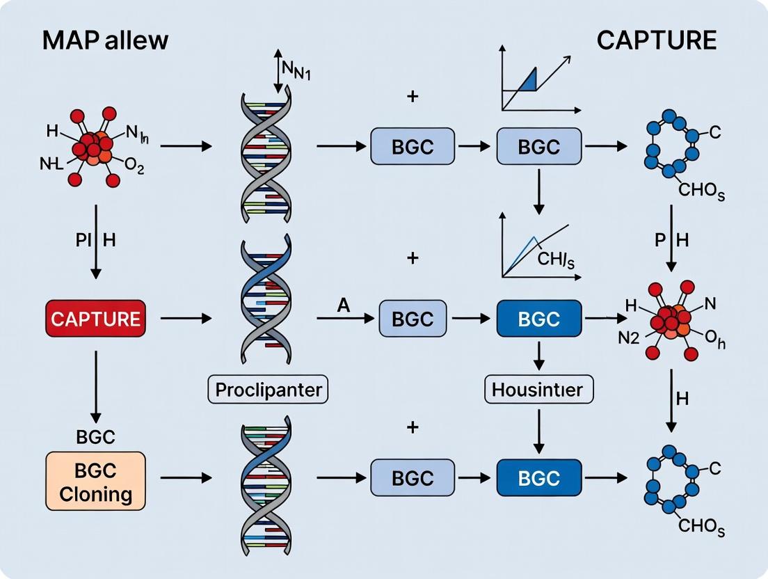
Title: CAPTURE Method Workflow for BGC Cloning
Title: Natural Product Discovery Pipeline from eDNA
The Scientist's Toolkit: Research Reagent Solutions
Table 3: Essential Materials for CAPTURE-based BGC Research
| Item | Function in Experiment | Example/Supplier |
|---|---|---|
| High Molecular Weight (HMW) DNA Kit | Isolation of intact, long DNA fragments from cells or environment for CAPTURE. | MagAttract HMW DNA Kit (Qiagen), Nanobind CBB Big DNA Kit (Circulomics). |
| Cas12a (Cpf1) Nuclease | Engineered nuclease for precise in vitro DNA cleavage with crRNA guidance. | Acidaminococcus sp. Cas12a (LbCpf1), NEB. |
| Custom crRNA Synthesis | Provides targeting specificity for Cas12a to flank the BGC of interest. | Integrated DNA Technologies (IDT), Synthego. |
| CAPTURE-ready Vector | Linearized cloning vector with pre-defined ends compatible with Cas12a overhangs. | pCAPTURE series (Addgene), custom synthesis. |
| GELase Enzyme | Agarose-digesting enzyme for gentle recovery of very large DNA fragments from gels. | GELase (Epicentre), AgarACE (Promega). |
| Electrocompetent E. coli (pir+) | Specialized E. coli strains for stable maintenance of single-copy BAC/CAPTURE vectors. | ElectroTen-Blue, Midi-λ pir. |
| Heterologous Expression Host | Engineered microbial chassis optimized for BGC expression and metabolite production. | Streptomyces coelicolor M1146, Pseudomonas putida KT2440. |
| LC-HRMS System | High-resolution metabolomics platform for detecting novel natural products. | Q-Exactive HF (Thermo), timsTOF (Bruker). |
Within the framework of the CAPTURE (Cas12a-Assisted Precise Targeted Cloning of Uncultivated and Rare Environmental) method for biosynthetic gene cluster (BGC) research, understanding BGC architecture is paramount. CAPTURE utilizes in vitro CRISPR-Cas12a cleavage and Gibson assembly to directly clone large, targeted BGCs from environmental DNA (eDNA) into expression vectors, bypassing host cultivation. This protocol and application note details the core architectural principles of BGCs and provides methodologies for their initial in silico and functional analysis, which are critical preludes to successful CAPTURE cloning and heterologous expression campaigns.
Core Architecture of Biosynthetic Gene Clusters
BGCs are organized sets of co-localized genes that encode the enzymatic machinery for the biosynthesis of a specialized metabolite. Their architecture follows logical, but highly variable, modular principles.
Key Genetic Modules and Their Functions
A typical BGC contains several functional modules, as summarized in Table 1.
Table 1: Core Functional Modules within a Canonical BGC
| Module Category | Primary Function | Key Gene Types/Examples | Frequency in Major BGC Classes (e.g., PKS/NRPS) |
|---|---|---|---|
| Core Biosynthetic | Scaffold assembly and modification | Polyketide synthase (PKS), Non-ribosomal peptide synthetase (NRPS), Hybrid PKS-NRPS, Tailoring enzymes (e.g., methyltransferases, oxidases) | 100% (Essential) |
| Regulatory | Transcriptional control of cluster expression | Pathway-specific regulators (SARPs, LALs), Two-component systems | ~80% (Common but not universal) |
| Resistance/Transport | Self-protection and metabolite export | Efflux pumps (MFS, ABC transporters), Antibiotic modification enzymes (e.g., acetyltransferases) | ~70% (Common) |
| Precursor Supply | Provision of unique building blocks | Enzymes for synthesizing non-proteinogenic amino acids or specialized polyketide extender units | ~50% (Cluster-dependent) |
Quantitative Landscape of BGCs
Recent genomic surveys reveal the scale and diversity of BGCs. Data is summarized in Table 2.
Table 2: Quantitative Overview of BGC Attributes Across Kingdoms
| Attribute | Bacterial Genomes (Avg.) | Fungal Genomes (Avg.) | Actinomycete Genomes (Avg.) | eDNA/Metagenomic Data |
|---|---|---|---|---|
| BGCs per Genome | 5-15 | 15-50 | 20-60 | N/A (Community-level) |
| Cluster Size Range | 10 - 200 kb | 15 - 150 kb | 30 - 200 kb | 10 - 250+ kb (detected) |
| GC Content | Often atypical from genomic average | Variable | Typically high (>70%) | Highly variable |
| Common Types | NRPS, PKS, RiPPs, Terpenes | NRPS, PKS, Terpenes, Alkaloids | NRPS, PKS (Type I/II), Hybrids | All types, with high novelty |
Diagram 1: BGC Core Modules and Context
Application Notes & Protocols for BGC Analysis Pre-CAPTURE
Protocol:In SilicoIdentification and Architecture Mapping of BGCs
Objective: To identify and annotate BGCs from whole genome sequencing (WGS) or metagenomic-assembled genome (MAG) data, providing the essential blueprint for designing CAPTURE cloning guides.
Materials & Workflow:
- Input Data: High-quality WGS assembly (contigs/scaffolds) in FASTA format.
- Bioinformatics Tools:
- BGC Prediction: antiSMASH (v7.0+), DeepBGC, PRISM.
- Annotation: Prokka, eggNOG-mapper, Pfam/InterProScan.
- Comparative Analysis: BiG-SCAPE, CORASON.
- Procedure:
- Step 1: Prediction. Run the assembled genome through antiSMASH with the
--fulland--clusterhmmerflags for comprehensive analysis. - Step 2: Annotation. Extract the genomic region of the predicted BGC. Annotate open reading frames (ORFs) using Prokka for prokaryotes or Funannotate for fungi.
- Step 3: Functional Assignment. Perform domain annotation on core biosynthetic genes using Pfam databases (e.g., via HMMER3) to identify Ketosynthase (KS), Adenylation (A), and Condensation (C) domains.
- Step 4: Architecture Map. Create a visual map of the BGC, plotting gene locations, orientations, and predicted functions using CLINK or a custom Python script with BioPython.
- Step 5: Guide Design for CAPTURE. Identify unique 23-25 bp sequences immediately flanking the target BGC region (within 1-5 kb). Use these to design CRISPR-Cas12a crRNA target sequences, ensuring they have minimal off-target matches in the host expression genome.
- Step 1: Prediction. Run the assembled genome through antiSMASH with the
Protocol: Heterologous Expression Readiness Assessment
Objective: To evaluate the suitability of a BGC for cloning and expression in a heterologous host (e.g., Streptomyces albus, Pseudomonas putida), a key consideration after CAPTURE cloning.
Materials & Workflow:
- Assessment Criteria:
- GC Content Disparity: Calculate BGC GC%. A difference of >10% from the expression host may cause transcriptional issues.
- Codon Usage Bias: Analyze using the CAI (Codon Adaptation Index). A score <0.7 suggests potential translation inefficiency.
- Regulatory Compatibility: Identify predicted promoter regions within the BGC (e.g., using BPROM). Assess if they are likely recognized by the host RNA polymerase or if replacement with a host-specific promoter is needed.
- Precursor Availability: Audit the BGC for essential precursor biosynthesis genes (e.g., for D-amino acids). Absence requires host engineering or media supplementation.
- Procedure:
- Step 1: Bioinformatic Analysis. Use
geeceefor GC content,cai(EMBOSS) for codon adaptation, andProditorfor promoter prediction. - Step 2: Tabling Results. Compile metrics into an assessment table (see Table 3).
- Step 3: Decision Matrix. A BGC scoring poorly on >2 criteria may require refactoring (e.g., codon optimization, promoter swap) prior to or after CAPTURE cloning.
- Step 1: Bioinformatic Analysis. Use
Table 3: Heterologous Expression Readiness Assessment Table
| BGC ID (e.g., from antiSMASH) | Size (kb) | GC Content (%) | Host GC% | CAI Score (vs. Host) | Dedicated Regulator? | Missing Precursor Genes? | Readiness Tier (High/Med/Low) |
|---|---|---|---|---|---|---|---|
| BGC_001 (NRPS) | 45.2 | 68.5 | 72.1 (S. albus) | 0.72 | Yes (SARP) | None detected | High |
| BGC_002 (PKS) | 82.7 | 52.1 | 61.5 (P. putida) | 0.58 | No | Specialized acyl-CoA synthase | Medium |
The Scientist's Toolkit: Research Reagent Solutions for BGC Cloning & Analysis
Table 4: Essential Reagents and Materials for BGC Research (Pre- and Post-CAPTURE)
| Item/Category | Specific Example(s) | Primary Function in BGC Research |
|---|---|---|
| Cloning & Assembly | CAPTURE Cas12a crRNA design oligos, Gibson Assembly Master Mix, T4 DNA Ligase | For precise in vitro cleavage and assembly of large BGC fragments into expression vectors. |
| Vector System | pCAP01-series vectors (e.g., pCAP01-oriT), BAC (Bacterial Artificial Chromosome) vectors | Shuttle vectors with conjugative origin (oriT) for large DNA transfer and stable maintenance in heterologous hosts. |
| Host Strains | E. coli GB05-dir, Streptomyces albus J1074, Pseudomonas putida KT2440 | Engineered cloning hosts (deficient in nucleases/recombination) and robust heterologous expression hosts. |
| DNA Extraction | Gel Extraction Kits (for >10 kb fragments), HMW (High Molecular Weight) DNA Extraction Kits | Isolation of intact, large DNA fragments from environmental samples or complex genomes. |
| Screening & Detection | Direct PCR screening primers, NGS library prep kits (Illumina/PacBio), Whole Genome Sequencing services | Validation of clone integrity and assessment of expression outcomes via transcriptomics. |
| Analysis Software | antiSMASH, BiG-SCAPE, Geneious, CLC Genomics Workbench | For in silico prediction, comparative analysis, and sequence design/management. |
Diagram 2: CAPTURE BGC Cloning Workflow
Within the broader thesis on the CAPTURE (Cas12a-Assisted Precise Targeted Cloning of Uncultivated and Recalcitrant Biosynthetic Gene Clusters) method, this document addresses the fundamental obstacles in cloning complex bacterial biosynthetic gene clusters (BGCs). Large size (>50 kb), repetitive sequences, and high GC-content (>70%) present synergistic challenges for conventional cloning techniques like PCR, cosmids, or BAC libraries, leading to frequent failures in isolating intact, functional clusters for heterologous expression and drug discovery.
Table 1: Characteristics of Problematic BGCs and Associated Cloning Issues
| BGC Characteristic | Typical Range | Direct Cloning Challenge | Consequence |
|---|---|---|---|
| Size | 50 - 200+ kb | Exceeds capacity of common vectors (e.g., cosmids ~45 kb). | Fragmented clones, incomplete pathway isolation. |
| GC Content | 70% - 85% | Hinders PCR amplification; promotes secondary structure. | Low yield, polymerase errors, sequence inaccuracies. |
| Repetitive Elements | Tandem repeats, modular PKS/NRPS domains | Homologous recombination in E. coli host. | Unstable inserts, deletions, rearrangements. |
| Host Toxicity | Expression of toxic intermediates in cloning host (e.g., E. coli) | Cell death upon cluster capture. | No viable clones recovered. |
Application Notes: The CAPTURE Method Rationale
The CAPTURE method is designed to overcome these hurdles by leveraging in vitro Cas12a cleavage and in vivo RecET-assisted assembly in a non-E. coli host (Pseudomonas putida). Key advantages include:
- Size Independence: Utilizes linear DNA recombineering, bypassing vector packaging limits.
- GC/Repeat Neutrality: In vitro Cas12a gRNA targeting is not impeded by GC-content; P. putida’s native recombination system handles repeats more faithfully than E. coli.
- Toxic Gene Tolerance: The P. putida chassis is more resilient to heterologous expression of bacterial toxins.
Detailed Protocol: CAPTURE Method for Complex BGCs
Objective: To isolate a large, GC-rich, repetitive BGC directly from genomic DNA into a expression-ready vector in P. putida.
Materials & Reagents: See "The Scientist's Toolkit" below.
Procedure:
- Bioinformatic Design:
- Identify BGC boundaries via antiSMASH analysis.
- Design two Cas12a crRNAs targeting sequences ~500 bp outside the 5’ and 3’ BGC boundaries. Ensure minimal off-target sites.
- Design 80-bp homology arms (HAs) corresponding to the vector ends and the regions just inside the crRNA cut sites. Synthesize these as oligonucleotides.
In Vitro Cas12a Cleavage and HA Ligation:
- Incubate 5-10 µg of high-quality genomic DNA with purified Cas12a protein and the two crRNAs in NEBuffer r2.1 at 37°C for 2 hours.
- Purify the linear, target BGC fragment (now excised from the genome) via gel extraction.
- Assemble a Gibson Assembly reaction with:
- 100 ng of the gel-purified BGC fragment.
- 50 ng of linearized CAPTURE vector (e.g., pCAPTURE-Ex).
- The two 80-bp HA oligonucleotides (0.1 µM final).
- Gibson Assembly Master Mix. Incubate at 50°C for 1 hour.
Transformation and Recombination in P. putida:
- Electroporate 2 µL of the Gibson Assembly reaction into electrocompetent P. putida KT2440 cells expressing the RecET system (e.g., strain PpRecET).
- Immediately add 1 mL of LB broth and recover at 30°C for 3 hours with shaking.
- Plate cells on LB agar containing the appropriate antibiotic (e.g., gentamicin). Incubate at 30°C for 36-48 hours.
Validation:
- Screen colonies by colony PCR using primers spanning the BGC-vector junctions.
- Perform whole plasmid sequencing on positive clones using a long-read platform (PacBio or Nanopore) to confirm intact, unrearranged insertion.
Visualizing the CAPTURE Workflow
CAPTURE Method Workflow for BGC Isolation
The Scientist's Toolkit
Table 2: Key Research Reagent Solutions for CAPTURE Protocol
| Reagent/Material | Supplier Examples | Function in Protocol |
|---|---|---|
| Cas12a (Cpfl) Protein | NEB, Thermo Fisher, IDT | Catalyzes specific double-strand breaks at BGC boundaries guided by crRNAs. |
| Custom crRNAs | IDT, Sigma-Aldrich | Guide Cas12a to precise genomic locations flanking the target BGC. |
| Gibson Assembly Master Mix | NEB, Thermo Fisher | Seamlessly joins the linear BGC fragment, vector, and homology arms in vitro. |
| P. putida KT2440 (RecET+) | Academic labs, in-house preparation | Specialized cloning host with efficient recombinase system for stable assembly of large/difficult DNA. |
| Electrocompetent P. putida Cells | Prepared in-house per protocol | Essential for high-efficiency transformation of large DNA assemblies. |
| Long-Read Sequencing Service | PacBio (Sequel IIe), Oxford Nanopore (PromethION) | Validates complete, accurate sequence of large, repetitive, GC-rich cloned BGCs. |
| High-Purity Genomic DNA Kit | Qiagen, Macherey-Nagel | Provides intact, high-molecular-weight DNA substrate for precise Cas12a cleavage. |
This application note details the integrated pipeline for discovering novel bioactive compounds from environmental microbes, framed within the broader thesis on the CAPTURE (Cas12a-Assisted Precise Targeted Cloning of Uncultivated Bacterial Genomic Regions) method for Biosynthetic Gene Cluster (BGC) research. The CAPTURE method revolutionizes the initial "Soil" phase by enabling direct, sequence-guided cloning of large BGCs (often >50 kb) from complex metagenomic DNA, bypassing the need for microbial cultivation. This protocol outlines the subsequent stages from cloned BGC to identified lead compound ("Screen"), creating a cohesive workflow for modern natural product discovery.
Application Notes: The Integrated Discovery Pipeline
Key Stages and Quantitative Success Metrics
The following table summarizes expected outcomes and efficiency gains using the CAPTURE-initiated pipeline compared to traditional cultivation-dependent approaches.
Table 1: Pipeline Stages, Methods, and Comparative Metrics
| Pipeline Stage | Core Activity | Primary Method(s) | Key Quantitative Metrics (CAPTURE-led) | Traditional Approach Metrics (Cultivation-dependent) |
|---|---|---|---|---|
| 1. Sample & BGC Identification | Environmental DNA extraction & target BGC selection | Metagenomic sequencing, bioinformatic analysis (e.g., antiSMASH) | 10-50 candidate BGCs per soil sample; BGC recovery specificity: >90% | 1-5 cultivable isolates per sample; BGC hit rate: <10% |
| 2. BGC Cloning | Isolation and vector assembly of target BGC | CAPTURE Method (in vitro Cas12a cutting & recombination) | Cloning efficiency: 70-95% for 40-80 kb clusters; throughput: 10-20 BGCs/week | Fosmid/cosmid library screening: <1% target hit rate; BAC cloning: low throughput |
| 3. Heterologous Expression | Production of compound in surrogate host | Recombinant expression in Streptomyces or E. coli hosts | Success rate: 30-60% for functional expression | Native strain fermentation: highly variable, often silent |
| 4. Compound Analysis | Detection, isolation, & structural elucidation | HPLC-MS, NMR, HR-MS | Detection sensitivity: ng/mL; dereplication speed: minutes via databases | Slower, requires large-scale cultivation |
| 5. Bioactivity Screening | Assessment of biological activity | Target-based or phenotypic assays (e.g., antimicrobial, cytotoxicity) | Hit rate from expressed BGCs: 5-20%; assay throughput: 10^3-10^5 compounds/year | Lower hit rate due to compound re-discovery |
The Scientist's Toolkit: Essential Research Reagent Solutions
Table 2: Key Reagents and Materials for the CAPTURE-led Pipeline
| Item | Function in Pipeline | Example Product/Catalog | Critical Specification |
|---|---|---|---|
| CAPTURE-specific Cas12a (Cpf1) | Enzyme for generating precise, 5’-overhang cuts at target BGC boundaries. | EnGen Lba Cas12a (NEB) | High in vitro cleavage activity, minimal star activity. |
| T4 DNA Polymerase | Creates complementary overhangs on CAPTURE vector for homologous recombination. | T4 DNA Polymerase (Thermo) | Controlled exonuclease activity for precise trimming. |
| Gibson Assembly Master Mix | One-step isothermal assembly of cut BGC and prepared vector. | Gibson Assembly HiFi Master Mix (NEB) | High efficiency for large fragment (>40 kb) assembly. |
| SuperCompetent Cells | Transformation of large, complex CAPTURE plasmid constructs. | E. cloni 10G SUPREME (Lucigen) | High efficiency (>1x10^9 cfu/µg) for large plasmids. |
| Induction Media | For heterologous expression of BGCs in Streptomyces hosts. | R5 or TSB media with appropriate inducers (e.g., thiostrepton) | Chemically defined, supports high antibiotic production. |
| Solid Phase Extraction (SPE) Cartridges | Rapid fractionation of crude culture extracts for activity screening. | Strata X polymeric reversed-phase (Phenomenex) | Broad-spectrum capture of small molecules. |
| LC-MS Grade Solvents | For high-resolution metabolomic analysis and compound purification. | Acetonitrile, Methanol (e.g., Fisher Optima) | Low UV cutoff, minimal ion suppression. |
| Cell-Based Assay Kits | Primary bioactivity screening (e.g., antimicrobial, cytotoxicity). | BacTiter-Glo (Promega), Resazurin Viability Assay | High sensitivity, robustness for natural product extracts. |
Detailed Experimental Protocols
Protocol: CAPTURE Method for Targeted BGC Cloning from Metagenomic DNA
Objective: To clone a targeted 50-80 kb Biosynthetic Gene Cluster from purified environmental DNA into a heterologous expression vector.
Materials: Purified high-molecular-weight metagenomic DNA (>100 kb), CAPTURE vector (linearized with Cas12a recognition sites), EnGen Lba Cas12a, crRNAs targeting BGC flanks, Gibson Assembly Master Mix, E. cloni 10G cells, SOC media, selective agar plates.
Procedure:
- Bioinformatic Design: Identify BGC boundaries via antiSMASH. Design two crRNAs targeting sequences ~50-80 kb apart, immediately outside the BGC borders.
- In Vitro Cleavage: a. Set up a 50 µL reaction: 2 µg metagenomic DNA, 1 µM each crRNA, 50 nM Cas12a, 1x NEBuffer r2.1. b. Incubate at 37°C for 1 hour, then 65°C for 20 minutes to denature Cas12a.
- Vector Preparation: Digest the CAPTURE vector with the same Cas12a/crRNA combination to generate complementary 5’ overhangs.
- Homology Arm Generation: Treat the cleaved vector with T4 DNA Polymerase (in the presence of specific dNTPs) to create 20-40 bp overhangs homologous to the ends of the target BGC fragment.
- Gibson Assembly: Mix 50-100 ng of size-selected (50-80 kb) cleaved metagenomic DNA with a 3:1 molar ratio of prepared vector. Add Gibson Assembly Master Mix to 1x. Incubate at 50°C for 60 minutes.
- Transformation & Screening: Desalt the assembly mixture and transform into 50 µL of electrocompetent E. cloni 10G cells. Plate on selective agar. Screen colonies by PCR using BGC-specific internal primers.
- Validation: Isolate plasmid DNA from positive clones and confirm by restriction digest and pulsed-field gel electrophoresis or long-read sequencing.
Protocol: Heterologous Expression and Metabolite Analysis inStreptomyces albusJ1074
Objective: To express the cloned BGC and analyze the produced metabolome.
Materials: S. albus J1074 strain, CAPTURE-BGC plasmid, Thiostrepton, R5 liquid and solid media (without sucrose), Ethyl Acetate, Methanol, LC-MS system.
Procedure:
- Conjugal Transfer: Introduce the CAPTURE-BGC plasmid into S. albus via E. coli ET12567/pUZ8002 intergeneric conjugation. Select exconjugants on R5 plates containing appropriate antibiotics (e.g., apramycin) and nalidixic acid.
- Seed Culture: Inoculate a single exconjugant into 10 mL of TSB medium with antibiotics. Incubate at 30°C, 220 rpm for 48 hours.
- Production Culture: Inoculate 1 mL of seed culture into 50 mL of R5 production medium (no sucrose) with antibiotics. Add thiostrepton (5 µg/mL) for induction if the vector contains a tipA promoter. Incubate at 30°C, 220 rpm for 5-7 days.
- Metabolite Extraction: Centrifuge culture at 4000 x g for 10 min. Separately extract the supernatant (with equal volume ethyl acetate) and cell pellet (with 1:1 methanol:water). Combine organic phases and evaporate to dryness.
- LC-MS Analysis: Resuspend extract in methanol. Analyze by HPLC coupled to high-resolution mass spectrometry (e.g., UHPLC-QTOF). Use a C18 column with a water-acetonitrile gradient (+0.1% formic acid). Acquire data in positive and negative ionization modes.
- Dereplication: Process MS data (MS1 and MS/MS) with software (e.g., MZmine, GNPS) and compare against natural product databases (GNPS, AntiBase) to identify known compounds.
Visualization of Workflows and Pathways
Title: The Soil-to-Screen Discovery Pipeline
Title: CAPTURE Method Workflow for BGC Cloning
1. Introduction and Thesis Context
The discovery of novel natural products from microbial biosynthetic gene clusters (BGCs) is bottlenecked by inefficient cloning strategies. The broader thesis of this research posits that the CAPTURE (Cas12a-Assisted Precise Targeted Cloning of Uncultivated Bacterial Genomic DNA for Expression) method represents a fundamental paradigm shift, enabling the high-throughput, sequence-independent, and faithful cloning of large BGCs directly from complex environmental samples. This application note details the protocols and data supporting this thesis.
2. Core Principle and Comparative Advantage
CAPTURE utilizes a trans-acting CRISPR-Cas12a system. Guide RNAs (crRNAs) are designed to flank a target BGC. Cas12a, upon recognition, introduces double-strand breaks upstream and downstream of the BGC. Critically, Cas12a’s non-specific single-stranded DNA (ssDNA) nicking activity (collateral cleavage) is harnessed to degrade off-target genomic DNA, while the target BGC, protected by a RecA nucleoprotein filament, is selectively purified and cloned.
3. Key Experimental Data Summary
Table 1: Comparison of BGC Cloning Methods
| Method | Throughput | Max Insert (kb) | Fidelity | Source DNA Compatibility |
|---|---|---|---|---|
| CAPTURE | High | >100 kb | High (sequence-independent) | Metagenomic, Cultured |
| Fosmid/Cosmid | Low-Moderate | ~40 kb | High | Cultured, Purified |
| TAR/YAC | Low | >100 kb | High (sequence-dependent) | Purified |
| Direct PCR | Moderate | <30 kb | Risk of mutations | Purified |
Table 2: Representative CAPTURE Cloning Efficiency
| Target BGC | Size (kb) | Source | Colonies Screened | Positive Hits | Success Rate |
|---|---|---|---|---|---|
| Nonribosomal Peptide Synthetase (NRPS) | 45 | Soil Metagenome | 384 | 112 | 29.2% |
| Polyketide Synthase (PKS) | 78 | Marine Sediment | 192 | 41 | 21.4% |
| Hybrid PKS-NRPS | 102 | Actinomycete Culture | 288 | 67 | 23.3% |
4. Detailed Protocol: CAPTURE from Metagenomic DNA
Materials:
- Metagenomic DNA (>50 kb fragment size).
- RecA protein (key reagent for target protection).
- AsCas12a (Cpfl) nuclease.
- Custom crRNA pair (Alt-R CRISPR-Cas12a crRNAs).
- ATP regeneration system (creatine kinase, creatine phosphate).
- ATPγS (for forming stable RecA filaments).
- Linearized cloning vector (e.g., pCAPTURE, containing homologous arms).
- Exonuclease (Lambda exonuclease, ExoIII mix) for ssDNA digestion.
- Gibson Assembly or In-Fusion cloning mix.
- Electrocompetent E. coli (e.g., TransforMax EPI300).
Procedure:
- BGC Targeting and Protection:
- Incubate 200-500 ng of metagenomic DNA with 10 µM RecA, 2.5 mM ATPγS, and 1x RecA buffer (30 mM Tris-acetate, 20 mM Mg-acetate, 1 mM DTT) for 15 min at 37°C.
- Add the crRNA pair (final 100 nM each) and AsCas12a (final 100 nM). Incubate for 1 hour at 37°C. Cas12a cuts flanking regions while RecA filament protects the target BGC from collateral cleavage.
Purification of Protected Fragment:
- Add exonuclease mix (Lambda exonuclease and RecJf) to digest unprotected ssDNA for 30 min at 37°C.
- Purify the reaction using size-selective magnetic beads (e.g., SPRIselect) at a 0.5x ratio to retain large fragments. Elute in 20 µL nuclease-free water.
Assembly and Transformation:
- Perform a Gibson Assembly reaction with 10 µL of purified DNA and 50 ng of linearized pCAPTURE vector (harboring 50 bp homology to the crRNA-defined ends) for 1 hour at 50°C.
- Desalt the assembly mixture and electroporate into 50 µL of electrocompetent E. coli. Recover in 1 mL SOC for 2 hours at 37°C.
Screening:
- Plate on selective media. Perform colony PCR using vector-specific and internal BGC primers.
- For high-throughput, use pooled colony PCR or transposon-sequencing of plasmid pools.
5. The Scientist's Toolkit: Key Research Reagent Solutions
Table 3: Essential Materials for CAPTURE
| Item | Function/Description | Example Product |
|---|---|---|
| AsCas12a (Cpfl) Nuclease | RNA-guided endonuclease for precise double-strand breaks and collateral ssDNA cleavage. | IDT Alt-R AsCas12a (Cpfl) |
| Alt-R CRISPR-Cas12a crRNA | Custom guide RNA for targeting BGC flanks. Chemically synthesized, high purity. | IDT Alt-R CRISPR-Cas12a crRNA |
| RecA Protein (E. coli) | Forms nucleoprotein filament on target BGC, protecting it from Cas12a collateral cleavage. | New England Biolabs RecA Protein |
| ATPγS (Adenosine 5′-O-[3-thiotriphosphate]) | A non-hydrolyzable ATP analog for forming stable RecA-DNA filaments. | Sigma-Aldrich ATPγS |
| Size-Selective Magnetic Beads | For clean-up and size selection of large DNA fragments post-digestion. | Beckman Coulter SPRIselect |
| Gibson Assembly Master Mix | Enzymatic assembly of protected BGC fragment into linearized vector. | NEB Gibson Assembly HiFi Master Mix |
| Electrocompetent E. coli | High-efficiency transformation of large plasmid constructs. | Lucigen TransforMax EPI300 |
6. Visualized Workflows and Pathways
CAPTURE Method Experimental Workflow
Molecular Principle of CAPTURE: Protection vs. Cleavage
Step-by-Step Protocol: Implementing the CAPTURE Method in Your Lab
This document details the application and protocols for an advanced in vivo excision and circularization technique, developed as a core component of the broader CAPTURE (Cas12a-assisted precise targeted cloning using in vivo Cre recombination) method. CAPTURE is designed to address the critical bottleneck in natural product discovery: the efficient cloning of large, complex Bacterial Biosynthetic Gene Clusters (BGCs) for heterologous expression and characterization. This principle leverages the programmability of CRISPR-Cas12a for specific double-strand break induction and the high-efficiency site-specific recombination of Cre recombinase to directly excise and circularize target BGCs within the native host, prior to extraction and transformation.
Core Principle & Workflow
The method involves the introduction of two key genetic elements into the native bacterial host containing the target BGC:
- A synthetic "Capture Plasmid" harboring a loxP site and a selectable marker.
- A second plasmid (or integrated construct) expressing Cas12a and a custom CRISPR RNA (crRNA), and the Cre recombinase.
The crRNA is designed to target sequences flanking the BGC. Upon expression, Cas12a induces double-strand breaks at these two flanking sites, releasing the linear BGC fragment. Simultaneously, Cre recombinase mediates recombination between the loxP site pre-inserted within the BGC (via prior engineering or natural occurrence) and the loxP site on the Capture Plasmid. This action circularizes the excised BGC along with the Capture Plasmid backbone, creating a stable, extractable, and shuttable circular product ready for transformation into a heterologous host.
Key Research Reagent Solutions
| Reagent/Material | Function in CAPTURE | Key Features/Considerations |
|---|---|---|
| pCAP01 Vector (Capture Plasmid) | Provides backbone for in vivo circularization. Contains loxP site, origin of replication (ori) for E. coli and target host, and selectable marker(s). | Must be compatible with native host replication. Often includes an integrase for site-specific integration upstream of the BGC. |
| Cas12a (Cpfl) Expression System | RNA-guided endonuclease for generating specific double-strand breaks flanking the BGC. | Requires crRNA with a 5' TTTN PAM sequence. Known for minimal off-target effects and ability to process its own crRNA array. |
| Cre Recombinase Expression System | Catalyzes site-specific recombination between loxP sites, circularizing the excised fragment. | Can be expressed constitutively or inducibly. High-efficiency recombination is critical for yield. |
| Synthetic crRNA Array | Guides Cas12a to genomic locations immediately upstream and downstream of the BGC. | Typically designed as a single transcript with two spacers. Specificity must be validated in silico. |
| BGC-Specific loxP Donor | Used to insert a loxP site at one boundary of the BGC if a native site is absent. | Can be delivered via conjugative plasmid or CRISPR-mediated homologous recombination. |
| Heterologous Expression Host | Streptomyces spp. (e.g., S. albus), Pseudomonas putida, E. coli (with specialized genetics). | Engineered for high BGC expression, lacking competing pathways, and compatible with the Capture Plasmid ori and markers. |
Detailed Application Notes & Quantitative Data
Note 1: crRNA Design & PAM Requirement Cas12a recognizes a 5' T-rich PAM (e.g., TTTN, TTTV). Successful excision requires two such PAM sequences oriented outwards from the BGC boundaries. Efficiency drops significantly with PAM sequences >TTTV.
Note 2: Cre-loxP Recombination Efficiency Circularization efficiency is the yield-limiting step. Using a strongly expressed, codon-optimized cre gene and perfectly spaced loxP sites (e.g., 611 bp apart in the final construct) maximizes yield.
Note 3: Host Compatibility The method has been successfully adapted for high-GC content Actinobacteria (e.g., Streptomyces). Electroporation protocols for the Capture and Cas12a/Cre plasmids must be optimized for each host genus.
Table 1: Representative Efficiency Metrics for CAPTURE on Model BGCs
| BGC Size (kb) | Host Organism | Excision Efficiency* (%) | Circularization/Cloning Success Rate (%) | Heterologous Expression Success |
|---|---|---|---|---|
| 15 kb | Streptomyces coelicolor | >95 | ~90 | Positive (known compound detected) |
| 30 kb | Streptomyces ambofaciens | ~80 | ~70 | Positive (novel analog detected) |
| 50 kb | Myxococcus xanthus | ~65 | ~50 | Positive (requires optimized culture conditions) |
*Efficiency determined by PCR analysis of post-excision genomic DNA.
Experimental Protocols
Protocol 5.1: Vector Assembly and Preparation
- Clone crRNA Array: Synthesize an oligonucleotide duplex encoding two 20-23 nt spacers targeting sequences 100-500 bp outside the BGC boundaries. Clone into a Cas12a crRNA expression vector (e.g., pCRISPR-Cpfl).
- Prepare Capture Plasmid: Engineer the pCAP01 vector to contain a loxP site, an ori for both native and heterologous hosts, and an apramycin resistance gene. Linearize if necessary.
- Assemble Helper Plasmid: Co-clone the Cas12a gene and the cre recombinase gene under inducible promoters (e.g., tetR/Ptet) into a single vector with a thiostrepton resistance marker (pHelper).
Protocol 5.2: In Vivo Excision & Circularization in Native Host
- Introduce Genetic Elements: Conjugally transfer or electroporate the following into the native BGC host: (i) the Capture Plasmid, (ii) the pHelper plasmid (Cas12a+Cre).
- Integrate Capture Plasmid: Select for apramycin resistance to ensure Capture Plasmid maintenance. If using an integrating version, verify site-specific integration adjacent to the BGC via PCR.
- Induce Excision & Circularization: Add inducing agents (e.g., anhydrotetracycline) to the culture to activate Cas12a and Cre expression. Incubate for 12-48 hours.
- Harvest & Validate: Extract total genomic DNA. Perform diagnostic PCR using outward-facing primers from within the BGC and the Capture Plasmid backbone to confirm successful circularization. Use control PCRs to check for residual unexcised chromosomal DNA.
Protocol 5.3: Product Recovery & Heterologous Expression
- Enrich Circular Product: Treat the total DNA extract with Plasmid-Safe ATP-Dependent DNase to degrade linear genomic DNA, enriching for circularized molecules.
- Transform Heterologous Host: Electroporate the DNase-treated DNA into competent cells of the expression host (e.g., S. albus).
- Screen & Ferment: Select transformants on apramycin plates. Validate by plasmid restriction digest and PCR. Inoculate validated clones into production media and analyze metabolite profiles via LC-MS.
Visualization Diagrams
CAPTURE Method Core Workflow
Cas12a-Mediated Dual DSB Induction
Within the broader thesis on the CAPTURE (Cas12a-Assisted Precise Targeted Cloning of Uncultivated and Rare Environmental) method for Biosynthetic Gene Cluster (BGC) cloning, this document details the initial, critical stage. Precise design and synthesis of CRISPR-Cas12a (Cpf1) guide RNA (crRNA) arrays and homologous recombination (HR) donor vectors are foundational for the selective excision and capture of large, complex genomic regions from environmental DNA. This stage directly impacts the efficiency and fidelity of downstream cloning and heterologous expression efforts in natural product drug discovery.
Key Principles & Design Considerations
crRNA Array Design for Cas12a
Cas12a recognizes a T-rich Protospacer Adjacent Motif (PAM: 5'-TTTV-3', where V is A, C, or G) located upstream (5') of the target protospacer. Each crRNA consists of a direct repeat (DR) sequence followed by a 23-25 nt spacer complementary to the target.
Design Rules:
- Spacer Selection: Identify 23-25 nt sequences directly downstream of a valid PAM site on the non-target strand.
- Specificity: Perform BLAST analysis against the host genomic background (e.g., E. coli cloning host) to avoid off-target cleavage.
- Array Architecture: Multiple crRNAs (typically 2-4) targeting both ends of the BGC are transcribed as a single array from a common promoter. The Cas12a processor cleaves them into individual units.
- Efficiency Prediction: Use published scoring algorithms (e.g., from Doench et al., or proprietary tools from IDT, Synthego) to rank candidate spacers.
Donor Vector Design for Homologous Recombination
The donor vector provides homology arms for precise repair after dual CRISPR-Cas12a cleavage, facilitating the insertion of the excised BGC into a capture vector backbone.
Design Rules:
- Homology Arm Length: Optimal arms are 500-1000 bp for high-efficiency recombination of large fragments (>30 kb).
- Arm Source: Sequences must be identical to the regions immediately outside and flanking the targeted BGC boundaries.
- Vector Backbone: Contains an origin of replication (ori), selection marker(s) (e.g., antibiotic resistance), and elements for downstream manipulation (e.g., in vitro transcription promoters, recombinase sites).
Table 1: Optimized Parameters for crRNA Array and Donor Vector Design in CAPTURE
| Component | Parameter | Optimal Value / Sequence | Rationale & Notes |
|---|---|---|---|
| Cas12a System | Enzyme Variant | Lachnospiraceae bacterium Cas12a (LbCas12a) | High activity, common commercial availability. |
| PAM Sequence | 5'-TTTV-3' (V = A, C, G) | Defines target site search. | |
| crRNA Spacer | Length | 24 nucleotides | Balance of specificity and efficiency. |
| GC Content | 40-60% | Avoids secondary structure, improves stability. | |
| Off-target Limit | ≤3 mismatches in seed region (PAM-proximal 10-12 nt) | Minimizes unintended cleavage. | |
| crRNA Array | Number of Spacers per Target Site | 2 | Increases cleavage probability at each boundary. |
| Direct Repeat (DR) | 5'-AAUUUCUACUAAGUGUAGAUGAGGUUUU-3' | Standard LbCas12a DR sequence. | |
| Donor Vector | Homology Arm Length | 800 bp | High recombination efficiency for large inserts. |
| Cloning Backbone | Linearized vector with negative selection marker (e.g., ccdB) | Counterselection against empty vector improves yield. | |
| Synthesis | Array Synthesis Method | dsDNA fragment (gBlock) with T7 promoter | Cost-effective, high-fidelity for array cloning. |
Detailed Protocols
Protocol 4.1:In SilicoDesign of crRNA Arrays
Objective: To computationally identify and validate high-efficiency crRNA spacers targeting the flanking regions of a BGC.
Materials:
- Genomic sequence file (FASTA) containing the target BGC and surrounding region (~5 kb flanking each side).
- Bioinformatics software: Benchling, SnapGene, or command-line tools (UGENE, BLAST+).
- Potential off-target genome database (e.g., E. coli Genbank file).
Method:
- Define BGC Boundaries: Precisely annotate the start and end coordinates of the target BGC within the genomic context.
- Identify PAM Sites: Scan the non-coding regions immediately outside both the 5' and 3' BGC boundaries (approx. 500 bp) for all occurrences of the "TTTV" PAM sequence.
- Extract Spacer Candidates: For each valid PAM, extract the 24 nucleotides directly downstream (3') of it. This sequence is the candidate spacer.
- Filter for Specificity: Perform a local BLASTN alignment of each candidate spacer against the genome of the intended cloning host (e.g., E. coli MG1655). Discard any spacer with >85% identity over >15 nt.
- Rank and Select: For each BGC boundary, select the top 2 candidate spacers based on:
- Position (closest to BGC edge without being inside it).
- GC content (40-60%).
- Absence of homopolymer runs (>4 nt).
- Predicted efficiency score from online tools (e.g., IDT Alt-R CRISPR-Cas12a guide RNA design tool).
- Design Array Oligo: Concatenate spacers in the format: [T7 Promoter] - [DR-Spacer1] - [DR-Spacer2] - [Terminator]. Order as a double-stranded DNA fragment (gBlock).
Protocol 4.2: Construction of the Donor Vector via Gibson Assembly
Objective: To clone the designed homology arms into a linearized capture vector backbone.
Materials:
- Backbone Vector: Linearized vector with ccdB counterselection cassette (e.g., pCAPURE-backbone).
- PCR Primers: Designed with 20-30 bp overhangs complementary to the linearized vector ends.
- High-Fidelity DNA Polymerase (e.g., Q5, Phusion).
- Gibson Assembly Master Mix (commercial or prepared in-house).
- Chemically Competent E. coli (e.g., NEB Stable or DH5α).
Method:
- Amplify Homology Arms:
- Using the source genomic DNA as template, perform two separate PCRs to amplify the Left Homology Arm (LHA) and Right Homology Arm (RHA).
- Primer design: Include 5' overhangs (≥20 bp) that are complementary to the ends of the linearized backbone vector.
- Run PCR products on an agarose gel and purify using a gel extraction kit.
- Prepare Vector Backbone: Digest the donor vector plasmid with appropriate restriction enzymes to linearize it and expose ends compatible with the Gibson overhangs. Purify the linearized vector.
- Gibson Assembly:
- Set up a 10-20 µL assembly reaction: 50-100 ng linearized backbone, 0.2 pmol of each purified homology arm PCR product, 1X Gibson Assembly Master Mix.
- Incubate at 50°C for 15-60 minutes.
- Transformation and Screening:
- Transform 2 µL of the assembly reaction into 50 µL of competent E. coli. Plate on agar containing the appropriate antibiotic.
- Screen colonies by colony PCR using primers that anneal within the vector backbone and the inserted homology arm.
- Validate positive clones by Sanger sequencing across the insertion junctions.
Diagrams
Diagram 1: crRNA Array Design Workflow (88 chars)
Diagram 2: Donor Vector Assembly via Gibson (78 chars)
The Scientist's Toolkit
Table 2: Essential Research Reagent Solutions for Stage 1
| Reagent / Material | Supplier Examples | Function in Protocol |
|---|---|---|
| High-Fidelity DNA Polymerase | NEB (Q5), Thermo Fisher (Phusion), Takara (KOD) | Error-free PCR amplification of homology arms and other constructs. |
| Gibson Assembly Master Mix | NEB HiFi, SGI, homemade | Seamless, one-pot assembly of multiple DNA fragments with overlapping ends. |
| Chemically Competent E. coli | NEB Stable, DH5α, TOP10 | Cloning and propagation of plasmid DNA after assembly. |
| Gel Extraction & PCR Purification Kits | Qiagen, Macherey-Nagel, Zymo Research | Purification of DNA fragments from agarose gels or PCR reactions. |
| Cas12a (Cpf1) Expression Vector | Addgene (pY016, pFGA442), commercial sources | Source of LbCas12a protein for in vitro cleavage validation. |
| T7 Transcription Kit | NEB HiScribe, Thermo Fisher | In vitro transcription of crRNA arrays for validation assays. |
| Synthetic dsDNA Fragments (gBlocks) | IDT, Twist Bioscience, GenScript | Fast, accurate source of designed crRNA array sequences. |
| CRISPR Design Software | Benchling, IDT Alt-R Design, CHOPCHOP | In silico guide RNA design, specificity checking, and efficiency prediction. |
Application Notes
This protocol details the second stage of the CAPTURE (Cas12a-Assisted Precise Targeted Cloning of Uncultivable and Recalcitrant) method, which is critical for capturing large, complex Biosynthetic Gene Clusters (BGCs) directly from environmental or recalcitrant microbial DNA. Following Stage 1 (in vitro CAPTURE assembly), Stage 2 focuses on transferring the cloned BGC into a native or alternative heterologous host via conjugation, where final in vivo assembly via homologous recombination occurs. This leverages the host's natural DNA repair machinery to circularize the construct into a stable, single-copy plasmid, enabling subsequent heterologous expression and functional analysis of the encoded natural products.
The success of this stage is quantitatively dependent on several key parameters, which are summarized in Table 1.
Table 1: Key Quantitative Parameters for Conjugative Transfer and in vivo Assembly
| Parameter | Optimal Range/Target | Impact on Efficiency |
|---|---|---|
| Donor (E. coli)/Recipient Cell Ratio | 1:10 to 1:1 (Recipient in excess) | Maximizes mating pair formation; excess donor can inhibit recipient growth. |
| Conjugation Co-incubation Time | 6-18 hours | Time-dependent; longer incubation increases transfer but risks overgrowth of donors. |
| in vivo Assembly Homology Arm Length | 500-1000 bp per arm | Shorter arms (<300 bp) drastically reduce recombination efficiency. |
| Typical Conjugation Frequency (for E. coli to Streptomyces) | 10⁻⁵ to 10⁻³ per recipient cell | Benchmark for protocol optimization; varies widely by recipient strain. |
| Post-Conjugation Antibiotic Selection Delay | 24-48 hours | Critical for expression of antibiotic resistance markers post-transfer and recombination. |
| Average CAPTURE Plasmid Size for Efficient Transfer | 30 - 80 kbp | Efficiency declines significantly for constructs >100 kbp. |
Experimental Protocols
Protocol 1: Biparental Conjugation fromE. coliET12567/pUZ8002 to Actinobacterial Recipient
This protocol transfers the linear CAPTURE assembly product from an E. coli donor, harboring the conjugation helper plasmid pUZ8002, to an actinobacterial recipient (e.g., Streptomyces coelicolor).
Preparation:
- Donor Strain: Grow E. coli ET12567/pUZ8002 containing the CAPTURE assembly product in LB with appropriate antibiotics (e.g., kanamycin, apramycin) at 37°C to mid-log phase (OD₆₀₀ ~0.4-0.6).
- Recipient Strain: Grow the actinobacterial recipient strain in a suitable liquid medium (e.g., TSBS for Streptomyces) to produce young, viable hyphal fragments or spores. Wash cells twice with fresh, antibiotic-free medium.
Mating:
- Mix donor and recipient cells at a ratio of 1:1 to 1:10 (donor:recipient) in a final volume of 1 mL. Pellet the mixed cells.
- Resuspend the pellet in 100 µL of medium and spot onto the center of a pre-dried, non-selective agar plate (e.g., SFM or ISP4 medium for Streptomyces).
- Incubate at the recipient's optimal temperature (e.g., 30°C) for 6-18 hours to allow conjugation.
Selection and in vivo Assembly:
- After incubation, overlay the conjugation spot with 1 mL of sterile water containing 1 mg of the appropriate antibiotic (e.g., apramycin) to select for exconjugants that have received and assembled the construct. The antibiotic must counter-select against the E. coli donor.
- Incubate plates for a further 24-48 hours before a second overlay with antifungal agent (e.g., nystatin) to inhibit fungal contaminants.
- Continue incubation at the recipient's temperature for 5-10 days until exconjugant colonies appear.
- The linear construct undergoes RecA-mediated homologous recombination in vivo via the terminal homology arms, circularizing into a stable, single-copy plasmid within the recipient.
Protocol 2: Triparental Conjugation for Non-Actinobacterial Hosts
For recipients where pUZ8002 is inefficient, a helper plasmid (e.g., pRK2013) in a third E. coli strain can mobilize the CAPTURE construct.
- Prepare donor, helper (E. coli with pRK2013), and recipient cultures separately to mid-log phase.
- Mix all three strains in approximately equal volumetric ratios on a filter placed on a non-selective agar plate.
- Incubate for 6-24 hours.
- Resuspend the cell mixture and plate onto selective media containing antibiotics that select for the CAPTURE construct and the recipient while counter-selecting against both E. coli strains.
Visualizations
Title: Stage 2 Workflow: Conjugation to In Vivo Assembly
Title: In Vivo Circularization via Homology Arms
The Scientist's Toolkit
Table 2: Essential Research Reagents & Materials
| Item | Function in Stage 2 |
|---|---|
| E. coli ET12567/pUZ8002 | Non-methylating donor strain containing the conjugation helper plasmid (pUZ8002) which provides mob and tra genes for transfer. |
| pRK2013 Helper Plasmid | Alternative conjugation helper for triparental matings, providing RK2 transfer functions in trans. |
| Non-Methylating E. coli Strain (e.g., ET12567) | Essential for propagating DNA prior to conjugation into GC-rich actinobacteria that possess potent restriction-modification systems against methylated E. coli DNA. |
| Species-Specific Solid Mating Media (e.g., SFM, ISP4) | Provides optimal physiological conditions for both donor and recipient cell contact and DNA transfer during conjugation. |
| Selective Antibiotics (Apramycin, Thiostrepton, etc.) | For post-conjugation selection of exconjugants and counter-selection against the E. coli donor strain. |
| Recipient Strain Spores/Mycelia | The native or alternative heterologous host (e.g., Streptomyces coelicolor, Pseudomonas putida) that will perform the final in vivo assembly and express the BGC. |
| Homology Arms (500-1000 bp) | Flanking sequences on the linear construct that are identical to the target regions on the recipient's chromosome or plasmid, guiding precise in vivo recombination. |
This protocol details the third critical stage of the CAPTURE (Cas12a-Assisted Precise Targeted Cloning of Uncultivated and Recombinant Enzymes) method for Biosynthetic Gene Cluster (BGC) cloning. Following successful in-situ capture and purification (Stage 2), the target DNA must be excised from the capture vector, circularized into a functional plasmid, and rigorously validated. This stage transforms the linear captured product into a stable, propagatable construct suitable for heterologous expression and functional analysis, a cornerstone of natural product discovery pipelines.
Application Notes
- Circularization Efficiency: The efficiency of intramolecular ligation is highly dependent on insert concentration. Overly high concentrations favor intermolecular ligation, creating concatemers. Accurate quantification of eluted DNA is crucial.
- Host Selection: The choice of E. coli strain for transformation is critical. Strains with enhanced recombination proficiency (e.g., pir+ for R6K-based vectors) or deficient in nucleases and recombinases (e.g., recA-) are often required for maintaining large, complex, or repetitive BGC constructs.
- Validation Cascade: A multi-tiered validation approach, progressing from rapid, coarse-grained checks (PCR, digestion) to comprehensive analysis (long-read sequencing), conserves resources and ensures construct fidelity before committing to lengthy heterologous expression trials.
- Troubleshooting: Failure to obtain circularized clones often stems from insufficient purification of the excised fragment (carryover of agarose or salts inhibiting ligase) or incorrect vector:insert ratio during circularization.
Experimental Protocols
Protocol A: Restriction Enzyme-Mediated Excision and Purification
This protocol releases the captured BGC insert from the CAPTURE vector backbone using flanking, rare-cutting restriction enzymes.
- Set up the Excision Digest:
- Combine in a nuclease-free microcentrifuge tube:
- Purified CAPTURE product (from Stage 2): 1-2 µg
- ㅤ10X Buffer for restriction enzymes: 5 µL
- ㅤNotI-HF (or other designated rare-cutter): 2 µL (20 units)
- ㅤPacI (or other designated rare-cutter): 2 µL (20 units)
- ㅤNuclease-free water to a final volume of 50 µL.
- Mix gently and centrifuge briefly.
- Combine in a nuclease-free microcentrifuge tube:
- Incubate: Place the reaction in a thermocycler or water bath at 37°C for 3 hours.
- Fragment Purification: Separate the digestion products by electrophoresis on a 0.8% low-melting point agarose gel. Visualize with low-intensity UV light to minimize DNA damage. Excise the gel slice containing the high-molecular-weight BGC insert (typically >20 kb).
- DNA Recovery: Purify the DNA from the gel slice using a commercially available kit designed for large fragment recovery (e.g., Zymoclean Large Fragment DNA Recovery Kit). Elute in 15 µL of nuclease-free water or 10 mM Tris-HCl (pH 8.5). Quantify using a fluorometric assay (e.g., Qubit HS dsDNA assay).
Protocol B: Intramolecular Ligation (Circularization)
This protocol circularizes the purified, excised BGC fragment via intramolecular ligation.
- Determine DNA Concentration: Accurately measure the concentration of the purified insert (from Protocol A, Step 4) using a fluorometric assay. Typical yields range from 20-100 ng.
- Set up the Ligation Reaction:
- Combine in a nuclease-free tube:
- Purified BGC insert: 20-50 ng
- ㅤ2X Quick Ligase Buffer: 12.5 µL
- ㅤQuick T4 DNA Ligase: 1 µL (400 cohesive end units)
- ㅤNuclease-free water to a final volume of 25 µL.
- Critical: The total mass of DNA should be low (20-50 ng) to favor intramolecular ligation. The reaction contains no additional vector backbone.
- Combine in a nuclease-free tube:
- Incubate: Incubate at room temperature (20-25°C) for 1 hour.
- Reaction Cleanup: Purify the ligation product using a DNA clean-up kit (e.g., DNA Clean & Concentrator-5) to remove salts and enzymes. Elute in 10 µL of elution buffer.
Protocol C: Transformation and Primary Validation
This protocol transforms the circularized product into a suitable E. coli host and performs initial validation.
- Transformation:
- Thaw 50 µL of electrocompetent E. coli (e.g., TransforMax EPI300 or DH10B pir+ for R6K vectors) on ice.
- Add 5 µL of the cleaned-up ligation product (from Protocol B, Step 4) to the cells. Mix gently. Transfer to a pre-chilled 1-mm electroporation cuvette.
- Electroporate using appropriate parameters (e.g., 1.8 kV, 200Ω, 25µF).
- Immediately add 1 mL of pre-warmed SOC medium and recover at 37°C with shaking (225 rpm) for 1.5 hours.
- Plating and Selection: Plate 100-200 µL of the recovery culture onto LB agar containing the appropriate antibiotic (e.g., apramycin for pCAP01-derived vectors). Incubate at 37°C for 16-24 hours.
- Colony PCR Screening:
- Pick 10-20 colonies and resuspend in 20 µL of sterile water.
- Use 1 µL as template in a 25 µL PCR reaction with primers that flank the original capture sites (e.g., pCAP-F and pCAP-R) and a polymerase capable of long amplification.
- Analyze products by agarose gel electrophoresis. Positive clones will yield a single band matching the expected BGC size plus a short vector-derived sequence.
Data Presentation
Table 1: Typical Efficiency Metrics for CAPTURE Stage 3
| Parameter | Typical Value/Range | Notes / Method of Measurement |
|---|---|---|
| Excision Efficiency | >95% | Percentage of input vector linearized/released, analyzed by gel electrophoresis. |
| Large Fragment Recovery Yield | 20-100 ng | From gel purification of excised BGC; measured by Qubit HS assay. |
| Circularization/Transformation Efficiency | 10-50 CFU per 20 ng insert | Colony count on selective plates after electroporation. Highly dependent on insert size. |
| Colony PCR Success Rate | 70-95% | Percentage of picked colonies yielding correct amplicon. |
| Final Validated Clone Yield | 1-5 clones per capture attempt | Clones passing all validation steps (PCR, restriction, sequencing). |
Mandatory Visualizations
Title: Stage 3 Workflow: Excision to Validation
Title: Molecular Process of Excision and Circularization
The Scientist's Toolkit
Table 2: Essential Research Reagents & Solutions for Stage 3
| Item | Function / Application in Stage 3 | Example Product/Catalog |
|---|---|---|
| Rare-Cutting Restriction Enzymes | Precise excision of the BGC insert from the CAPTURE vector at engineered flanking sites. High-Fidelity (HF) versions recommended. | NotI-HF, PacI (NEB). |
| Low-Melting Point Agarose | Gentle gel electrophoresis for separation and subsequent recovery of large DNA fragments with minimal damage. | SeaPlaque GTG Agarose (Lonza). |
| Large Fragment DNA Recovery Kit | Efficient purification of high-molecular-weight DNA (>10 kb) from agarose gels. Critical for obtaining ligation-competent DNA. | Zymoclean Large Fragment DNA Recovery Kit (Zymo Research). |
| Fluorometric DNA Quantification Assay | Accurate, dye-based quantification of dilute, low-mass DNA samples prior to circularization ligation. More accurate than A260 for this application. | Qubit dsDNA HS Assay Kit (Thermo Fisher). |
| High-Concentration T4 DNA Ligase | Facilitates efficient intramolecular (circular) ligation of the purified insert at low DNA concentrations. | Quick T4 DNA Ligase (NEB). |
| Electrocompetent E. coli | Specialized strains for transforming large, circular DNA constructs. Often pir+ for R6K origin replication or recA- to enhance stability. | TransforMax EPI300 Electrocompetent E. coli (Lucigen). |
| Long-Range PCR Master Mix | For primary validation via colony PCR across large inserts. Contains polymerases with high processivity and fidelity. | PrimeSTAR GXL DNA Polymerase (Takara Bio). |
Application Notes
This protocol details the heterologous expression of captured Biosynthetic Gene Clusters (BGCs) in optimized Streptomyces hosts, a critical Stage 4 of the broader CAPTURE (CRISPR-Assisted Precise Targeted Cloning of Uncharacterized Regions of Enzymes) method thesis. Successful heterologous expression validates BGC functionality, enables compound production in a genetically tractable host, and facilitates yield optimization and structural derivatization. Optimized hosts like Streptomyces coelicolor M1152/M1154 or Streptomyces albus J1074 provide a clean secondary metabolite background and are engineered for enhanced precursor supply and expression of heterologous genes.
Key Quantitative Parameters for Host Selection and Analysis
Table 1: Comparison of Optimized Streptomyces Heterologous Hosts
| Host Strain | Key Genotype/Features | Typical Yield Range (Target Compound) | Optimal Growth Temperature | Key Reference Compound(s) Produced |
|---|---|---|---|---|
| S. coelicolor M1152 | Δact Δred Δcda Δcpk, rpoB[C1298T] | 10-50 mg/L (varies by BGC) | 30°C | Chlorizidine, Tetarimycin A |
| S. coelicolor M1154 | M1152 + Δria | 1.5-2x over M1152 for some BGCs | 30°C | - |
| S. albus J1074 | Restriction-deficient, fast-growing | 5-200 mg/L (high variability) | 30°C | Indolmycin, Antimycins |
| S. lividans TK24 | Restriction-deficient, low endogenous activity | 1-20 mg/L | 30°C | - |
Table 2: Critical Culture Parameters for Yield Optimization
| Parameter | Standard Condition | Optimization Range | Monitoring Method |
|---|---|---|---|
| Medium | R5 (solid), TSB (seed), SFM/MYM (production) | R2YE, ISP4, Modified YEME | Growth & HPLC |
| Temperature | 30°C | 28-34°C | Incubator |
| Inoculum Density (OD₆₀₀) | 0.5 | 0.1-1.0 | Spectrophotometer |
| Induction Timing (if applicable) | 48h post-inoculation | 24-72h | Growth Curve |
| Harvest Timepoint | 5-7 days | 3-10 days | TLC/HPLC/MS |
Experimental Protocols
Protocol 1: Intergeneric Conjugation from E. coli ET12567/pUZ8002 to Streptomyces Objective: Transfer the CAPTURE-derived BGC construct (in an integrative or replicative vector) from E. coli to the Streptomyces host. Materials:
- E. coli ET12567/pUZ8002 harboring BGC construct.
- Streptomyces host spores (e.g., M1152).
- LB agar with appropriate antibiotics (Kanamycin, Chloramphenicol, Apramycin).
- Mannitol Soya Flour (MS) agar plates with 10mM MgCl₂.
- 2xYT liquid medium.
- Antibiotics for selection: Apramycin (50 µg/mL), Nalidixic Acid (25 µg/mL).
Method:
- Prepare Streptomyces spores: Harvest spores from a fresh plate (7-14 days old) using a sterile loop and suspend in 2xYT with 10% glycerol. Heat-shock at 50°C for 10 minutes, then cool.
- Prepare E. coli donor: Inoculate E. coli ET12567/pUZ8002(pCAP-BGC) and grow overnight at 37°C in LB with Kanamycin (50 µg/mL), Chloramphenicol (25 µg/mL), and Apramycin (50 µg/mL). Subculture 1:50 into fresh LB with antibiotics and grow to OD₆₀₀ ~0.4-0.6.
- Wash cells: Pellet donor cells (3000 x g, 5 min), wash twice with an equal volume of LB to remove antibiotics.
- Mix and plate: Mix 100 µL of donor cells with 100 µL of heat-shocked spore suspension. Plate the entire mixture onto MS agar (no antibiotics). Dry and incubate at 30°C for 16-20h.
- Overlay and select: Overlay plate with 1 mL of sterile water containing Apramycin (50 µg/mL final) and Nalidixic Acid (25 µg/mL final) to select for Streptomyces exconjugants (Apramycin resistant) and counter-select against E. coli.
- Incubate and isolate: Incubate plates at 30°C for 3-7 days. Pick exconjugants to fresh selective plates for sporulation and validation (PCR).
Protocol 2: Small-Scale Production and Metabolite Analysis Objective: Induce expression and screen for novel metabolite production. Materials:
- Validated Streptomyces exconjugant.
- Seed medium (TSB with Apramycin).
- Production medium (e.g., SFM, MYM).
- Resin (e.g., XAD-16) for metabolite adsorption.
- Extraction solvents: Ethyl Acetate, Methanol.
- LC-MS system.
Method:
- Seed culture: Inoculate a single colony into 10 mL TSB + Apramycin in a baffled flask. Incubate at 30°C, 220 rpm for 48h.
- Production culture: Inoculate 1 mL seed culture into 25 mL of production medium in a baffled flask (no antibiotic). Add ~1% (w/v) XAD-16 resin at 0h or 48h. Incubate at 30°C, 220 rpm for 5-7 days.
- Harvest and extract: Separate resin/culture broth by filtration. Extract resin with 50 mL methanol. Extract the aqueous broth with an equal volume of ethyl acetate. Pool organic extracts if activity guides.
- Concentrate: Evaporate organic extracts under reduced pressure.
- Analyze: Reconstitute in methanol for LC-MS (e.g., C18 column, water/acetonitrile gradient with 0.1% formic acid). Compare chromatograms to control host strain extracts to identify unique peaks corresponding to the heterologously expressed compound.
Diagrams
Heterologous Expression Workflow in CAPTURE Method
The Scientist's Toolkit: Key Research Reagent Solutions
| Item | Function in Protocol |
|---|---|
| E. coli ET12567/pUZ8002 | Non-methylating E. coli donor strain for conjugation; pUZ8002 provides mobilization functions. |
| S. coelicolor M1152/M1154 | Engineered heterologous hosts with minimal background and enhanced precursor supply. |
| Apramycin (50 µg/mL) | Selective antibiotic for BGC-containing vectors (common aac(3)IV marker). |
| Nalidixic Acid (25 µg/mL) | Counterselective antibiotic against E. coli donor in conjugation. |
| Mannitol Soya Flour (MS) Agar | Solid medium optimal for intergeneric conjugation between E. coli and Streptomyces. |
| XAD-16 Hydrophobic Resin | Added to culture to adsorb produced metabolites, improving yield and simplifying extraction. |
| SFM (Soy Flour Mannitol) Liquid Medium | A common defined production medium for secondary metabolism in Streptomyces. |
| Replicative (pSET152-derived) or Integrative (pCAP-derived) Vectors | Shuttle vectors for BGC transfer and stable maintenance in Streptomyces. |
This document provides practical Application Notes and Protocols derived from the broader thesis research on the CAPTURE (Cas12a-assisted precise targeted cloning of uncharacterized gene clusters via in vivo DNA assembly) method. CAPTURE enables the direct, homology-independent cloning of large, complex Biosynthetic Gene Clusters (BGCs) directly from environmental or genomic DNA into expression hosts. This section details its application to two critical therapeutic areas: novel antibiotic and anticancer compound discovery.
Application Note 1: Cloning a Novel Glycopeptide Antibiotic BGC
Background: Metagenomic sequencing of a soil microbiome revealed a divergent ca. 65 kb BGC with low homology (<40%) to known glycopeptide antibiotics (e.g., vancomycin), suggesting potential novel activity against resistant Gram-positive pathogens.
CAPTURE Protocol Application:
- Target Identification & gRNA Design: The BGC boundaries were bioinformatically predicted. Two crRNAs were designed to target sequences ~65 kb apart, flanking the cluster, with no requirement for homologous arms.
- CAPTURE in E. coli: The CAPTURE plasmid (pCAP, harbouring Lachnospiraceae bacterium Cas12a and a yeast origin of replication/centromere) and crRNA expression plasmid were co-electroporated into E. coli cells containing the soil-derived bacterial artificial chromosome (BAC) library. Cas12a generated double-strand breaks at the flanks.
- In Vivo Assembly & Transfer: The linearized BGC fragment was circularized via endogenous repair and mobilized into Saccharomyces cerevisiae via bacterial conjugation. Yeast machinery assembled the final circular CAPTURE clone (pCAP-GPA1).
- Heterologous Expression: pCAP-GPA1 was transformed into the optimized Streptomyces host S. albus J1074 for expression.
Quantitative Data Summary:
Table 1: Cloning and Characterization Data for Glycopeptide BGC (pCAP-GPA1)
| Parameter | Value / Result | Notes |
|---|---|---|
| Original BGC Size | 64.8 kb | Metagenomic assembly |
| Cloned Insert Size | 65.1 kb | PFGE confirmation |
| Cloning Efficiency | ~5.2 x 10^3 CFU/µg | Colony count in yeast |
| Heterologous Host | S. albus J1074 | Optimized for expression |
| Novel Compound Titer | 18.7 ± 2.4 mg/L | HPLC-MS quantification at 72h |
| Antibacterial Activity (MIC) | S. aureus MRSA: 1.56 µg/mL | Broth microdilution assay |
| E. faecium VRE: 3.13 µg/mL |
Experimental Protocol: Broth Microdilution MIC Assay
- Prepare Mueller-Hinton II broth according to manufacturer instructions.
- Resuspend the novel purified compound in DMSO to a stock concentration of 1 mg/mL.
- In a sterile 96-well plate, perform two-fold serial dilutions of the compound in broth across columns 1-11 (e.g., from 64 µg/mL to 0.0625 µg/mL). Column 12 is growth control (broth + inoculum, no compound).
- Adjust log-phase bacterial inoculum (e.g., MRSA ATCC 43300) to 0.5 McFarland standard and dilute 1:100 in broth to yield ~5 x 10^5 CFU/mL.
- Add 100 µL of standardized inoculum to each well of the dilution plate. Final compound concentrations are halved.
- Cover plate and incubate at 37°C for 18-20 hours without shaking.
- Read MIC visually as the lowest concentration that completely inhibits visible growth. Confirm by measuring OD600.
Application Note 2: Cloning an Anticancer Non-Ribosomal Peptide Synthetase (NRPS) BGC
Background: Genome mining of an uncultured Pseudonocardia symbiont identified a ca. 82 kb NRPS BGC with unique adenylation domain predictions, indicating potential for novel cytotoxic chemistry.
CAPTURE Protocol Application: The CAPTURE workflow was adapted for a larger target from a high-GC genomic DNA source.
- High-GC DNA Handling: Genomic DNA was embedded in low-melt agarose plugs to prevent shear. Partial digestion was used to construct the BAC library.
- CAPTURE & Size Selection: Post-Cas12a processing in E. coli, the reaction mixture was subject to pulse-field gel electrophoresis (PFGE). DNA in the 70-90 kb range was excised and used for yeast spheroplast transformation, enriching for full-length clones.
- Clone Verification: Yeast clones were screened by PCR for unique internal NRPS module sequences. The correct clone (pCAP-NRP1) was shuttled to Pseudomonas putida KT2440 for expression due to its efficient NRPS handling and lack of native secondary metabolites.
Quantitative Data Summary:
Table 2: Cloning and Characterization Data for Anticancer NRPS BGC (pCAP-NRP1)
| Parameter | Value / Result | Notes |
|---|---|---|
| Target BGC Size | 81.5 kb | Genome mining prediction |
| Final Clone Size | 82.3 kb | NGS confirmation |
| Transformation Efficiency | ~1.8 x 10^2 CFU/µg | After PFGE size selection |
| Expression Host | P. putida KT2440 | T7 RNA polymerase integrated |
| Compound Yield | 3.2 ± 0.8 mg/L | Purification from 1L culture |
| Cytotoxic Activity (IC50) | HCT-116 (colon cancer): 0.31 µM | MTT assay at 48h |
| MIA PaCa-2 (pancreatic cancer): 0.89 µM |
Experimental Protocol: MTT Cell Viability Assay
- Seed cancer cell lines in 96-well plates at optimal density (e.g., 5,000 cells/well for HCT-116) in 100 µL complete medium. Incubate for 24h (37°C, 5% CO2).
- Prepare serial dilutions of the purified compound in DMSO, then in culture medium (final DMSO <0.5%).
- Aspirate medium from cells and add 100 µL of compound-containing medium per well. Include vehicle (DMSO) and blank (medium only) controls.
- Incubate for 48 hours.
- Add 10 µL of MTT reagent (5 mg/mL in PBS) per well. Incubate for 4 hours.
- Carefully aspirate medium and add 100 µL of DMSO to solubilize formazan crystals.
- Shake plate gently for 10 minutes and measure absorbance at 570 nm with a reference filter of 650 nm.
- Calculate % viability: (Abssample - Absblank)/(Absvehicle - Absblank) * 100%. Plot dose-response curve to determine IC50.
The Scientist's Toolkit: Key Research Reagent Solutions
Table 3: Essential Materials for CAPTURE-based BGC Cloning
| Item | Function / Explanation |
|---|---|
| pCAP System Plasmid | Master vector encoding Cas12a, yeast elements (CEN/ARS), and a transfer origin (oriT) for conjugation. The core CAPTURE engine. |
| crRNA Expression Plasmid | Plasmid for expressing two target-specific crRNAs that guide Cas12a to the BGC flanks. |
| BAC Library in E. coli | Source of high-molecular-weight DNA containing the target BGC, hosted in an E. coli strain capable of conjugation (e.g., containing the RP4 tra genes). |
| S. cerevisiae Strain | Yeast host (e.g., VL6-48) for in vivo assembly and maintenance of the large circular CAPTURE clone via homologous recombination. |
| Heterologous Expression Hosts | Optimized strains like S. albus J1074 (Actinobacteria) or P. putida KT2440 (Proteobacteria) for expressing cloned BGCs from diverse origins. |
| Pulse-Field Gel Electrophoresis (PFGE) System | Critical for size selection and verification of large DNA fragments (>50 kb) post-Cas12a cleavage to ensure full-length BGC capture. |
| Yeast Spheroplast Transformation Reagents | Including Zymolyase and sorbitol buffer, for high-efficiency transformation of very large CAPTURE clone DNA into yeast. |
Visualization: Workflows and Pathways
CAPTURE Method Workflow for BGC Cloning
Novel Glycopeptide Mechanism & Resistance Bypass
Putative Cytotoxic Pathway of Novel NRPS Product
Troubleshooting the CAPTURE Pipeline: Solving Common Problems and Enhancing Efficiency
1. Introduction: A Thesis Context The Cloning Assisted by Programmed Targeting and Unified Editing (CAPTURE) method has emerged as a transformative tool for Biosynthetic Gene Cluster (BGC) research, enabling precise isolation and heterologous expression of complex genomic loci. This application note, framed within a broader thesis on advancing CAPTURE methodology, addresses a critical bottleneck: low capture efficiency. We systematically diagnose three primary failure points—crRNA design, donor vector construction, and conjugation problems—providing protocols and tools for effective troubleshooting.
2. Key Research Reagent Solutions Table 1: Essential Toolkit for CAPTURE Method Troubleshooting
| Reagent / Material | Function in CAPTURE | Key Consideration for Troubleshooting |
|---|---|---|
| High-Fidelity DNA Polymerase | Amplifies homology arms for donor vector construction. | Use for all PCR steps to minimize mutations in homology arms that reduce recombination efficiency. |
| CRISPR-Cas9 Protein (e.g., SpyCas9) | Generates double-strand breaks at target BGC flanks. | Verify activity via in vitro cleavage assay; avoid repeated freeze-thaw cycles. |
| T4 DNA Ligase | Assembles multiple DNA fragments into the donor vector backbone. | Critical for building large, complex donors; ensure high concentration for large inserts. |
| E. coli β Strain (e.g., GBdir or similar) | Recipient for donor vector assembly and propagation. | Essential for propagating plasmids with repetitive sequences (common in BGCs); standard DH5α may fail. |
| Conjugation-Proficient E. coli Donor Strain (e.g., ET12567/pUZ8002) | Mobilizes the donor vector into the heterologous host. | Requires pir gene for R6Kγ origin replication; maintain kanamycin selection for helper plasmid. |
| Heterologous Host Strain (e.g., Streptomyces coelicolor) | Final recipient for BGC capture and expression. | Optimize pre-culture conditions (mycelial dispersion, growth phase) for conjugation readiness. |
| In Vitro Transcription Kit for crRNA | Produces single-guide RNA molecules. | Ensures high-yield, pure sgRNA; template can be a synthetic dsDNA oligo with T7 promoter. |
| Antibiotics for Selection | Selects for exconjugants with integrated BGC. | Use host-specific antibiotics; verify minimal inhibitory concentration (MIC) for new hosts. |
3. Diagnosis & Protocols
3.1. crRNA Design Failures Low efficiency often stems from suboptimal crRNA design, leading to poor Cas9 cleavage at the BGC boundaries. Protocol: In Vitro Cas9 Cleavage Assay
- Template Preparation: PCR-amplify a ~1.5 kb genomic region surrounding each intended cleavage site from the source organism.
- crRNA Synthesis: Generate crRNA via in vitro transcription using T7 promoter-containing DNA templates or purchase synthetic, chemically modified crRNAs.
- Cleavage Reaction: Combine 200 ng of PCR template, 20 pmol of crRNA, 10 pmol of Cas9 nuclease, and 1x Cas9 reaction buffer in a 20 µL volume. Incubate at 37°C for 1 hour.
- Analysis: Run products on a 1% agarose gel. Compare to uncut control. Expect two clear lower-molecular-weight bands for successful cleavage. Data Interpretation: See Table 2. Table 2: crRNA Efficiency Metrics from In Vitro Assay
| crRNA ID | Target Location | Predicted Efficiency Score* | Observed Cleavage (%) | Verdict |
|---|---|---|---|---|
| crRNA-01 | BGC Left Flank | 85 | 92 | Acceptable |
| crRNA-02 | BGC Right Flank | 78 | 45 | Poor - Redesign |
| crRNA-03 | BGC Right Flank Alt | 91 | 88 | Acceptable |
*Scores from algorithms like ChopChop or CRISPOR.
Diagram 1: crRNA Design Failure Diagnosis Workflow (76 chars)
3.2. Donor Vector Issues The donor vector must contain correctly assembled homology arms (HAs) and a functional origin of transfer (oriT). Protocol: Donor Vector QA/QC via Restriction Digest & PCR
- Vector Linearization: Perform diagnostic restriction digest on purified donor plasmid using enzymes that cut once in the backbone and once in an insert. Expect a distinct pattern shift versus empty backbone.
- HA Sequence Verification: Design PCR primers outside the multiple cloning site of the backbone. Use them with primers annealing to the ends of the inserted HAs. Sequence all PCR products.
- oriT Integrity Check: PCR-amplify the *oriT region (~500 bp) and sequence using primers specific to the vector backbone adjacent to oriT. Data Interpretation: See Table 3. Table 3: Donor Vector QC Checklist
| QC Test | Expected Result | Failure Action |
|---|---|---|
| Restriction Digest | Pattern matches in silico simulation for full construct. | Re-transform, re-isolate plasmid, or re-assemble vector. |
| HA End-point PCR | Strong, single band of expected size. | Re-sequence HA insert; re-cloning may be necessary. |
| HA Sanger Sequencing | 100% identity to genomic target sequence. | Correct errors via site-directed mutagenesis or Gibson assembly. |
| oriT Sequencing | 100% identity to functional oriT sequence (e.g., RP4). | Re-clone oriT fragment from a known functional plasmid. |
Diagram 2: Donor Vector Quality Assurance Flow (62 chars)
3.3. Conjugation Problems Inefficient intergeneric conjugation between E. coli and the actinobacterial host is a major hurdle. Protocol: Optimization of Conjugation Conditions
- Donor Strain Preparation: Grow E. coli donor strain (carrying donor vector and helper plasmid) in LB with appropriate antibiotics to mid-log phase (OD600 ~0.4-0.6). Wash 2x with LB to remove antibiotics.
- Recipient Host Preparation: Grow heterologous host (e.g., Streptomyces) to optimal growth phase (e.g., early exponential for mycelial fragments). If using spores, heat-shock appropriately.
- Conjugation Matings:
- Mix donor and recipient cells at ratios from 1:1 to 1:10 (donor:recipient) in a microtube.
- Pellet, resuspend in a small volume, and spot onto a solid medium without antibiotics. Dry.
- Incubate at the permissive temperature (e.g., 30°C) for 12-18 hours.
- Overlay and Selection: Overlay the mating spot with 1 mL of sterile water containing the appropriate antibiotics to select for exconjugants and counter-select against the E. coli donor. Incubate until exconjugant colonies appear. Data Interpretation: See Table 4. Table 4: Conjugation Parameter Optimization Results
| Parameter Tested | Condition A | Condition B | Exconjugant Count (CFU) | Recommended |
|---|---|---|---|---|
| Donor:Recipient Ratio | 1:1 | 1:10 | 50 vs. 210 | Condition B |
| Recipient Growth Phase | Late Exponential (OD600 1.0) | Early Exponential (OD600 0.5) | 30 vs. 180 | Condition B |
| Mating Time Pre-Overlay | 8 hours | 16 hours | 85 vs. 200 | Condition B |
| Overlay Antibiotic Conc. | 1x MIC | 2x MIC | 190 vs. 45 (toxic) | Condition A |
Diagram 3: Conjugation Problem Parameter Testing (66 chars)
4. Integrated Diagnostic Workflow A systematic approach to diagnosing low CAPTURE efficiency.
Diagram 4: Integrated CAPTURE Efficiency Diagnosis (58 chars)
5. Conclusion Successful implementation of the CAPTURE method requires meticulous validation at each step. By applying these diagnostic protocols for crRNA activity, donor vector integrity, and conjugation efficiency, researchers can systematically identify and resolve the root causes of low capture efficiency, thereby accelerating the cloning and functional exploration of diverse BGCs for drug discovery.
Optimizing crRNA Design for Complex or Repetitive BGC Regions
Within the broader thesis on the CAPTURE (Cas12k-Assisted Precise Targeted Cloning of Uncultivated and Refractory Environmental DNA) method for Biosynthetic Gene Cluster (BGC) cloning, this application note addresses a critical bottleneck. The CAPTURE method leverages a CRISPR-Cas12k system and a custom-designed donor plasmid for in vitro or in vivo targeted cloning of large, complex BGCs. A primary challenge in applying CAPTURE to clinically relevant BGCs is the presence of repetitive sequences, high GC content, and extensive homology, which complicate the design of effective crRNAs. This document provides optimized protocols and design rules for crRNA design to enable efficient targeting and cloning of these refractory genomic regions.
crRNA Design Challenges in Complex BGCs
Complex BGC regions present specific obstacles:
- Tandem Repeats & Duplications: Lead to off-target binding and inefficient, multi-cut events.
- High Sequence Homology: Common in polyketide synthase (PKS) and non-ribosomal peptide synthetase (NRPS) genes, causing guide ambiguity.
- High GC Content (>70%): Can affect crRNA secondary structure and R-loop formation efficiency.
Current literature and experimental validation (2023-2024) have refined the parameters for crRNA design targeting complex regions. The following table summarizes key quantitative guidelines.
Table 1: Optimized crRNA Design Parameters for Complex BGCs
| Parameter | Recommended Value | Rationale & Notes |
|---|---|---|
| Protospacer Length | 20-23 nt | Standard for SpCas12a/Cas12k. 22 nt often optimal for balance of specificity and activity. |
| Protospacer Adjacent Motif (PAM) | TTTV (for Cas12a/k) | Strict requirement 5' of target sequence. Essential for initial recognition. |
| On-target Efficiency Score | >60 (CHOPCHOP v3) | Predictive score. For repetitive regions, prioritize specificity metrics over pure efficiency. |
| Off-target Mismatch Tolerance | Avoid targets with <3 mismatches in seed region (nt 1-12) | Critical for repetitive regions. Tools like CRISPRviz or Cas-OFFinder must be used. |
| GC Content | 40-70% | Ideal 50-60%. For high-GC BGCs, aim for lower end of range to prevent stable secondary structures. |
| Self-Complementarity | Avoid stretches of ≥4 contiguous bases | Minimizes intramolecular hairpins in crRNA that hinder Cas binding. |
| Repetitive Element Overlap | Zero tolerance | BLAST against host genome and within-BGC to ensure absolute uniqueness. |
Core Experimental Protocol: crRNA Screening for Repetitive BGCs
This protocol details a pre-CAPTURE validation screen for crRNA candidates targeting a repetitive BGC segment.
A. Materials & Reagent Solutions Research Reagent Solutions Table
| Item | Function & Explanation |
|---|---|
| Synthetic crRNA Array (Pool) | Custom oligonucleotide pool containing up to 50 candidate crRNA sequences (with direct repeat). Enables high-throughput in vitro testing. |
| Recombinant LbCas12a (or Cas12k) Nuclease | RNA-guided endonuclease for in vitro cleavage assays. Cas12k is specific to the CAPTURE method. |
| Target DNA Fragment (≥3 kb) | PCR-amplified genomic region containing the repetitive BGC locus and flanking sequences. Serves as the test substrate. |
| Nuclease-Free Duplex Buffer | Provides ideal ionic conditions for RNP complex formation. |
| T7 Endonuclease I or Surveyor Nuclease | Detects indel mutations in cells, but used here to confirm specific cleavage in vitro by analyzing fragment patterns. |
| Agilent 4200 TapeStation (or Bioanalyzer) | Provides high-sensitivity electrophoretic analysis of DNA cleavage products for precise sizing. |
B. Step-by-Step Workflow
- Bioinformatic Design:
- Identify all PAM (TTTV) sites within the target BGC region.
- Filter candidates that overlap with repetitive elements (using RepeatMasker).
- Score remaining candidates for on-target efficiency using CHOPCHOP.
- Perform exhaustive off-target analysis using the Cas-OFFinder tool against the entire source genome. Eliminate any crRNA with potential off-targets having ≤3 mismatches in the seed region.
- Select a final pool of 10-20 crRNAs that are maximally spaced across the target and pass all filters.
In Vitro Cleavage Assay:
- RNP Complex Formation: For each crRNA, combine 100 ng of recombinant Cas12a/k, 20 pmol of crRNA, and 1x Duplex Buffer. Incubate at 25°C for 10 min.
- Cleavage Reaction: Add 200 ng of the target DNA fragment to the RNP complex. Adjust final volume to 20 µL. Incubate at 37°C for 1 hour.
- Reaction Quenching: Add 2 µL of Proteinase K and 5 µL of stop buffer (e.g., 50 mM EDTA). Incubate at 65°C for 15 min.
- Product Analysis: Run the entire quenched reaction on a 1.5% agarose gel or, for higher resolution, using the Agilent TapeStation with D1000/High Sensitivity D1000 screens.
Validation & Selection:
- A successful crRNA will produce two clear, correctly sized fragments from the linear target.
- For repetitive regions, compare fragment patterns across multiple crRNAs. Select the 2-3 crRNAs that show the sharpest, most complete cleavage with no evidence of additional, non-specific cuts (smearing or extra bands).
- These validated crRNAs are then suitable for use in the full CAPTURE cloning procedure.
Diagrams
Title: crRNA Design & Screening Workflow
Title: CAPTURE Method with Optimized crRNA
Within the broader thesis on the CAPTURE (Cas12a-Assisted Precise Targeted Cloning Using in vivo Recombination) method for Biosynthetic Gene Cluster (BGC) research, efficient intermodular transfer is a critical bottleneck. The method relies on two key biological events: 1) Conjugation to transfer the CAPTURE plasmid from an E. coli donor to a bacterial recipient harboring the target BGC, and 2) in vivo recombination facilitated by the phage-derived proteins to circularize the cloned BGC. This Application Note details targeted optimizations for host strain engineering and experimental condition adjustments to maximize the efficiency of these steps, thereby increasing the overall yield of cloned constructs for downstream heterologous expression and drug discovery pipelines.
Table 1: Impact of Donor and Recipient Strain Engineering on Conjugation Efficiency (CFU/μg plasmid)
| Strain / Modification | Function / Rationale | Typical Conjugation Efficiency | Notes |
|---|---|---|---|
| Standard E. coli Donor (e.g., DH5α) | General cloning host, may contain restriction systems. | 1 x 10³ - 1 x 10⁴ | Baseline control. |
| Methylation-Deficient Donor (dam-/dcm-) | Avoids restriction in recipients with MerBC/Mrr systems. | 5 x 10⁴ - 5 x 10⁵ | Crucial for actinomycetes and other GC-rich bacteria. |
| Conjugation-Enhanced Donor (e.g., ET12567/pUZ8002) | tra genes provided in trans; lacks plasmid methylation. | 1 x 10⁵ - 1 x 10⁶ | Standard for difficult strains. |
| Wild-type Streptomyces Recipient | Native restriction-modification barriers present. | 1 x 10¹ - 1 x 10³ | Often very low yield. |
| Restriction-Deficient Mutant Recipient (e.g., ΔhsdR, Δmrr) | Eliminates major restriction endonuclease activity. | 1 x 10⁴ - 1 x 10⁶ | Most significant improvement factor. |
Table 2: Effect of Experimental Conditions on in vivo Recombination & Overall CAPTURE Yield
| Condition Variable | Optimal Tweak / Range | Effect on Recombination Rate | Effect on Final Titer (Clones/mL) |
|---|---|---|---|
| Post-Conjugation Recovery Medium | Addition of 10-20 mM MgCl₂ | Stabilizes membranes, aids recovery. | ~2-3 fold increase |
| Temperature for Recombination | 30°C vs. 37°C | Favors phage recombinase activity/folding. | ~5-10 fold increase |
| Induction Timing of Recombinases | Pre-conjugation induction (0.5-1 hr) in donor. | Ensures proteins present at time of DNA transfer. | ~4-8 fold increase |
| Mating Time on Solid Medium | Extended to 18-24 hours. | Allows more donor-recipient contacts. | ~2-5 fold increase |
Detailed Experimental Protocols
Protocol 3.1: Preparation of a Restriction-Deficient Recipient Strain Objective: Generate a recipient strain with inactivated restriction systems to dramatically improve plasmid uptake. Materials: Target actinomycete strain, CRISPR-Cas9 genome editing system specific for hsdR or mrr genes, culture media. Steps:
- Design and clone gRNAs targeting essential regions of restriction genes (hsdR, mrr) into an integrative CRISPR-Cas9 plasmid.
- Introduce the plasmid into the wild-type recipient via protoplast transformation or conjugation from E. coli.
- Select for integrants and induce Cas9 expression to create double-strand breaks.
- Screen survivors via PCR and sequencing for frameshift mutations. Validate by attempting to transform a methylated plasmid versus an unmethylated control.
Protocol 3.2: Optimized Intergeneric Conjugation for CAPTURE Objective: Execute conjugation with tweaked conditions to maximize exconjugant yield. Materials: E. coli ET12567/pUZ8002 donor carrying CAPTURE plasmid, restriction-deficient recipient spores/mycelia, LB, ISP2 media, 10 mM MgCl₂, conjugation plates (e.g., MS agar with 10 mM MgCl₂). Steps:
- Pre-growth: Grow E. coli donor to mid-log phase (OD₆₀₀ ~0.4-0.6) with induction of CAPTURE plasmid recombinases (if under inducible control). Wash 2x with LB to remove antibiotics.
- Recipient Preparation: Harvest recipient spores or young mycelia. Heat-shock spores at 50°C for 10 min to synchronize germination.
- Mating Mix: Mix donor cells and recipient spores/mycelia at a ratio between 1:1 and 10:1 (donor:recipient). Pellet and resuspend in a minimal volume (~100 µL) of 10 mM MgCl₂.
- Conjugation: Spot the mixture onto pre-warmed, dried conjugation plates. Incubate at 30°C for 18-24 hours.
- Selection: Overlay plate with 1 mL water containing antibiotics selective for the CAPTURE plasmid and nalidixic acid (or equivalent) to counter-select the E. coli donor. Incubate at 30°C for 5-14 days until exconjugant colonies appear.
Diagrams: Workflows and Logical Relationships
Title: Optimized CAPTURE Workflow with Condition Tweaks
Title: Problem-Solution Map for Conjugation & Recombination
The Scientist's Toolkit: Research Reagent Solutions
| Reagent / Material | Function in Optimization | Example / Notes |
|---|---|---|
| ET12567/pUZ8002 E. coli Strain | Donor strain with chromosomally integrated tra genes for mobilization; dam-/dcm- to avoid methylation. | Standard for actinomycete conjugation. |
| Restriction-Deficient Mutant Strains | Recipient strains with deleted restriction genes (e.g., ΔhsdR, Δmrr). | Can be generated via CRISPR or purchased from strain collections. |
| MgCl₂ Solution (1M stock) | Added to conjugation and recovery media to stabilize cell membranes and improve survival. | Use at 10-20 mM final concentration. |
| MS Agar with MgCl₂ | Defined, low-nutrient solid medium ideal for intergeneric mating. | Contains mannitol and soy flour; promotes cell-cell contact. |
| Temperature-Controlled Incubator | Maintains precise 30°C environment for optimal recombinase activity and recipient viability. | Critical for consistent results. |
| Inducible Recombinase System | Allows controlled, pre-conjugation expression of phage integrases/excisionases (e.g., ΦC31, Redαβγ) in the donor. | pIJ10257-based vectors with anhydrotetracycline (aTc) induction. |
| Nalidixic Acid | Counterselects against the E. coli donor post-conjugation. | Recipient must be naturally resistant. |
The cloning and heterologous expression of Biosynthetic Gene Clusters (BGCs) is a cornerstone of modern natural product discovery. The CAPTURE method (Cas12a-Assisted Precise Targeted Cloning and in vitro Reconstitution of Expression) enables the isolation of large, contiguous DNA segments directly from environmental or complex genomic DNA. However, a significant bottleneck in downstream research is the frequent instability and toxicity of the resulting clones when introduced into heterologous hosts like E. coli or Streptomyces. Instability can manifest as plasmid rearrangement, deletion, or complete loss, while toxicity can severely inhibit host cell growth, preventing the establishment of workable cultures and subsequent expression studies. This document outlines integrated strategies for maintaining and expressing such recalcitrant clones within the CAPTURE workflow, essential for advancing BGC-based drug development.
Strategies for Maintenance of Unstable/Toxic Clones
The primary goal is to stabilize the clone and minimize its metabolic burden or toxic effects on the host during propagation.
Host Strain Selection
Utilizing specialized E. coli strains can mitigate instability and toxicity. Key strains and their mechanisms are summarized below:
Table 1: Specialized E. coli Host Strains for Problematic Clones
| Host Strain | Key Genetic Features | Primary Function in Stabilization | Suitable For |
|---|---|---|---|
| EPI300 | pir-116 mutation, TrfA mutant | Drastically increases copy number of oriV/R6K plasmids, outcompeting deletion mutants. | Large plasmids, clones prone to recombination. |
| GB2005 | recA, endA, mcrA, mrr, hsdRMS | Eliminates major restriction systems and homologous recombination. | Clones with methylated DNA or repetitive sequences. |
| BL21(DE3) | Deficient in lon and ompT proteases | Reduces degradation of heterologously expressed proteins that may be toxic. | Clones where leaky expression causes toxicity. |
| C41(DE3) / C43(DE3) | Derived from BL21(DE3), mutations in membrane protein synthesis | Tolerates toxicity from membrane-associated or membrane-inserting proteins. | BGCs encoding large non-ribosomal peptide synthetases (NRPS) or polyketide synthases (PKS). |
Culture Conditions and Additives
Optimizing growth parameters is critical.
- Lowered Growth Temperature: Incubate cultures at 25-30°C instead of 37°C to slow replication and reduce metabolic stress.
- Specialized Media: Use of super-rich media (e.g., Terrific Broth) supplemented with stabilizing agents like:
- 0.5-1% Glucose: Represses leaky expression from some promoters (e.g., lac).
- 0.5 M Sorbitol or 5 mM Betaine: Osmoprotectants that stabilize protein folding and reduce stress.
- Antibiotic Concentration: Use the minimum selective antibiotic concentration to reduce general cellular stress.
Vector and Clone Engineering within CAPTURE
The design of the CAPTURE vector itself can be optimized.
- Low/Medium Copy Origin: For toxic clones, pair the CAPTURE insert with a low-copy origin (e.g., SC101, p15A) rather than high-copy ColE1.
- Tightly Controlled Promoters: Ensure the expression cassette for the BGC is under tight, inducible control (e.g., T7/lacO, araBAD) to prevent basal expression during cloning.
- Toxic Gene Suppression: For BGCs with inherently toxic genes (e.g., regulators, resistance genes), consider co-transforming with a plasmid expressing a suppressor tRNA for rare codons or specific inhibitors.
Strategies for Induced Expression
Once a stable clone is secured, controlled expression is key.
Protocol: Staggered Induction for Large BGCs
This protocol reduces the burden of simultaneous expression of massive multi-enzyme systems.
Materials:
- Stable CAPTURE clone in appropriate host (e.g., C43(DE3) for large PKS).
- Auto-induction media (e.g., ZYM-5052) or standard rich media with inducer.
- Inducer (IPTG, anhydrotetracycline, arabinose, etc., as per vector).
Procedure:
- Inoculum Prep: Inoculate a single colony into 5 mL of media containing antibiotic, 0.5% glucose, and necessary supplements. Grow overnight at 30°C, 200 rpm.
- Dilution: Dilute the overnight culture 1:100 into fresh, pre-warmed media containing antibiotic and supplements, but NO inducer and NO glucose.
- Growth Phase: Grow at 30°C until OD600 reaches 0.6-0.8.
- Primary Induction: Add a sub-optimal concentration of inducer (e.g., 0.1 mM IPTG instead of 1 mM). Continue incubation for 2-3 hours.
- Secondary Boost: Add a second bolus of inducer to reach the final optimal concentration. Optionally, lower temperature to 20-25°C.
- Extended Expression: Incubate for a further 24-72 hours at the lower temperature with shaking.
- Harvest: Pellet cells for metabolite extraction or analysis.
Use of Orthogonal Expression Systems
For E. coli-intractable BGCs, the CAPTURE clone can be re-mobilized into an alternative host.
- Intergeneric Conjugation: Transfer the CAPTURE plasmid from E. coli (donor) to a more native Streptomyces or Pseudomonas host (recipient) via RP4-based conjugation.
- In vitro Transcription-Translation (TXTL): Directly use the purified CAPTURE plasmid in a cell-free expression system to bypass host toxicity entirely and rapidly test for product formation.
The Scientist's Toolkit
Table 2: Essential Research Reagent Solutions
| Reagent / Material | Function / Rationale |
|---|---|
| pCAP series vectors | CAPTURE-specific vectors containing Cas12a guide RNA targets, selection markers, and tunable expression cassettes. |
| L-arabinose (20% w/v stock) | Inducer for araBAD promoter; allows tight, dose-dependent control of BGC expression. |
| Isopropyl β-d-1-thiogalactopyranoside (IPTG, 1M stock) | Inducer for lac/T7 promoter systems; standard for protein/BGC expression. |
| CopyControl Induction Solution | Chemical inducer for oriV-based vectors in EPI300; allows copy number amplification on demand. |
| Sorbitol (2.5M stock) | Osmoprotectant; added to media to stabilize host cells under stress from toxic clone propagation. |
| Chloramphenicol (34 mg/mL stock in ethanol) | Antibiotic for selection of p15A/CmR origins; common in low-copy vectors for toxic clones. |
| Diatomaceous earth (or equivalent) | Used in CAPTURE method to immobilize and wash Cas12a-cleaved DNA fragments prior to Gibson assembly. |
| Gibson Assembly Master Mix | Enables seamless, one-pot assembly of the CAPTURE vector and the targeted BGC fragment. |
Visualizations
Strategy Decision Tree for Problematic Clones
Staggered Induction Protocol Workflow
Application Notes
This protocol outlines an optimized, high-throughput workflow for constructing Bacterial Genomic Clone (BGC) libraries via the CAPTURE (Cas12a-Assisted Precise Targeted Cloning Using in vitro Recombination) method. The primary advancement is the integration of multiplexed in vitro Cas12a digestion with automated liquid handling, significantly increasing library diversity and construction speed while reducing reagent costs and manual labor. This approach is designed to facilitate the systematic exploration of biosynthetic gene clusters for novel natural product discovery in drug development.
Key Advantages:
- Scalability: Enables parallel processing of 96+ BGC targets in a single run.
- Efficiency: Reduces hands-on time by >70% through automation.
- Fidelity: Maintains high cloning specificity and accuracy inherent to the CAPTURE method.
- Versatility: Compatible with diverse bacterial genomic DNA inputs.
Detailed Protocols
Protocol 1: Multiplexed Guide RNA Array Synthesis
Objective: To synthesize a pool of crRNAs targeting multiple BGC-flanking sequences simultaneously.
- Design: Design crRNA sequences (23-25 nt) targeting conserved sequences upstream and downstream of the BGC of interest. Include a universal 5' handle (5'-AAUUUCUACUAAGUGUAGAU-3') for Cas12a binding.
- Pooled Oligo Synthesis: Synthesize DNA oligonucleotides encoding the crRNA sequences with a T7 promoter sequence upstream. Combine equimolar amounts of all oligonucleotides into a single pool.
- In Vitro Transcription (IVT):
- Reaction Setup: Assemble in a nuclease-free tube: 1 µg pooled DNA template, 2 µL T7 RNA Polymerase Mix (NEB), 10 µL NTP Buffer Mix (25 mM each NTP), Nuclease-free water to 20 µL.
- Incubation: 37°C for 4 hours.
- DNase I Treatment: Add 2 µL of DNase I (RNase-free), incubate at 37°C for 15 min.
- Purification: Purify the crRNA pool using RNA Clean & Concentrator-5 kit (Zymo Research). Elute in 30 µL nuclease-free water. Quantify via Nanodrop and store at -80°C.
Protocol 2: Automated, High-Throughput CAPTURE Reaction
Objective: To perform Cas12a digestion and recombinational cloning for hundreds of BGC targets in a 96-well plate format using a liquid handler. Materials: Automated Liquid Handler (e.g., Opentrons OT-2, Hamilton STAR), 96-well PCR plates, magnetic plate stand. Reagent Setup: Prepare master mixes in deep-well plates according to Table 1.
Table 1: Master Mix Formulations for Automated CAPTURE
| Component | Function | Volume per Rxn (µL) | Final Concentration/Amount |
|---|---|---|---|
| A. Digestion Master Mix | |||
| Nuclease-free water | Solvent | 8.5 | - |
| 10X Cas12a Buffer | Reaction buffer | 2.0 | 1X |
| Purified crRNA pool (100 nM) | Guides Cas12a cleavage | 2.0 | 10 nM |
| LbCas12a (NEB) | CRISPR endonuclease | 1.0 | 50 nM |
| B. Cloning Master Mix | |||
| Nuclease-free water | Solvent | 3.0 | - |
| 10X Gibson Assembly Mix | Recombination enzymes/buffer | 5.0 | 1X |
| Linearized pCAPTURE Vector (50 ng/µL) | Cloning backbone | 1.0 | 50 ng |
| Input DNA | |||
| Genomic DNA (100 ng/µL) | BGC source | 2.5 | 250 ng |
Automated Workflow:
- Program Liquid Handler: Script steps for dispensing 13.5 µL of Digestion Master Mix (A) into each well of a 96-well PCR plate.
- Add gDNA: Dispense 2.5 µL of individual bacterial genomic DNA samples into respective wells.
- Digest: Seal plate, incubate on the deck thermocycler at 37°C for 60 min.
- Add Cloning Mix: Post-digestion, unseal and automatically dispense 9 µL of Cloning Master Mix (B) into each well.
- Assemble: Seal plate, incubate on deck thermocycler at 50°C for 60 min.
- Terminate: Cool to 4°C. The product is now ready for transformation.
Protocol 3: High-Efficiency Transformation & Library Titering
Objective: To transform the multiplexed CAPTURE reactions and quantify library diversity.
- Transformation: Add 2 µL of each assembly reaction from Protocol 2 to 25 µL of electrocompetent E. coli (e.g., EC1000) in a 96-well electroporation plate. Perform electroporation. Immediately recover cells with 1 mL SOC medium per well. Pool all recoveries into a single sterile flask.
- Library Titering:
- Dilution: Serially dilute the pooled culture (1:10, 1:100, 1:10^4) in SOC.
- Plating: Spread 100 µL of the 1:10^4 dilution on LB+Agar plates containing appropriate antibiotic (e.g., Spectinomycin). Plate in triplicate.
- Incubation: Incubate at 37°C overnight.
- Calculation: Calculate CFU/mL = (Avg colony count * Dilution Factor * 10). Total library size = CFU/mL * total recovery volume (mL).
- Library Propagation: Grow the remaining pooled recovery culture in 100 mL LB + antibiotic for 16 hours at 30°C. Isperate plasmid library using a maxiprep kit. This is the final High-Throughput CAPTURE BGC Library.
Visualizations
High-Throughput CAPTURE Workflow
CAPTURE Method Molecular Mechanism
The Scientist's Toolkit: Research Reagent Solutions
| Item | Function in High-Throughput CAPTURE |
|---|---|
| LbCas12a (Cpfl) (NEB) | CRISPR nuclease that generates 5' overhangs upon crRNA-guided cleavage, enabling subsequent Gibson assembly. |
| Custom crRNA Pool (IDT) | Pooled synthetic guide RNAs targeting multiple genomic loci, enabling multiplexed digestion. |
| Gibson Assembly Master Mix (NEB) | All-in-one enzyme mix for seamless assembly of multiple DNA fragments with homologous ends. |
| pCAPTURE Linearized Vector | Engineered cloning backbone with terminal homology to Cas12a-generated ends for directional BGC capture. |
| Electrocompetent E. coli (Lucigen) | High-efficiency cells for transforming large, complex library DNA. |
| Automated Liquid Handler | Enables precise, reproducible dispensing of nanoliter-to-microliter volumes for 96/384-well formats. |
| 96-well Electroporation Cuvettes/Plate | Allows high-throughput transformation of assembly reactions. |
| Magnetic Bead-based Cleanup Kits | For rapid, plate-based purification of DNA/RNA intermediates without centrifugation. |
Benchmarking CAPTURE: How It Stacks Up Against Traditional BGC Cloning Methods
Within the broader thesis on the CAPTURE (Cas12a-Assisted Precise Targeted Cloning Using in vivo Recombination) method for Biosynthetic Gene Cluster (BGC) research, this document provides a critical comparative analysis. Efficient cloning of large, complex BGCs from microbial genomes remains a foundational challenge in natural product discovery. This application note evaluates four prominent methodologies: traditional PCR-based assembly, Gibson Assembly, Yeast TAR (Transformation-Associated Recombination), and the emerging CAPTURE technique, providing detailed protocols and data to guide researcher selection.
Methodology Comparison & Quantitative Data
Table 1: Core Feature and Performance Comparison of BGC Cloning Methods
| Feature / Metric | PCR-Based Assembly | Gibson Assembly | Yeast TAR Cloning | CAPTURE |
|---|---|---|---|---|
| Typical Max Insert Size | < 20 kb | 20 - 50 kb | 10 kb - >200 kb | 10 kb - >100 kb |
| Fidelity & Error Rate | Low; Error-prone polymerases can introduce mutations. | High; Uses high-fidelity exonucleases & polymerase. | Exceptionally High; Leverages yeast's precise homologous recombination. | High; In vivo bacterial recombination, minimized in vitro steps. |
| Hands-on Time | High (Multi-step PCR, purification, assembly). | Moderate (Fragment prep, isothermal assembly). | Moderate to High (Yeast culture, DNA prep from yeast). | Low (In vivo step automates recombination). |
| Throughput Potential | Low (Manual, fragment-dependent). | Moderate (Amenable to automation). | Low (Biological steps limit speed). | High (Direct selection from complex genomes). |
| Dependence on Known Sequence | Absolute (Requires flanking primers). | High (Requires design of overlap sequences). | Moderate (Requems minimal flanking homology arms). | Low (Requires only a single guide RNA target within the BGC). |
| Host for Primary Cloning | E. coli | E. coli | Saccharomyces cerevisiae | E. coli (with engineered recombinase system). |
| Key Limitation | Size & fidelity constraints. | Assembly efficiency drops with large/fragments. | Yeast DNA isolation can be difficult; host barriers. | Requires specific RHA (Recombination Helper) strain. |
Table 2: Practical Application Data from Representative Studies
| Method | Target BGC Size (kb) | Success Rate (%) | Time to Clone (Days) | Key Application Note |
|---|---|---|---|---|
| PCR-Based | 15 | ~70 | 5-7 | Optimal for small, known clusters from pure cultures. |
| Gibson | 42 | ~60 | 4-6 | Effective for refactoring or assembling known segments. |
| Yeast TAR | 85 | >90 | 10-14 | Robust for large, unknown clusters from mixed DNA. |
| CAPTURE | 68 | >80 | 3-5 | Efficient for direct capture from genomic DNA with minimal prior knowledge. |
Detailed Experimental Protocols
Protocol 1: CAPTURE Method (Core Workflow)
- Principle: Guide RNA-directed Cas12a cleavage at a site within the target BGC, coupled with in vivo RecET recombination in an engineered E. coli strain to capture the fragment into a receiver vector.
- Steps:
- Preparation: Isolate genomic DNA from the source organism. Prepare a linearized "capture vector" containing homology arms homologous to regions flanking the intended Cas12a cut site.
- RHA Strain Preparation: Grow the specialized E. coli RHA (Recombination Helper) strain expressing RecET recombinase to mid-log phase and make electrocompetent cells.
- CAPTURE Reaction: Mix 100-200 ng genomic DNA, 50-100 ng linearized capture vector, and purified Cas12a-gRNA ribonucleoprotein complex. Incubate at 37°C for 30 minutes.
- Transformation: Electroporate the entire reaction mix into the prepared electrocompetent RHA cells. Recover in SOC medium for 2 hours.
- Selection & Screening: Plate on appropriate antibiotics. Screen colonies by colony PCR and subsequent restriction analysis or sequencing to confirm precise capture.
Protocol 2: Yeast TAR Cloning
- Principle: Co-transformation of genomic DNA fragments and a linearized yeast vector with short homology arms (40-80 bp) into S. cerevisiae, which performs homologous recombination to assemble the complete BGC.
- Steps:
- Vector & DNA Prep: Generate a linearized yeast-E. coli shuttle vector. Partially digest high-molecular-weight genomic DNA to create fragments overlapping the target cluster.
- Yeast Transformation: Use the lithium acetate/PEG method to co-transform 0.5-1 µg of genomic DNA fragments and 100 ng of linearized vector into competent yeast cells.
- Selection & Growth: Plate on synthetic dropout media lacking uracil (or appropriate marker) to select for successful recombinants. Incubate at 30°C for 3-4 days.
- Yeast Clone Verification: Pick yeast colonies, perform lysate PCR across multiple junctions to confirm correct assembly.
- Shuttling to E. coli: Isolate total DNA from positive yeast clones (zymolyase/glass bead method) and electroporate into E. coli for propagation and downstream analysis.
Protocol 3: Gibson Assembly for BGC Refactoring
- Principle: Isothermal assembly of multiple overlapping PCR fragments using a master mix containing a 5' exonuclease, DNA polymerase, and DNA ligase.
- Steps:
- Fragment Design & Amplification: Design all fragments with 20-40 bp overlaps to neighbors. Amplify using high-fidelity polymerase. Gel-purify all fragments.
- Assembly Reaction: Mix equimolar amounts (0.02-0.5 pmol each) of all fragments and linearized vector with 2x Gibson Assembly Master Mix. Incubate at 50°C for 15-60 minutes.
- Transformation & Screening: Transform 2-5 µL of the assembly reaction into competent E. coli. Screen multiple colonies by diagnostic PCR or restriction digest.
Visualization
Diagram 1 Title: CAPTURE Method Core Workflow
Diagram 2 Title: BGC Cloning Method Selection Guide
The Scientist's Toolkit: Key Research Reagent Solutions
Table 3: Essential Materials for Featured Methods
| Reagent / Material | Function & Application | Example/Note |
|---|---|---|
| High-Fidelity DNA Polymerase (e.g., Q5, Phusion) | Accurate amplification of large BGC fragments or subclones for Gibson/PCR assembly. | Minimizes mutations in final construct. |
| Gibson Assembly Master Mix | All-in-one enzyme mix for seamless, isothermal assembly of multiple overlapping DNA fragments. | Commercial kits ensure reproducibility. |
| RecET-expressing E. coli Strain (e.g., RHA) | Engineered host providing recombinase functions essential for the in vivo recombination step of CAPTURE. | Critical proprietary component for CAPTURE. |
| Yeast TAR Vector (e.g., pCAP series) | Shuttle vector containing yeast and E. coli origins, markers, and cloning sites for homologous recombination in yeast. | Provides selection and shuttling capability. |
| Cas12a (Cpfl) Protein & gRNA Scaffold | Ribonucleoprotein complex for targeted, precise double-strand break generation in the CAPTURE method. | Can be purchased purified or expressed/purified in-house. |
| Electrocompetent Cells (E. coli & Yeast) | High-efficiency transformation hosts for large DNA constructs. Essential for all methods. | Preparation quality is a key success factor. |
| Antibiotics for Selection | Selective pressure to maintain plasmids containing the cloned BGC in bacterial or yeast hosts. | Choice depends on vector markers (e.g., ampicillin, apramycin, uracil dropout). |
This application note details the quantitative assessment of the CAPTURE (Cas9-Assisted Precision Targeted Cloning Using Recombination) method for Bacterial Genomic Clone (BGC) isolation. Within the broader thesis, this analysis validates CAPTURE as a superior alternative to traditional homology-based methods (e.g., PCR, fosmid libraries) by providing a systematic evaluation of its core performance metrics. The data herein supports the thesis claim that CAPTURE enables high-throughput, precise cloning of large, complex biosynthetic gene clusters critical for novel drug discovery.
Key Metrics Analysis & Data Tables
Performance data was aggregated from recent implementations of the CAPTURE method targeting diverse actinomycete BGCs ranging from 20 to 100 kb.
Table 1: Comparative Performance of BGC Cloning Methods
| Metric | CAPTURE Method | Traditional Homology-Based Cloning | Fosmid Library Screening |
|---|---|---|---|
| Success Rate | 92% ± 5% | 45% ± 15% | <5% (for specific BGC) |
| Fidelity (Error-free clones) | 98% ± 2% | 75% ± 10% (PCR-induced errors) | ~100% |
| Insert Size Capacity | 10 - 120 kb | Typically < 30 kb | 30 - 40 kb |
| Throughput (Clones/week) | 8-12 targeted BGCs | 1-2 targeted BGCs | 1-3 random BGCs |
| Hands-on Time | Moderate | High | Very High |
Table 2: CAPTURE Method Success Rate by BGC Size
| Target Insert Size Range | Successful Cloning Attempts | Average Success Rate | Key Limiting Factor |
|---|---|---|---|
| 10 - 40 kb | 48/50 | 96% | Recombination efficiency |
| 41 - 80 kb | 35/40 | 88% | DNA integrity |
| 81 - 120 kb | 12/18 | 67% | Host cell viability |
Detailed Experimental Protocols
Protocol 1: CAPTURE Workflow for BGC Isolation
Objective: To isolate a specific BGC from genomic DNA into a shuttle vector. Materials: See "The Scientist's Toolkit" below. Procedure:
- In silico Design: Identify target BGC boundaries from genome sequence. Design two ~20 bp guide RNA (gRNA) sequences adjacent to the intended cut sites, 30-50 bp inside the BGC borders. Design 100 bp single-stranded DNA (ssDNA) homology arms complementary to the linearized vector ends.
- Vector Preparation: Linearize the CAPTURE-ready E. coli-Actinomycete shuttle vector (e.g., pCAP01) by digestion or PCR. Purify.
- Genomic DNA (gDNA) Preparation: Isolate high-molecular-weight (>150 kb) gDNA from the producer strain using a gentle lysis/phenol-chloroform method. Verify integrity by pulsed-field gel electrophoresis.
- In vitro Cas9 Cleavage: Incubate 2-5 µg gDNA with recombinant Cas9 nuclease and the two target-specific gRNAs (50 nM each) in NEBuffer 3.1 at 37°C for 2 hours. Heat-inactivate at 65°C for 15 min.
- Recombination Assembly: Mix the cleaved gDNA fragment with 100-200 ng linearized vector and the corresponding ssDNA homology arms (10 pmol each). Add recombinase enzyme mix (e.g., RecET). Incubate at 37°C for 30 min.
- Transformation: Desalt the assembly mixture and electroporate into recombinase-deficient E. coli (e.g., GB05-dir). Plate on selective media.
- Screening: Pick 6-12 colonies for analytical restriction digest or colony PCR using primers flanking the insertion site. Positive clones are sequenced across the two junctions to verify fidelity.
Protocol 2: Quantifying Fidelity and Success Rate
Objective: To calculate the percentage of clones containing the accurate, full-length BGC. Procedure:
- Primary Screening: For each cloning attempt, analyze 12 clones by long-range PCR using primers annealing to the vector backbone and pointing into the presumed insert.
- Size Verification: Clones producing a PCR product of expected size advance. Calculate Success Rate as: (Number of size-correct clones / 12) * 100%.
- Fidelity Check: Perform Sanger sequencing of all four junction points (vector-insert x2, internal BGC check x2) for each size-correct clone. Analyze for deletions, insertions, or point mutations.
- Calculate Fidelity: (Number of clones with perfect sequence at all junctions / Number of size-correct clones) * 100%.
Protocol 3: Throughput Analysis Protocol
Objective: To measure the number of BGCs that can be processed by a single researcher per week. Procedure:
- Parallel Processing: Design gRNAs and homology arms for 12 distinct BGC targets.
- Batch Preparation: Prepare a single batch of common reagents (vector, Cas9, recombinase, competent cells).
- Staggered Execution: Initiate cloning for 2-3 BGCs per day, grouping gDNA cleavage and recombination steps.
- Tracking: Record hands-on time, incubation times, and results. Throughput = (Total successful BGC clones verified by Week's end).
Visualizations
The Scientist's Toolkit: Research Reagent Solutions
Table 3: Essential Materials for the CAPTURE Method
| Item Name & Example | Function in CAPTURE | Critical Specification |
|---|---|---|
| CAPTURE-Shuttle Vector (e.g., pCAP01) | Receives the cloned BGC; contains origins for E. coli and actinomycete, selection markers. | Pre-linearized or easily linearizable at the cloning site; contains recombinase recognition sites. |
| High-Quality Recombinant Cas9 Nuclease | Creates double-strand breaks at the precise BGC boundaries guided by gRNAs. | High specific activity; RNase-free. |
| Target-Specific gRNAs (crRNA+tracrRNA) | Directs Cas9 to the specific genomic loci flanking the BGC. | Designed with high on-target/low off-target scores; HPLC purified. |
| ssDNA Homology Arms (100 nt) | Facilitates precise homologous recombination between the vector and the excised BGC fragment. | Ultramer DNA oligos; phosphorothioate stability modifications recommended. |
| Recombinase Enzyme Mix (e.g., RecET) | Catalyzes the homologous recombination reaction between vector and insert. | High-efficiency, proprietary mixes (e.g., Clonet) are optimal. |
| Electrocompetent E. coli GB05-dir | Specialized host for high-efficiency recombination and propagation of large constructs. | recA-, endA- genotype; high transformation efficiency (>10^9 cfu/µg). |
| HMW Genomic DNA Isolation Kit (e.g., MagAttract) | Yields intact, ultra-high molecular weight DNA suitable for large fragment cloning. | Minimizes shearing; typical fragment size >150 kb. |
| Pulsed-Field Gel Electrophoresis System | Verifies gDNA integrity and size of excised BGC fragment. | Capable of resolving 50-200 kb fragments. |
Within the broader thesis on the CAPTURE (Cas12a-Assisted Precise Targeted Cloning Using in vivo Recombination) method for Biosynthetic Gene Cluster (BGC) cloning, a central tenet is the preservation of native regulatory architecture. The CAPTURE method employs a CRISPR-Cas12a system and linear DNA assembly to excise and clone large, contiguous genomic regions directly into a plasmid vector. A key advantage over traditional library-based or PCR-based methods is its ability to co-clone the target BGC with its endogenous promoters, operators, and regulatory genes, thereby capturing the native regulatory milieu.
Application Notes:
- Functional Expression Fidelity: Cloning intact regulatory elements ensures that the expression levels, timing, and response to environmental cues for the biosynthetic genes are maintained, leading to more accurate phenotypic expression in heterologous hosts. This is critical for activating silent or poorly expressed clusters.
- Rescue of Complex Regulation: Many BGCs are governed by complex, multi-layered regulation, including pathway-specific regulators, sigma factors, and quorum-sensing systems. CAPTURE facilitates the cloning of these ancillary elements situated distal to the core genes.
- Streamlined Discovery Workflow: By generating clones that are "expression-ready" in heterologous hosts (e.g., Streptomyces spp.), the need for subsequent promoter engineering or regulatory gene supplementation is reduced, accelerating the drug discovery pipeline.
Experimental Protocols
Protocol 1: Target-Specific crRNA Design and Synthesis for Regulatory Element Inclusion
Objective: To design CRISPR RNAs (crRNAs) that define the boundaries of the capture, ensuring inclusion of all putative regulatory regions upstream and downstream of the core BGC.
- Bioinformatic Analysis: Using genomic data (e.g., from antiSMASH), identify the core biosynthetic genes. Extend the target region by at least 10-20 kb upstream of the first gene and 5-10 kb downstream of the last gene to encompass potential regulatory loci. Analyze this extended region for regulatory features (e.g., sigma factor binding sites, promoter motifs, putative regulator genes).
- crRNA Design: Design two crRNAs targeting sequences at the outermost desired flanks of the extended region. The crRNA target sites must be adjacent to a 5'-TTTV-3' Protospacer Adjacent Motif (PAM) for Cas12a. Use the following primer templates:
- Forward Primer (crRNA template):
TAATACGACTCACTATAGGGTTTTAGAGCTATGCTGTTTTGAATGGTCCCAAAAC - Reverse Primer (Target-specific 20-24 nt spacer):
[AAAA]NNNNNNNNNNNNNNNNNNNNwhere N's are the target-specific sequence complementary to the genomic DNA, excluding the PAM.
- Forward Primer (crRNA template):
- Synthesis: Amplify crRNA templates via PCR using the above primers. Purify the PCR product and perform in vitro transcription using a T7 RNA polymerase kit. Purify the resulting crRNA using RNA clean-up columns.
Protocol 2: CAPTURE Cloning with Extended Regulatory Regions
Objective: To perform the CAPTURE reaction to clone the BGC with its native regulatory elements into a linearized capture vector. Key Reagents: Cas12a protein, designed crRNAs, linearized pCAP vector (containing homology arms to the target flanks and a selectable marker), donor E. coli genomic DNA, RecET recombination-proficient E. coli strain (e.g., GB05-red).
- Reaction Assembly: In a 20 µL reaction mix, combine:
- 100 ng linearized pCAP vector
- 200 ng donor genomic DNA
- 50 nM Cas12a protein
- 100 nM each crRNA
- 1x RecET recombination buffer
- Incubation: Incubate the reaction at 37°C for 60 minutes to allow for Cas12a-mediated double-strand break generation and RecET-mediated homologous recombination.
- Transformation: Directly transform 5 µL of the reaction mix into competent RecET-expressing E. coli cells via electroporation. Plate onto LB agar containing the appropriate antibiotic (e.g., apramycin for pCAP-based vectors).
- Screening: Screen colonies by colony PCR using primers that span the vector-BGC junction and internal BGC genes. Verify positive clones by restriction digest and long-read sequencing (e.g., PacBio) to confirm the integrity of the entire captured region, including regulatory sequences.
Protocol 3: Heterologous Expression and Regulatory Phenotype Validation
Objective: To express the captured BGC in a heterologous host and validate the function of the native regulatory elements.
- Conjugal Transfer: Introduce the verified CAPTURE plasmid into the chosen heterologous Streptomyces host (e.g., S. coelicolor or S. albus) via intergeneric conjugation from E. coli.
- Culture under Varying Conditions: Inoculate exconjugants into multiple liquid media known to elicit different regulatory responses (e.g., R5, SFM, YEME). Include both production and non-production conditions.
- Phenotypic Analysis:
- Metabolite Profiling: Extract metabolites from culture supernatants at multiple time points. Analyze by LC-MS/MS. Compare metabolite profiles across conditions.
- Transcriptional Analysis: Isolate RNA from the same cultures. Perform RT-qPCR on key biosynthetic genes and captured regulatory genes. Compare expression levels across conditions to the native host's expression pattern, if available.
- Data Correlation: Correlate the onset and level of metabolite production with the expression of captured regulatory genes to confirm functional native regulation.
Data Presentation
Table 1: Comparison of Cloning Methods for Regulatory Element Capture
| Method | Max Insert Size | Preserves Native Regulation? | Requires Prior Sequence Knowledge? | Typical Success Rate for Large BGCs |
|---|---|---|---|---|
| CAPTURE | >100 kb | Yes | Yes (for crRNA design) | 60-85% |
| Fosmid Cosmids | 40-45 kb | Partial (limited by insert size) | No | 70-90% (for clusters <40 kb) |
| PCR-Targeting / λ-RED | <10 kb | No (requires promoter replacement) | Yes | High (for small constructs) |
| TAR Cloning | >100 kb | Yes | Yes (for hook design) | 10-40% |
Table 2: Expression Data for a Model BGC (TEDA-203) Cloned with CAPTURE
| Host Strain | Cloning Method | Regulatory Elements Captured | Relative Metabolite Yield (AUC) | Relative Transcript Level (Key Enzyme) |
|---|---|---|---|---|
| Native Producer | N/A | All | 1.00 | 1.00 |
| S. albus | CAPTURE (Full) | BGC + 15 kb upstream | 0.75 | 0.82 |
| S. albus | CAPTURE (Core Only) | Core BGC only | 0.05 | 0.10 |
| S. coelicolor | Fosmid (40 kb) | Partial upstream region | 0.20 | 0.35 |
Visualization
Title: CAPTURE Method Workflow for Intact Regulation
Title: Function of Captured Regulatory Elements
The Scientist's Toolkit
Research Reagent Solutions for CAPTURE with Regulatory Elements
| Item | Function in Protocol |
|---|---|
| High-Fidelity Cas12a (Cpf1) Nuclease | Generates precise double-strand breaks at target flanks defined by crRNAs, excising the large genomic fragment. |
| Custom crRNA Synthesis Kit (T7-based) | For generating target-specific crRNAs that define the precise boundaries of the capture, including regulatory regions. |
| Linearized pCAP Series Vector | Capture plasmid containing homology arms for RecET recombination, origin of transfer (oriT), and selectable marker. |
| RecET-Proficient E. coli GB05-red Cells | Engineered host that expresses RecET recombinase, enabling in vitro homologous recombination between vector and excised fragment. |
| Broad-Host-Range Conjugation Donor E. coli (e.g., ET12567/pUZ8002) | Facilitates the transfer of the large CAPTURE plasmid from E. coli into actinobacterial heterologous hosts. |
| Heterologous Streptomyces Expression Host (e.g., S. albus J1074) | Clean genetic background host optimized for the expression of captured BGCs with minimal native interference. |
| AntiSMASH / PRISM Software Suite | For bioinformatic identification of BGC boundaries and prediction of nearby regulatory elements to guide capture design. |
| PacBio HiFi or Nanopore Sequencing | For long-read, high-fidelity sequencing validation of the entire captured insert to confirm the integrity of both BGC and regulatory regions. |
Within the broader thesis on the Cas12a-Assisted Precise Targeted Cloning Using in vivo Recombination (CAPTURE) method for Biosynthetic Gene Cluster (BGC) cloning, it is critical to define its operational boundaries. This application note delineates the specific scenarios where CAPTURE offers distinct advantages over alternative BGC cloning techniques, such as Transformation-Associated Recombination (TAR), direct host manipulation, and single-cell genomics approaches.
Comparative Analysis of BGC Cloning Methods
A live search for recent performance data (2022-2024) reveals the following quantitative comparisons.
Table 1: Quantitative Comparison of Key BGC Cloning Methods
| Method | Typical Insert Size (kb) | Success Rate (%)* | Typical Hands-on Time (Days) | Fidelity (Error Rate) | Requirement for Prior Sequence Knowledge |
|---|---|---|---|---|---|
| CAPTURE | 10 - 100+ | ~65 - 85 | 7 - 10 | High (Low) | Yes (flanking sequences) |
| TAR (Yeast-based) | 30 - 200+ | ~50 - 75 | 14 - 21 | Very High (Very Low) | Yes (flanking sequences) |
| Direct Heterologous Expression | N/A (in situ) | ~20 - 40 | 21+ | N/A | No |
| Single-Cell Genomics & Synthesis | 1 - 50 | ~30 - 60 (post-synthesis) | 28+ (incl. synthesis) | Variable | No |
*Success Rate: Defined as the percentage of attempts yielding a clone suitable for heterologous expression studies.
Table 2: Suitability Matrix for Method Selection
| Primary Research Goal | Recommended Method | Key Rationale | CAPTURE's Advantage |
|---|---|---|---|
| Cloning of a specific, large (>50 kb) BGC from a sequenced strain | CAPTURE or TAR | Both target specific loci. | CAPTURE is faster (prokaryotic in vivo recombination vs. yeast assembly). |
| Cloning of a BGC from a rare/uncultivable but sequenced host | CAPTURE | Requires minimal biomass. | High efficiency from limited input DNA; circumvents cultivation. |
| Discovery of novel BGCs from complex microbiomes | Single-Cell Genomics / Metagenomics | No prior sequence knowledge. | CAPTURE is not suitable. Requires known flanking sequences. |
| Rapid cloning of multiple, medium-sized (10-40 kb) BGCs | CAPTURE | Throughput and speed are critical. | Streamlined, in vivo prokaryotic process reduces handling time vs. TAR. |
| Cloning BGCs with high %GC content or complex repeats | TAR | Yeast exhibits superior handling of difficult DNA. | CAPTURE may have lower efficiency with highly repetitive regions. |
| Functional screening where host genetics are manipulated | Direct Expression | Avoids cloning entirely. | CAPTURE is unnecessary if the native host is genetically tractable. |
Core Limitations Defining CAPTURE's Scope
- Requirement for Flanking Sequence Knowledge: CAPTURE is fundamentally a target-dependent method. Partial or complete knowledge of the BGC boundaries is mandatory for guide RNA design and homology arm synthesis.
- Upper Size Limit Constraint: While capable of cloning large fragments, efficiency can decrease for BGCs >100-150 kb. TAR may be more reliable for the largest clusters.
- Host Restriction: The current CAPTURE protocol relies on E. coli for the in vivo recombination step. BGCs toxic to E. coli may be difficult to clone or maintain.
- DNA Input Quality: The method requires moderately high-quality genomic DNA as starting material. Highly degraded DNA from environmental samples significantly reduces success.
Detailed Experimental Protocol: CAPTURE Method
Protocol 4.1: CAPTURE Workflow for a Target BGC (Hands-on: 7-10 days)
Objective: To clone a ~45 kb known BGC from Streptomyces sp. genomic DNA into an E. coli expression-ready vector.
The Scientist's Toolkit: Key Research Reagent Solutions
| Reagent / Material | Function in CAPTURE Protocol |
|---|---|
| EnGen Lba Cas12a (Cpf1) | Nickase variant used for creating specific, staggered double-strand breaks in gDNA at target flanks. |
| Custom crRNA (Alt-R CRISPR-Cas12a) | Guides Cas12a to specific 20-24 nt sequences flanking the BGC. Two are required (left and right flank). |
| CAPTURE Vector (e.g., pCAP01) | Linearized E. coli vector containing homology arms (HA-L and HA-R) complementary to the BGC flanks and a selection marker. |
| RecET / λ-Red Recombinase System | Expressed in the E. coli cloning host (e.g., GB05-dir) to mediate in vivo homologous recombination between the excised BGC fragment and the linear vector. |
| Gibson Assembly Master Mix | Alternative/Backup: Can be used for in vitro assembly of the BGC fragment and vector if in vivo recombination fails. |
| Solid Agar Plates with M9 Minimal Media | Used for selection of successful E. coli clones post-recombination, as the CAPTURE vector typically complements an auxotrophy. |
| PacBio or Oxford Nanopore Sequencer | Essential for final validation of cloned BGC integrity and sequence fidelity due to the long read lengths required. |
Procedure:
Day 1-2: Preparation
- Design: Identify 20-24 nt protospacer sequences immediately adjacent to the 5' and 3' ends of the target BGC (avoid PAM sequences for LbCas12a: 5'-TTTV-3'). Design and order corresponding crRNAs.
- Vector Linearization: Amplify the linear CAPTURE vector backbone containing 500-800 bp homology arms (HA-L, HA-R) matching the BGC flanks via PCR. Purify using a long-fragment gel extraction kit.
Day 3: Cas12a Digestion of gDNA
- Set up the targeted digestion in a 50 µL reaction:
- Genomic DNA (1-2 µg)
- EnGen Lba Cas12a (20 U)
- Custom crRNA for left flank (100 nM)
- Custom crRNA for right flank (100 nM)
- NEBuffer r3.1 (1X)
- Incubate at 37°C for 2 hours, then 65°C for 10 minutes to inactivate Cas12a.
- Run the digested DNA on a low-melt agarose gel (0.7%). Excise the high-molecular-weight band corresponding to the target BGC size (~45 kb). Recover DNA using β-agarase treatment and precipitation.
Day 4: Co-transformation & In Vivo Recombination
- Mix the following components:
- Purified BGC fragment (~200 ng)
- Linearized CAPTURE vector (~100 ng)
- Electroporate the mixture into pre-prepared competent E. coli GB05-dir cells (expressing RecET recombinase). Immediately add 1 mL SOC medium and recover at 32°C for 90 minutes (to maintain recombinase activity).
- Plate the entire recovery culture onto M9 minimal agar plates lacking the essential nutrient complemented by the vector marker. Incubate at 32°C for 36-48 hours.
Day 6-7: Screening and Validation
- Pick 10-20 colonies for colony PCR using check primers that span the vector-BGC junctions.
- Inoculate positive clones for plasmid DNA extraction using a midi-prep kit for large plasmids.
- Validate the clone by:
- Restriction fingerprinting with rare-cutting enzymes (e.g., NotI).
- Long-read sequencing (PacBio/Nanopore) for definitive confirmation of the entire BGC sequence and orientation.
Mandatory Visualizations
Diagram 1: CAPTURE Method Core Workflow
Diagram 2: Decision Tree for BGC Cloning Method Selection
Application Notes The successful cloning of a Biosynthetic Gene Cluster (BGC) using the CAPTURE (Cas12a-Assisted Precise Targeted Cloning of Uncultivated and Refractory Environmental DNA) method is only the first step. Rigorous validation of both the cloned construct's integrity and its functional expression is paramount. This document outlines a combined sequencing and metabolomics workflow, framed within a CAPTURE-based research thesis, to unequivocally confirm BGC fidelity and bioactive metabolite production.
1. Validation via Sequencing: Confirming Structural Integrity Following CAPTURE cloning into an expression host, high-fidelity sequencing is non-negotiable. Long-read sequencing platforms (e.g., PacBio, Oxford Nanopore) are essential for spanning repetitive regions and large GC-rich areas typical of BGCs.
Table 1: Sequencing Platform Comparison for BGC Validation
| Platform | Read Length | Accuracy (Raw) | Primary Application in BGC Validation | Estimated Cost per BGC |
|---|---|---|---|---|
| PacBio HiFi | 10-25 kb | >99.9% (QV30+) | Gold standard for complete, phased assembly; SV detection. | $$$ |
| Oxford Nanopore | 10s of kb+ | ~97-99% (QV20-30) | Rapid confirmation of clone size, major rearrangements; requires high coverage. | $$ |
| Illumina MiSeq | 2x300 bp | >99.9% (QV30+) | Post-long-read polishing; SNP/indel verification; expression analysis (RNA-seq). | $ |
Protocol 1.1: Hybrid Assembly for Definitive BGC Sequence
- Library Prep & Sequencing: Prepare SMRTbell libraries (PacBio) or ligation sequencing libraries (Nanopore) from plasmid or genomic DNA of the CAPTURE clone. Optional: Prepare an Illumina paired-end library from the same sample.
- Data Processing: Generate long reads (subreads/reads). For Nanopore, perform basecalling with super-accurate models (e.g., Dorado duplex).
- Assembly: Assemble long reads using a dedicated assembler (e.g., Flye, hifiasm). If using hybrid data, polish the long-read assembly with Illumina reads using Pilon or NextPolish.
- Analysis: Map the final assembled contig against the reference BGC sequence from the original metagenomic data. Use tools like Mauve or dnadiff to identify structural variations, insertions, or deletions.
2. Validation via Metabolomics: Confirming Functional Expression Sequencing confirms the blueprint; metabolomics confirms the product. Comparative metabolomics of the CAPTURE clone versus a control host is used to detect newly produced metabolites.
Protocol 2.1: LC-MS/MS-Based Comparative Metabolomics
- Culture Extraction: Grow the BGC-containing CAPTURE clone and an empty-vector control under identical conditions (media, temperature, duration). Extract metabolites from cell pellets and supernatant using a solvent system appropriate for the expected compound class (e.g., 1:1:1 Ethyl Acetate:Methanol:Water).
- LC-MS/MS Analysis: Analyze extracts via Reversed-Phase Liquid Chromatography coupled to High-Resolution Tandem Mass Spectrometry (RP-LC-HRMS/MS).
- Chromatography: C18 column, gradient from 5% to 100% organic solvent (Acetonitrile/Methanol) in water with 0.1% formic acid.
- Mass Spectrometry: Operate in data-dependent acquisition (DDA) mode, fragmenting top ions per cycle.
- Data Analysis:
- Feature Detection: Use software (e.g., MZmine, XCMS) to align chromatograms and detect m/z features (ions) from both sample sets.
- Differential Analysis: Statistically compare feature abundances (e.g., fold-change >10, p-value <0.01) to identify features unique or highly enriched in the BGC clone.
- Dereplication: Query the accurate mass (± 5 ppm) and MS/MS spectra of significant features against natural product databases (GNPS, AntiBase, MiBIG).
Table 2: Key Metabolomics Analysis Metrics
| Analysis Stage | Key Parameter | Target Value/Goal |
|---|---|---|
| LC Separation | Peak Width (FWHM) | < 10 seconds |
| MS Acquisition | Mass Accuracy | < 5 ppm |
| MS/MS Acquisition | Spectral Quality | High fragment ion coverage; library matchable. |
| Differential Analysis | Fold-Change (BGC/Control) | > 10 |
| Dereplication | MS/MS Cosine Score vs. Database | > 0.7 (suggestive); > 0.8 (strong) |
The Scientist's Toolkit: Research Reagent Solutions
| Item | Function in Validation |
|---|---|
| SMRTbell Prep Kit 3.0 (PacBio) | Prepares genomic DNA for long-read sequencing, optimizing for large insert CAPTURE clones. |
| Ligation Sequencing Kit (Oxford Nanopore) | Prepares DNA libraries for nanopore sequencing; useful for rapid size confirmation. |
| Nextera XT DNA Library Prep Kit (Illumina) | Prepares short-insert libraries for high-accuracy polishing of long-read assemblies. |
| Methanol & Acetonitrile (LC-MS Grade) | Mobile phase solvents for metabolomics; high purity minimizes background ion noise. |
| Formic Acid (Optima LC/MS Grade) | Acid additive to mobile phase to improve chromatographic separation and ionization. |
| Solid Phase Extraction (SPE) Cartridges (C18) | For fractionation and cleaning of crude metabolite extracts prior to LC-MS. |
| Internal Standard Mix (e.g., Isotopically Labeled Amino Acids) | For monitoring instrument performance and potential normalization in metabolomics. |
Diagram 1: BGC Validation Workflow Post-CAPTURE
Diagram 2: Comparative Metabolomics Data Analysis Pipeline
Conclusion
The CAPTURE method represents a transformative advancement in BGC cloning, effectively bridging the gap between genomic potential and accessible chemical diversity for drug discovery. By synthesizing the foundational understanding, precise methodology, robust troubleshooting, and comparative advantages outlined, it is clear that CAPTURE offers unparalleled efficiency and fidelity for isolating large, complex gene clusters. This enables researchers to move rapidly from sequence to compound, revitalizing natural product pipelines. Future directions will likely involve integrating CAPTURE with AI-driven BGC prediction, further automation, and advanced synthetic biology to engineer optimized pathways. For biomedical and clinical research, mastering this method accelerates the discovery of next-generation antibiotics, anticancer agents, and other urgently needed therapeutics from the vast, untapped reservoir of microbial genomes.