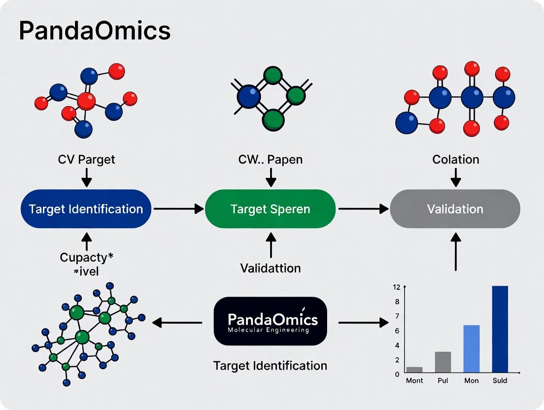
How PandaOmics is Revolutionizing Drug Target Discovery: An AI-Powered Platform for Faster Target Identification & Validation
This article provides a comprehensive guide for researchers, scientists, and drug development professionals on leveraging the AI-driven platform PandaOmics for target identification and validation.
How PandaOmics is Revolutionizing Drug Target Discovery: An AI-Powered Platform for Faster Target Identification & Validation
Abstract
This article provides a comprehensive guide for researchers, scientists, and drug development professionals on leveraging the AI-driven platform PandaOmics for target identification and validation. We begin by exploring the platform's foundational AI architecture and multi-omics data integration capabilities. We then detail its methodological workflows for hypothesis generation, candidate prioritization, and experimental design. The guide addresses common troubleshooting scenarios and optimization strategies to enhance discovery outcomes. Finally, we examine validation frameworks and comparative analyses against traditional methods, demonstrating PandaOmics's impact on accelerating and de-risking early-stage drug discovery.
What is PandaOmics? Exploring the AI Engine Behind Modern Target Discovery
PandaOmics is an AI-powered bioinformatics platform designed to accelerate target discovery and validation in drug development. It integrates multi-omics data, scientific literature, and proprietary business intelligence to prioritize novel therapeutic targets and biomarkers. This document provides application notes and protocols for researchers within the context of a thesis on AI-driven target identification.
Table 1: Core Data Sources Integrated into PandaOmics
| Data Type | Estimated Scale (as of 2023) | Update Frequency |
|---|---|---|
| Transcriptomics (e.g., TCGA, GTEx) | >30,000 samples | Quarterly |
| Genomics & Genome-Wide Association Studies (GWAS) | >5,000 studies | Monthly |
| Proteomics & Metabolomics Datasets | >1,000 studies | Monthly |
| Scientific Literature (PubMed, Patents) | >35 million documents | Real-time |
| Clinical Trial Records (ClinicalTrials.gov) | >450,000 studies | Daily |
| AI Models for Target Scoring | >20 unique algorithms | Continuously trained |
Table 2: Typical Target Identification Output Metrics
| Metric | Description | Example Range |
|---|---|---|
| iTTS (AI-derived Novelty Score) | Identifies novel, less explored targets (low score = more novel) | 0.0 (Novel) to 1.0 (Established) |
| TDL (Target Development Level) | Classification from Tclin (clinical) to Tdark (unknown) | Tdark, Tbio, Tchem, Tclin |
| Disease Association Score | Strength of AI-predicted link to disease biology | 0 to 100 |
| Tractability Scores | Druggability (e.g., small molecule, antibody feasibility) | Low, Medium, High |
Application Notes & Protocols
Protocol 1: Initial Target Identification for a Disease of Interest
Objective: To generate a ranked list of novel and tractable candidate targets for a specific disease using PandaOmics.
Materials & Workflow:
- Platform Access: Log in to the PandaOmics web interface (https://pandaomics.com/).
- Project Creation: Create a new "Target Identification" project. Name it (e.g., "ALSFibroblastAnalysis").
- Disease/Cohort Definition:
- Select "Analysis Type" as "Disease vs. Control".
- Under "Disease/Case Group," use the search function to select relevant public datasets (e.g., from GEO, TCGA) or upload processed transcriptomics data from your own experiments (normalized counts matrix).
- Define matched control samples from healthy tissue or unrelated conditions.
- Analysis Configuration:
- Differential Expression: Set thresholds (e.g., |log2FC| > 1, adjusted p-value < 0.05).
- Pathway Analysis: Select enrichment methods (e.g., KEGG, GO, Reactome).
- AI Prioritization: Enable "iTTS" and "Disease Association" scoring models. Apply filters for desired TDL level (e.g., prioritize "Tdark" or "Tbio" for novelty).
- Execution & Review: Run the analysis. Review results in the interactive dashboard. The primary output is a sortable table of genes/proteins ranked by the composite AI priority score.
Diagram Title: PandaOmics Target ID Workflow
Protocol 2: Multi-Omics Validation & Biological Context Analysis
Objective: To validate and contextualize a shortlist of candidate targets using integrated omics layers and literature mining.
Materials & Workflow:
- Target Shortlist Input: From Protocol 1, select 5-10 top candidate genes.
- Multi-Omics Correlation:
- Navigate to the "Multi-Omics" module.
- For each candidate, examine correlation between mRNA expression and proteomics or phosphoproteomics data across available cohorts to assess translational relevance.
- Pathway Network Visualization:
- Use the "Pathway Map" tool.
- Input candidate genes to generate an interactive signaling network, highlighting their positions within disease-relevant pathways (e.g., apoptosis, inflammation).
- Literature & Clinical Trial Validation:
- Open the "Insights" panel for each gene.
- Review AI-extracted relationships from literature, noting supporting evidence, contradictory findings, and co-mention trends.
- Check the "Trials" tab to see if the target has any ongoing or past clinical investigations.
Diagram Title: Multi-Modal Target Validation Strategy
Protocol 3:In SilicoCompound Screening & Tractability Assessment
Objective: To identify potential existing compounds or novel modalities for a prioritized target.
Materials & Workflow:
- Target Input: Select a single, high-priority target from previous protocols.
- Druggability Assessment:
- Review the "Tractability" scores for modalities like small molecule (SM), antibody (Ab), or PROTAC.
- Examine the 3D structure viewer if a protein structure is available, noting binding pockets.
- Compound Identification:
- Use the "AI Compound Matching" feature.
- The platform will suggest known drugs, clinical candidates, or screening hits predicted to interact with the target, based on chemical structure and bioactivity models.
- Repurposing Analysis: For suggested known drugs, review the "Indications" panel to assess potential for drug repurposing across different diseases.
The Scientist's Toolkit: Key Research Reagent Solutions
Table 3: Essential Materials for Downstream Experimental Validation
| Reagent/Material | Function in Target Validation | Example Vendor/Catalog |
|---|---|---|
| siRNA or shRNA Libraries | Gene knockdown to assess target loss-of-function phenotypes in cellular disease models. | Dharmacon, Sigma-Aldrich |
| cDNA ORF Clones | Gene overexpression to study gain-of-function effects and rescue experiments. | GenScript, Origene |
| Validated Antibodies | For Western Blot, IHC, or flow cytometry to detect protein expression and modification changes. | Cell Signaling Technology, Abcam |
| CRISPR-Cas9 Knockout Kits | Complete gene knockout to create stable cell lines for phenotypic and mechanistic studies. | Synthego, ToolGen |
| Recombinant Target Protein | For in vitro binding assays (SPR, ITC) or high-throughput screening (HTS). | R&D Systems, Sino Biological |
| Patient-Derived Cells/IPSCs | Biologically relevant models for validating target role in human disease biology. | ATCC, Fujifilm Cellular Dynamics |
The identification and validation of novel therapeutic targets is a complex, high-risk, and data-intensive foundation of drug discovery. Within the PandaOmics platform, this process is augmented by an integrated core architecture leveraging generative artificial intelligence (AI) and large language models (LLMs). This architecture synthesizes vast, disparate datasets—from multi-omics to scientific literature and clinical trial data—to generate and prioritize high-confidence target hypotheses. These AI-driven systems move beyond correlation to infer causal biology, propose novel mechanisms, and significantly de-risk the early research pipeline by providing actionable, evidence-rich insights for researchers and drug development professionals.
Core Architectural Components & Data Flow
The hypothesis generation engine is built upon a synergistic pipeline of specialized models.
Table 1: Core AI Components in Target Hypothesis Generation
| Component | Primary Function | Key Input Data | Output |
|---|---|---|---|
| Foundation LLM (Biomedically-Tuned) | Semantic understanding of biological entities, relationships, and mechanisms. | Unstructured text: Full-text papers, patents, grants, clinical protocols. | Structured knowledge graphs; embedded relationships (e.g., Gene-X inhibits Pathway-Y in Disease-Z). |
| Multi-Omics Analysis AI | Identifies differentially expressed genes, proteins, metabolites; infers pathway activity. | Structured data: Transcriptomics, proteomics, epigenomics, GWAS data from public/private cohorts. | Ranked lists of dysregulated biological entities; differential activity scores for pathways and processes. |
| Causal Inference & Generative Model | Distinguishes causal drivers from correlative signals; generates novel target ideas. | Integrated outputs from LLM & Multi-Omics AI; known drug-target-disease networks. | Hypothesized causal targets with proposed mechanisms of action (e.g., inhibition of Gene-A to restore Pathway-B). |
| Validation Evidence Scorer | Prioritizes targets by synthesizing feasibility, novelty, and confidence metrics. | Generated hypotheses; real-time data from clinicaltrials.gov, bio-banks, competitive intelligence. | Consolidated PandaOmics Score (0-100), detailing novelty, tractability, safety, and commercial potential. |
AI-Powered Target Hypothesis Generation Pipeline
Application Notes & Experimental Protocols
Protocol: LLM-Driven Knowledge Graph Construction for Novel Association Mining
Objective: To extract implicit relationships between genes, diseases, and biological processes from literature to seed hypothesis generation.
Workflow:
- Corpus Curation: Assemble a domain-specific corpus (e.g., neurodegenerative disease literature) from PubMed, PMC, and proprietary repositories.
- Entity Recognition & Linking: Utilize a fine-tuned BioNER model to identify and map mentions to standard identifiers (e.g., NCBI Gene, MONDO, GO).
- Relationship Extraction: Apply a relation extraction LLM (e.g., tuned on BioRel tasks) to triples (Subject, Predicate, Object) from sentences (e.g., "GSK3B phosphorylation inhibits Wnt signaling in Alzheimer's models").
- Graph Embedding & Completion: Encode the constructed graph using a model like TransE or ComplEx. Use the model to predict novel, plausible links (e.g., "What gene is most associated with tauopathy and synaptic loss?").
Table 2: Sample Output from Knowledge Graph Query
| Query | Top Predicted Gene | Predicted Relationship | Confidence Score | Supporting Paths in Graph |
|---|---|---|---|---|
| "Find genes that cause neuronal death when overexpressed and are upregulated in ALS." | RPS6KA3 | overexpression leads to neuronal death | 0.92 | Linked to FASLG expression, p38 MAPK activation. |
| "Identify novel inhibitors of the NLRP3 inflammasome pathway." | RNF125 | negatively regulates NLRP3 activity | 0.87 | Connected to ubiquitination of ASC; found in COPD contexts. |
Protocol: Generative AI for Novel Target & Mechanism Proposal
Objective: To generate testable hypotheses for understudied ("dark") genes within a disease-associated genomic locus.
Methodology:
- Input Definition: Provide the generative model with: a) A seed gene from a GWAS hit, b) The disease phenotype, c) Relevant pathway context (e.g., "immune cell infiltration").
- Conditional Generation: Use a transformer-based generative model (e.g., fine-tuned GPT) conditioned on the inputs to propose:
- Novel Gene Candidates: In the same pathway or protein family as the seed.
- Mechanism of Action (MOA): A short description of how modulation might alter disease biology (e.g., "Allosteric inhibition of Gene-X to reduce pro-inflammatory cytokine release without affecting homeostatic function").
- In-silico Validation: Cross-reference generative outputs against the proprietary knowledge graph for indirect evidence (e.g., does a proposed gene co-express with known markers?).
Generative AI Workflow for Novel Target Proposal
Protocol: Integrated Validation Evidence Scoring
Objective: To assign a comprehensive PandaOmics Score to AI-generated target hypotheses.
Procedure:
- Multi-Metric Calculation: For each target hypothesis, compute scores (0-1) across dimensions:
- Novelty: Inverse frequency in literature, patents, and clinical trials.
- Biological Confidence: Concordance of multi-omics signals (genomic, transcriptomic, proteomic).
- Tractability: Predicted druggability (small molecule/biologics), presence of crystal structures, assay feasibility.
- Safety: Genetic constraint (pLI), knockout mouse phenotypes, tissue expression specificity.
- Commercial Potential: Competitive landscape, unmet need size, biomarker strategy.
- Weighted Aggregation: Combine dimension scores using a dynamically weighted model (weights adjustable by research strategy: first-in-class vs. fast-follower).
- Evidence Dossier Assembly: Automatically compile supporting data points for each score into a report.
Table 3: Sample PandaOmics Scoring for Hypothetical Targets in Fibrosis
| Target Gene | Novelty (0.4) | Bio. Confidence (0.3) | Tractability (0.2) | Safety (0.1) | Overall Score | Suggested Next Step |
|---|---|---|---|---|---|---|
| TGFBR1 | 0.2 | 0.9 | 0.8 | 0.7 | 67 | Repurposing opportunity; check chemical matter. |
| PKD1L3 | 0.9 | 0.7 | 0.4 | 0.6 | 72 | High novelty; requires assay development for HTS. |
| LOXL2 | 0.5 | 0.8 | 0.7 | 0.5 | 65 | Clinical failures exist; investigate new MOA. |
The Scientist's Toolkit: Research Reagent Solutions
Table 4: Essential Resources for Experimental Validation of AI-Generated Targets
| Reagent / Solution | Provider Examples | Primary Function in Validation |
|---|---|---|
| CRISPR-Cas9 Knockout/Knockdown Kits | Synthego, Horizon Discovery | Functional validation of target gene necessity in disease-relevant cellular phenotypes. |
| siRNA/sgRNA Libraries | Dharmacon, Sigma-Aldrich | High-throughput screening of target gene families or pathway members identified by AI. |
| Recombinant Proteins | R&D Systems, Sino Biological | For binding assays, structural studies, and in vitro functional characterization of novel targets. |
| Phospho-Specific & Total Antibodies | Cell Signaling Technology, Abcam | Detecting pathway activity changes (e.g., phosphorylation) upon target modulation. |
| Patient-Derived Organoid Co-Culture Systems | STEMCELL Technologies, proprietary biobanks | Testing target hypotheses in a physiologically relevant human tissue microenvironment. |
| Phenotypic Screening Assay Kits (e.g., apoptosis, cytokine release) | Thermo Fisher, Promega | Quantifying the functional outcome of target perturbation in disease models. |
Within the thesis framework of PandaOmics for AI-driven target identification and validation, the "Integrated Data Universe" is the foundational paradigm. It posits that the convergence and computational integration of disparate, large-scale data modalities—multi-omics, scientific text, and structured clinical trial data—generates synergistic insights far exceeding the sum of individual parts. This integrated approach powers the identification of novel, high-confidence, and druggable targets with strong disease association and clinical tractability.
The following table summarizes the primary data layers integrated within the PandaOmics platform to construct the target discovery universe.
Table 1: Core Data Modalities in the Integrated Data Universe
| Data Modality | Primary Sources | Key Metrics/Volume | Role in Target Identification |
|---|---|---|---|
| Genomics & GWAS | dbGaP, UK Biobank, GWAS Catalog | ~100M genetic variants; >5,000 published studies | Identifies disease-associated loci and candidate causal genes. |
| Transcriptomics | GTEx, TCGA, GEO, ArrayExpress | >1M RNA-seq samples across >50 tissues and diseases | Pinpoints differentially expressed genes and expression quantitative trait loci (eQTLs). |
| Proteomics & Phosphoproteomics | CPTAC, PRIDE, HPA | ~20,000 proteins; >200,000 phosphorylation sites | Validates protein-level dysregulation and identifies signaling hubs. |
| Scientific Literature (Text) | PubMed, PubMed Central, Patent DBs | >35M abstracts; full-text articles | Contextualizes gene-disease relationships, extracts novel associations. |
| Clinical Trial Data | ClinicalTrials.gov, WHO ICTRP | ~450,000 registered studies | Informs on target druggability, safety profiles, and competitive landscape. |
| Knowledge Graphs | STRING, DisGeNET, Reactome | >20,000 genes; >1M curated interactions | Provides mechanistic and pathway-level context for candidate targets. |
Application Notes
Note 1: Multi-Omics Convergence for Novel Target Discovery
- Objective: Identify high-confidence oncology targets by intersecting genomic, transcriptomic, and proteomic dysregulation.
- Procedure: Load a TCGA cancer cohort (e.g., Glioblastoma). Execute a multi-omics query filtering for: 1) Genes with significant copy number amplification (CNV > 2), 2) Concurrent mRNA up-regulation (log2FC > 1, adj. p < 0.01), and 3) Protein overexpression (z-score > 2) in CPTAC data. The resulting shortlist represents genes with coherent multi-omics evidence supporting their role as oncogenic drivers.
- Outcome: A ranked list of candidates like EGFR, PDGFRA, and novel, less-characterized kinases, prioritized for further validation.
Note 2: AI-Powered Text Mining for Indication Expansion
- Objective: Repurpose a known target for a new disease by mining implicit relationships in literature.
- Procedure: Using natural language processing (NLP) models trained on biomedical corpora, query the text universe for co-mentions of a target gene (e.g., IL6) and a disease of interest (e.g., Alzheimer's) where a mechanistic link (e.g., "signaling," "inflammation," "pathway") is present but not the primary focus of the paper. The system scores and ranks publications by semantic relevance.
- Outcome: Discovery of under-appreciated mechanistic links, generating hypotheses for novel therapeutic indications supported by existing biological evidence.
Note 3: Clinical Trial Intelligence for De-risking
- Objective: Assess the development landscape and potential safety signals for a candidate target.
- Procedure: For a target (e.g., TREM2), query structured clinical trial data for all interventional studies. Analyze phase distribution, termination reasons, and frequently reported adverse events. Cross-reference with the integrated knowledge graph to identify potential mechanistic explanations for observed safety signals.
- Outcome: A comprehensive development profile informing go/no-go decisions, competitive positioning, and potential safety monitoring requirements.
Detailed Experimental Protocols
Protocol 1: Multi-Omics Target Prioritization Workflow
Title: In silico Target Identification via Multi-Omics Integration.
1. Data Acquisition & Curation:
* Download RNA-seq (counts), CNV (segmented), and clinical data for your disease cohort from a repository like TCGA or GEO using the TCGAbiolinks (R) or GEOfetch (Python) packages.
* Download matching normal tissue or control cohort data.
* Curate data: normalize RNA-seq counts (e.g., DESeq2 median ratio), log2-transform. Align patient identifiers across omics layers.
2. Differential Analysis & Intersection:
* Differential Expression: Using DESeq2 (R) or limma-voom, identify differentially expressed genes (DEGs). Filter: |log2FoldChange| > 1 & adj. p-value < 0.01.
* CNV Analysis: Using GISTIC2.0 or a simple threshold (e.g., CNV > 0.3 for amp, < -0.3 for del), identify genes with recurrent amplifications/deletions.
* Intersection: Perform a Venn or UpSet plot analysis to identify genes that are both amplified and overexpressed (for oncogenes) or deleted and underexpressed (for tumor suppressors).
3. Functional Enrichment & Pathway Mapping: * Input the intersected gene list into enrichment tools like Enrichr or clusterProfiler (R). * Perform Gene Ontology (GO), KEGG, and Reactome pathway analysis (FDR < 0.05). * Visualize top enriched pathways and construct a protein-protein interaction network using STRING or Cytoscape.
4. AI-Driven Scoring & Ranking: * Feed the candidate list into PandaOmics' "iTAR" or similar AI scoring engine, which incorporates the integrated data universe (text, clinical trials, etc.) to generate a composite "novelty" and "confidence" score. * Generate a final ranked target list.
Protocol 2: Literature-Based Hypothesis Generation & Validation
2. Automated Literature Retrieval & NLP Processing:
* Use PubMed E-utilities API (biopython.Entrez) to fetch abstracts.
* Process text: Sentence splitting, tokenization, part-of-speech tagging using spaCy with the en_core_sci_md model.
* Apply a named entity recognition (NER) pipeline to identify genes, diseases, drugs, and biological processes.
3. Relationship Extraction & Scoring: * Use a rule-based or deep learning relation extraction model (e.g., BioBERT fine-tuned on BC5CDR) to classify sentences as describing a direct "INHIBITS", "ACTIVATES", or "ASSOCIATED_WITH" relationship. * Score each extracted relationship by the confidence of the model and the journal impact factor of the source.
4. Triangulation with Experimental Data: * Cross-reference top literature-derived links with expression correlations in relevant transcriptomic datasets. * Validate if the relationship is supported by shared pathway membership in knowledge graphs (e.g., Reactome).
Diagrams
Title: Integrated Data Universe for Target Discovery Workflow
Title: IL-6/JAK/STAT3 Signaling Pathway
The Scientist's Toolkit: Key Research Reagent Solutions
Table 2: Essential Reagents for Target Validation Experiments
| Reagent/Category | Example Product/Kit | Primary Function in Validation |
|---|---|---|
| Gene Silencing | siRNA pools (Dharmacon), CRISPR-Cas9 Lentiviral Particles (Sigma) | Knockdown/knockout of candidate target gene to assess phenotypic impact (viability, migration). |
| Antibodies for Immunoblotting | Phospho-STAT3 (Tyr705) (Cell Signaling #9145), Total STAT3 (CST #12640) | Confirm protein expression and activation status of target and downstream pathway components. |
| Cell Viability/Proliferation Assay | CellTiter-Glo 3D (Promega), MTT Assay Kit (Abcam) | Quantify changes in cell growth and metabolic activity upon target modulation. |
| qRT-PCR Assays | TaqMan Gene Expression Assays (Thermo Fisher), SYBR Green Master Mix (Bio-Rad) | Validate mRNA expression changes of the target and its downstream effectors. |
| High-Content Imaging Reagents | HCS CellMask Deep Red Stain (Invitrogen), Nuclear Stains (Hoechst/DAPI) | Enable multiplexed, automated analysis of cell morphology, proliferation, and signaling in situ. |
| Proteomics Sample Prep | TMTpro 16-plex Kit (Thermo), S-Trap Micro Columns (Protifi) | For deep, quantitative profiling of protein expression and phosphorylation changes post-target modulation. |
Within the thesis of the PandaOmics platform's role in modern drug discovery, the central challenge of target identification is reframing biological complexity into testable, tractable hypotheses. This document provides application notes and detailed protocols to operationalize this approach, leveraging multi-omic data, artificial intelligence, and systematic validation to transition from disease biology to high-confidence therapeutic targets.
Application Notes: A Multi-Omic & AI-Driven Workflow
Foundational Data Acquisition and Integration
The initial phase involves the aggregation and normalization of heterogeneous data types to construct a comprehensive disease model.
Table 1: Core Multi-Omic Data Types for Target Identification
| Data Type | Key Metrics | Primary Source | Role in Hypothesis Generation |
|---|---|---|---|
| Transcriptomics | Differentially Expressed Genes (DEGs), p-value, Log2FC, TPM/FPKM | RNA-Seq, Microarrays | Identifies gene expression dysregulation in disease states. |
| Proteomics | Protein Abundance Fold Change, p-value, AUC | LC-MS/MS, SOMAscan | Confirms transcriptional changes at protein level; reveals PTMs. |
| Genomics | Variant Frequency (SNPs, Indels), Odds Ratio, p-value | Whole Genome/Exome Sequencing | Identifies inherited or somatic genetic drivers of disease. |
| Epigenomics | Methylation Beta Value, Chromatin Accessibility Peaks | ChIP-Seq, ATAC-Seq, WGBS | Uncovers regulatory mechanisms influencing gene expression. |
| Pharmaco-Omics | IC50, AUC, Gene Essentiality Scores (CERES, DepMap) | CRISPR Screens, Drug Sensitivity Databases | Informs on druggability and potential resistance mechanisms. |
AI-Powered Target Prioritization with PandaOmics
The integrated data is processed through the PandaOmics platform, which applies AI models to score and rank potential targets.
Table 2: Exemplary AI-Generated Target Scores for Alzheimer's Disease
| Gene Symbol | "iTAP" Score | Novelty Score | Druggability Score | Confidence Tier |
|---|---|---|---|---|
| APOE | 98 | Low (Established) | High | Tier 1 (Known) |
| TREM2 | 85 | Medium | Medium | Tier 1 |
| HKDC1 | 76 | High | Medium | Tier 2 (Novel) |
| PYRL1 | 72 | High | Low | Tier 2 |
| C3 | 68 | Medium | High | Tier 1 |
Note: Scores are illustrative. The "iTAP" (integrated Target Assessment Profile) is a composite metric weighing causal evidence, tractability, and safety.
Title: AI-Driven Target Identification Workflow
Pathway and Network Analysis
Top-ranked targets are contextualized within biological pathways and interaction networks to understand mechanism and identify co-targets or biomarkers.
Title: TREM2 Signaling Pathway in Microglia
Experimental Protocols for Target Validation
Protocol: CRISPR-Cas9 Knockout for Functional Validation in a Cell Line Model
Objective: To validate the essentiality of a novel target gene (e.g., HKDC1) for cell proliferation/survival in a disease-relevant cell line.
I. Materials & Reagents (The Scientist's Toolkit)
| Item | Function | Example Product/Catalog # |
|---|---|---|
| sgRNA Design Tool | Designs specific guide RNAs for target gene knockout. | CHOPCHOP, IDT Custom Alt-R CRISPR-Cas9 sgRNA |
| Lipofectamine 3000 | Transfection reagent for delivering RNP complexes into cells. | Thermo Fisher Scientific, L3000015 |
| Alt-R S.p. Cas9 Nuclease V3 | High-fidelity Cas9 enzyme for precise genome editing. | Integrated DNA Technologies, 1081058 |
| Target-specific sgRNA | Guides Cas9 to the genomic locus of interest. | Synthesized as crRNA+tracrRNA or as single guide. |
| Cell Titer-Glo 2.0 | Luminescent assay to quantify viable cells based on ATP. | Promega, G9242 |
| Genomic DNA Extraction Kit | Isolates DNA for validation of editing efficiency. | Qiagen, DNeasy Blood & Tissue Kit, 69504 |
| T7 Endonuclease I | Detects indel mutations via surveyor nuclease assay. | New England Biolabs, M0302S |
II. Procedure
- sgRNA Design & Complex Formation:
- Design two sgRNAs targeting early exons of HKDC1 using a validated web tool.
- Reconstitute and complex 10 pmol of each sgRNA with 20 pmol of Cas9 nuclease in duplex buffer to form Ribonucleoprotein (RNP) complexes. Incubate at 25°C for 10 min.
Cell Culture & Transfection:
- Culture disease-relevant cells (e.g., glioblastoma line U87-MG) in appropriate medium.
- At 70-80% confluence, dissociate cells. For each transfection, mix 2e5 cells with the pre-formed RNP complex in buffer. Electroporate using a Neon system (1100V, 20ms, 2 pulses) or use lipofection according to manufacturer protocol.
Post-Transfection Culture:
- Seed transfected cells into 96-well plates for viability assay and 6-well plates for genomic analysis.
- Allow recovery and gene editing for 72 hours.
Validation of Knockout Efficiency:
- Extract genomic DNA from cells in the 6-well plate.
- Amplify the target region by PCR (~500 bp amplicon).
- Perform T7 Endonuclease I assay on purified PCR products: Denature/reanneal, digest with T7E1, analyze fragments via gel electrophoresis. Calculate indel percentage.
Phenotypic Assessment (Viability):
- At 72h and 120h post-transfection, equilibrate plates to room temperature.
- Add equal volume of Cell Titer-Glo 2.0 reagent to each well of the 96-well plate.
- Shake, incubate for 10 min, and record luminescence. Normalize signal to non-targeting sgRNA control.
Protocol: High-Content Imaging Analysis of Phenotypic Changes
Objective: To quantify morphological changes (e.g., phagocytosis, synapse loss) upon target modulation in a cellular assay.
I. Key Reagents
| Item | Function |
|---|---|
| iPSC-derived Microglia | Disease-relevant primary-like cell model. |
| pHrodo Red Aβ Conjugates | Fluorescent amyloid-beta that fluoresces upon phagocytosis. |
| Hoechst 33342 | Nuclear counterstain for cell segmentation. |
| CellMask Deep Red | Cytoplasmic stain for cell morphology. |
| Opera Phenix or ImageXpress | High-content screening microscope. |
| Harmony/Columbus Software | Image analysis software for phenotypic quantification. |
II. Procedure
- Cell Preparation & Treatment: Seed iPSC-derived microglia in a 384-well imaging plate. Treat cells with a small molecule modulator of the target (e.g., TREM2 agonist) or relevant controls for 24 hours.
- Phagocytosis Assay: Add pHrodo Red-labeled Aβ fibrils to the medium. Incubate for 4-6 hours.
- Staining & Fixation: Stain nuclei with Hoechst (1 µg/mL), then stain cytoplasm with CellMask (1:2000). Fix cells with 4% PFA for 15 min.
- Image Acquisition: Image plates using a 40x water objective on a high-content imager. Acquire fields in channels: DAPI (nuclei), Cy5 (cytoplasm), TRITC (pHrodo Red Aβ).
- Image Analysis:
- Segmentation: Identify nuclei (DAPI), then expand region to define cytoplasm (Cy5).
- Quantification: Within each cell mask, measure mean TRITC fluorescence intensity (phagocytosed Aβ). Calculate per-cell and per-well metrics: % of phagocytosing cells, average cargo intensity.
The transition from biological complexity to tractable hypotheses is systematized through the integration of multi-omic data, AI-powered prioritization as exemplified by the PandaOmics thesis, and rigorous, standardized experimental validation. The provided protocols offer a roadmap for researchers to functionally deconvolute and validate next-generation therapeutic targets.
Application Note: Oncology Target Identification
Context & Rationale
Within the PandaOmics platform for target identification and validation, oncology remains a primary application. The integration of multi-omics data (genomics, transcriptomics, proteomics) with AI-driven analysis enables the discovery of novel therapeutic targets and biomarkers in complex tumor microenvironments. Current research emphasizes immune checkpoint modulation, synthetic lethality, and oncogene dependency.
Table 1: High-Confidence Novel Oncology Targets from PandaOmics Analysis
| Target Gene | Disease Indication (Cancer Type) | AI Score (PandaOmics) | Supporting Evidence Type (Omics) | Druggability Level (PandaOmics) |
|---|---|---|---|---|
| CDK11B | Triple-Negative Breast Cancer | 0.94 | CRISPR screen, Transcriptomics | High |
| P4HA2 | Glioblastoma Multiforme | 0.89 | Proteomics, Metabolomics | Medium |
| KIF18A | Pancreatic Ductal Adenocarcinoma | 0.92 | Genomics, Clinical Survival Data | High |
| NSD3 | Squamous Cell Carcinoma (Lung) | 0.87 | Methylomics, Chromatin Profiling | High |
| RIPK2 | Colorectal Cancer | 0.85 | Phosphoproteomics, Cytokine Data | Medium |
AI Score: Composite score (0-1) generated by PandaOmics AI models integrating novelty, druggability, and scientific confidence.
Protocol:In SilicoTarget Identification & Validation Workflow for Oncology
Protocol 1: Multi-Omics Data Integration and Target Prioritization.
Objective: To identify and prioritize novel oncology targets using PandaOmics.
Materials:
- PandaOmics software platform (access to database).
- Publicly available or proprietary patient tumor multi-omics datasets (e.g., TCGA, CPTAC).
- High-performance computing environment.
Procedure:
- Data Curation: Upload or select disease-specific transcriptomic, genomic (mutations, CNV), and proteomic datasets within PandaOmics. Define case (tumor) and control (normal adjacent tissue) groups.
- Differential Analysis: Execute differential expression, mutation enrichment, and pathway activation analysis (using built-in algorithms).
- AI-Powered Ranking: Initiate the "Target Identification" module. The AI engine (incorporating knowledge graphs, NLP on publications/patents/grant abstracts, and multi-omics correlations) will generate a ranked list of targets.
- Filtering & Prioritization: Apply filters for novelty (low bibliography count), druggability (presence of known domains or homologs), safety (essentiality scores from DepMap), and association with patient survival.
- Pathway Enrichment: For top candidates, run pathway and network enrichment analysis to elucidate mechanistic context.
- Output: Generate a final report with top candidates, supporting evidence, and suggested validation experiments.
Pathway Diagram: CDK11B in TNBC Cell Cycle & Transcription
Diagram 1: Proposed CDK11B Role in TNBC Survival.
The Scientist's Toolkit: Oncology Target Validation
Table 2: Key Reagents for Validating Novel Oncology Targets In Vitro
| Reagent / Solution | Function in Validation | Example Vendor/Catalog |
|---|---|---|
| siRNA/shRNA Pool (Human) | Gene knockdown to assess target essentiality for cancer cell proliferation. | Horizon Discovery, Sigma-Aldrich |
| Recombinant Human Protein (Target) | For in vitro kinase/activity assays and binding studies with candidate compounds. | Sino Biological, R&D Systems |
| Phospho-Specific Antibody (Downstream Marker) | Detect modulation of target activity in cell-based assays via Western Blot/IHC. | Cell Signaling Technology |
| Cell Titer-Glo 3D Assay | Measure 3D tumor spheroid viability post-target perturbation. | Promega, Cat# G9683 |
| Human Primary Cancer Cells (Relevant indication) | Validate target role in physiologically relevant models. | ATCC, StemCell Technologies |
Application Note: Neurodegeneration Target Identification
Context & Rationale
PandaOmics addresses the complexity of neurodegenerative diseases (e.g., Alzheimer's, Parkinson's, ALS) by integrating central and peripheral omics data to uncover targets involved in proteostasis, neuroinflammation, and neuronal survival. The platform's ability to analyze data from induced pluripotent stem cell (iPSC)-derived neurons and cerebrospinal fluid (CSF) proteomics is critical.
Table 3: AI-Prioritized Targets for Neurodegenerative Diseases
| Target Gene | Associated Disease | Modality Suggestion (from AI) | Novelty Score (1-10) | Link to Hallmark Pathway |
|---|---|---|---|---|
| USP12 | Alzheimer's Disease | Small Molecule Inhibitor | 8.5 | Protein Clearance, Tauopathy |
| SYT11 | Parkinson's Disease | Gene Therapy / ASO | 9.1 | Synaptic Vesicle Recycling |
| KIF5A | Amyotrophic Lateral Sclerosis | ASO | 7.8 | Axonal Transport |
| LRRK2 | Parkinson's Disease (Sporadic) | Small Molecule Inhibitor | 4.2 (Known) | Neuroinflammation |
| MARCKS | Frontotemporal Dementia | Peptide Therapeutic | 8.7 | Membrane Stability, Glial Activation |
Protocol: Target Discovery Using iPSC-Derived Neuron Omics Data
Protocol 2: Integrating iPSC Transcriptomics and Phosphoproteomics for Target Discovery.
Objective: To identify dysregulated signaling nodes in a disease-relevant neuronal model.
Materials:
- PandaOmics platform.
- Transcriptomic (RNA-seq) and phosphoproteomic (LC-MS/MS) data from patient-derived iPSC neurons (e.g., with disease mutation vs. isogenic control).
- List of known disease-associated genes from curated databases (e.g., AlzGene, GWAS).
Procedure:
- Data Upload: Import normalized gene expression and phosphopeptide abundance matrices into PandaOmics.
- Multi-Omic Correlation: Use the "Multi-Omics Correlation" tool to identify genes whose expression is strongly correlated with phosphorylation changes in key disease pathways (e.g., autophagy, synaptic signaling).
- Crosstalk Analysis: Overlay transcriptomic and phosphoproteomic pathway analysis results to identify convergent, dysregulated signaling axes.
- Network Proximity Analysis: Run a network analysis to measure the proximity of differentially expressed genes to known disease genes in a human protein-protein interaction network. Short proximity suggests shared function or pathway.
- AI Filtering: Feed the convergent gene list into the AI prioritization engine, weighting "neuro-specific druggability" and "blood-brain barrier penetrance" predictors.
- Output: A shortlist of targets that are central to the dysregulated signaling network and have predictive druggability for the CNS.
Pathway Diagram: USP12 in Alzheimer's Proteinopathy
Diagram 2: USP12 Interferes with Protein Clearance.
The Scientist's Toolkit: Neurodegeneration Research
Table 4: Essential Reagents for Neuronal Target Validation
| Reagent / Solution | Function in Validation | Example Vendor/Catalog |
|---|---|---|
| iPSC Line (Patient-derived & Isogenic Control) | Physiologically relevant human neuronal model for target perturbation. | Cedars-Sinai Biomanufacturing Center, FUJIFILM Cellular Dynamics |
| Neuronal Differentiation Kit | Generate consistent cultures of glutamatergic or dopaminergic neurons. | StemCell Technologies, Cat# 05835 |
| Phospho-Tau (Ser396/404) Antibody | Readout for Tauopathy pathway modulation in AD models. | Thermo Fisher Scientific, Cat# 44-752G |
| - Synapsin I Antibody (Alexa Fluor 488) | Visualize neuronal synapses and assess synaptic density changes. | Synaptic Systems, Cat# 106 011AF488 |
| - Caspase-3/7 Glo Assay | Quantitate apoptosis in neuronal cultures post-target modulation. | Promega, Cat# G8091 |
Application Note: Rare Disease Target Identification
Context & Rationale
For rare diseases, where patient data is scarce, PandaOmics leverages cross-disease analytics and model organism data to identify gene-disease associations and repurposable targets. The platform's strength lies in analyzing transcriptomic signatures from limited patient biopsies and linking them to perturbational databases (e.g., LINCS) to find potential therapeutic compounds.
Table 5: Example Output for a Fibrodysplasia Ossificans Progressiva (FOP) Study
| Analysis Step | Method in PandaOmics | Key Finding | Implication for Target ID | ||
|---|---|---|---|---|---|
| Differential Expression | ACES2 Algorithm (FOP vs. control muscle) | 345 DEGs (FDR<0.05) | Defines disease-specific signature. | ||
| Pathway Enrichment | iPathway Guide | TGF-beta, BMP, Hypoxia pathways activated (p<0.001) | Confirms known biology; identifies active signaling context. | ||
| Upstream Regulator Analysis | Causality Analysis | SMAD1/5, HIF1A, mTOR predicted as active ( | z-score | >2) | Points to key regulatory nodes as potential targets. |
| AI-Powered Target Suggestion | Deep Learning on Knowledge Graph | mTORC1 (AI Score: 0.88), ALKBH5 (AI Score: 0.91) | Prioritizes novel, druggable targets within the active pathways. | ||
| Drug Repurposing Screen | Signature Matching to LINCS | Rapamycin (mTOR inhibitor) signature inversely correlates with FOP signature (p<0.01) | Suggests immediate repurposing candidate. |
Protocol: Target & Drug Candidate Identification for a Rare Disease
Protocol 3: From Patient Signature to Repurposing Candidate.
Objective: To identify a novel target and a repurposable drug candidate for a rare disease with limited patient samples.
Materials:
- PandaOmics platform with LINCS connectivity module.
- RNA-seq data from a small cohort of patient biopsies (n=5-10) and matched healthy controls.
- List of FDA-approved drugs with transcriptomic signatures in the LINCS L1000 database.
Procedure:
- Signature Generation: Perform differential expression analysis in PandaOmics to create a consolidated "disease signature" (list of up- and down-regulated genes).
- Target Inference: Use the disease signature in the "Target Discovery" module. The AI will identify genes whose perturbation (knockdown/overexpression) in public databases best reverses the disease signature.
- Drug Signature Matching: Input the disease signature into the "Drug Repurposing" tool. The platform will compute connectivity scores against the LINCS L1000 drug perturbation signatures. Negative scores indicate a drug that reverses the disease signature.
- Mechanistic Link Analysis: For top drug candidates, examine the predicted target(s) of the drug. Cross-reference with the AI-prioritized target list from Step 2 to establish a mechanistic hypothesis.
- Safety & Feasibility Check: Use integrated databases to review the safety profile and pharmacokinetics of the repurposing candidate for potential application in the rare disease population.
- Output: A report detailing: a) A novel, high-priority target (e.g., ALKBH5), and b) A repurposable drug candidate (e.g., Rapamycin) with its predicted mechanism linked to the target/disease pathway.
Diagram: FOP Target & Drug Discovery Workflow
Diagram 3: Rare Disease Discovery Pipeline.
The Scientist's Toolkit: Rare Disease Research
Table 6: Key Solutions for Rare Disease Mechanistic Studies
| Reagent / Solution | Function in Validation | Example Vendor/Catalog |
|---|---|---|
| CRISPRa/i Pooled Library (Human) | Activate or inhibit gene expression to screen for modifiers of a disease phenotype in a relevant cell line. | Twist Bioscience, Santa Cruz Biotechnology |
| - Recombinant Mutant Protein (Disease variant) | Study altered biochemistry and test target engagement of candidate inhibitors. | Custom synthesis (e.g., GenScript) |
| - RNAscope Assay (for low-abundance targets) | Detect and visualize target mRNA expression in limited patient tissue samples. | ACD Bio, Cat# 323100 |
| - Organoid Culture Kit (Disease-relevant tissue) | Create 3D patient-derived tissue models for functional studies. | STEMCELL Technologies, Corning |
| - Compound Library (FDA-Approved Drugs) | Rapidly test repurposing hypotheses in high-throughput in vitro assays. | Selleckchem, MedChemExpress |
Step-by-Step Guide: Using PandaOmics for Target Prioritization and Workflow Integration
Application Notes: Foundational Concepts for Disease Area Definition
Defining a disease area with precision is the critical first step in any target discovery pipeline, such as within the PandaOmics platform. A well-scoped disease area ensures that subsequent AI-driven analyses, including natural language processing of biomedical literature and multi-omics data integration, are focused and biologically relevant. This phase involves moving from a broad clinical phenotype to a specific molecular and pathological understanding.
Key Considerations:
- Phenotypic vs. Mechanistic Definitions: A disease can be defined by its clinical manifestations (e.g., "heart failure with preserved ejection fraction") or by its underlying pathophysiology (e.g., "cardiac fibrosis driven by TGF-β signaling"). The latter is more productive for target identification.
- Disease Heterogeneity: Most diseases consist of multiple subtypes with distinct molecular drivers. Defining parameters to capture this heterogeneity (e.g., specific genetic mutations, biomarker-positive populations) is essential for identifying patient-stratified targets.
- Temporal Dynamics: Disease progression (early vs. late stage) can dramatically alter molecular profiles. Search filters should account for the disease stage of interest.
The initial definition directly influences the composition of the "Knowledge Graph" and "Data Universe" in PandaOmics, which aggregates findings from thousands of omics datasets, patents, grants, and publications.
Protocol: Establishing Initial Search Parameters
This protocol outlines a systematic approach to defining a disease area for entry into a target identification platform.
Materials & Software Requirements
- Access to biomedical databases (e.g., PubMed, ClinVar, DisGeNET, MONDO).
- PandaOmics platform or similar AI-driven research suite.
- Reference terminology resources (e.g., MeSH, SNOMED CT, ICD-11).
Procedure
Step 1: Core Disease Concept Identification
- Start with a broad disease term of interest (e.g., "Alzheimer's Disease").
- Perform a preliminary literature scan to identify:
- Official Nomenclature: Standardized names and acronyms.
- Key Pathological Hallmarks: (e.g., amyloid plaques, neurofibrillary tangles).
- Associated Genes/Proteins: High-level players (e.g., APP, PSEN1, MAPT, APOE).
- Document these core identifiers.
Step 2: Expansion via Ontologies and Related Terms
- Query disease ontology databases (e.g., MONDO, DOID) using the core term.
- Map the disease to its ontological hierarchy, noting:
- Parent Terms: Broader categories (e.g., "Neurodegenerative Disease").
- Child Terms: Specific subtypes or closely related disorders (e.g., "Early-Onset Alzheimer's Disease", "Familial Alzheimer's Disease").
- Related Synonyms: Alternative names used in literature or clinics.
- Compile a comprehensive list of terms for data retrieval.
Step 3: Definition of Inclusion and Exclusion Filters
- Based on the ontology mapping, define explicit filters to bound the disease area.
- Molecular Filters: Include specific genes, proteins, pathways, or biomarkers. Exclude those strongly associated with differential diagnoses.
- Phenotypic Filters: Include relevant symptoms, anatomical locations, and histopathological findings. Exclude overlapping phenotypes from other disorders.
- Etiological Filters: Specify germane genetic, environmental, or infectious triggers if applicable.
Step 4: Validation of Disease Area Scope
- Use the compiled term list and filters to conduct a test query within PandaOmics or a major literature database.
- Manually review a sample (e.g., top 50 abstracts) of the returned results.
- Assess precision (are most results truly relevant?) and recall (are key seminal studies captured?).
- Iteratively refine the term list and filters until the query accurately represents the intended disease biology.
Data Presentation: Example Disease Area Parameters for "Non-Alcoholic Steatohepatitis (NASH)"
Table 1: Defined Search Parameters for NASH Target Identification Project
| Parameter Category | Included Terms & Concepts | Excluded Terms & Concepts | Rationale |
|---|---|---|---|
| Core Disease Terms | Non-alcoholic steatohepatitis, NASH, steatohepatitis, metabolic dysfunction-associated steatohepatitis (MASH) | Alcoholic hepatitis, hepatitis C, autoimmune hepatitis | Focus on metabolic etiology. |
| Ontological Hierarchy | Parent: Non-alcoholic fatty liver disease (NAFLD), Metabolic Liver Disease. Child: NASH with fibrosis (F2-F4), Pre-cirrhotic NASH. | NAFL (simple steatosis), Alcoholic Liver Disease, Cirrhosis (as a primary term) | Isolate inflammatory & fibrotic stage; cirrhosis is an endpoint. |
| Key Pathologies | Hepatic steatosis (>5%), lobular inflammation, hepatocyte ballooning, fibrosis | Isolated macrovesicular steatosis without inflammation | Define histological requirements. |
| Molecular Hallmarks | Insulin resistance, de novo lipogenesis, TLR4 signaling, NLRP3 inflammasome, TGF-β1, COL1A1 | Viral replication proteins (HCV NS3, HBV surface antigen) | Specify core perturbed pathways. |
| Associated Biomarkers | Increased ALT/AST (ALT > AST), CK-18 (M30/M65), Pro-C3, FIB-4 score | Markers of primary biliary cholangitis (e.g., AMA) | Focus on NASH-specific serum markers. |
| Disease Comorbidities | Type 2 Diabetes, Obesity, Metabolic Syndrome | Chronic alcohol use, Wilson's disease, Alpha-1 antitrypsin deficiency | Account for common comorbidities while excluding other liver disease causes. |
Visualization: Disease Area Definition Workflow
Diagram 1: Disease area definition and refinement workflow.
Diagram 2: Application of filters to scope disease for knowledge graph input.
The Scientist's Toolkit: Research Reagent Solutions for Disease Area Validation
Table 2: Key Reagents for Experimental Validation of NASH Disease Models In Vitro
| Reagent / Material | Provider Examples | Function in Disease Area Context |
|---|---|---|
| Primary Human Hepatocytes (PHHs) | Lonza, Thermo Fisher | Gold-standard in vitro model for studying human-specific hepatic metabolism, steatosis, and inflammatory responses. |
| HepG2 or Huh-7 Cell Line | ATCC | Immortalized human liver carcinoma cell lines; widely used for mechanistic studies on lipogenesis and signaling pathways. |
| Palmitic Acid/Oleic Acid (PA/OA) Mixture | Sigma-Aldrich | Used to induce lipotoxicity and intracellular lipid accumulation (steatosis) in hepatocyte cultures. |
| Recombinant Human TGF-β1 | R&D Systems, PeproTech | Key cytokine to activate pro-fibrotic signaling pathways in hepatic stellate cells (LX-2 cells) co-cultured with hepatocytes. |
| LPS (Lipopolysaccharide) | InvivoGen | TLR4 agonist used to trigger innate immune and inflammatory responses mimicking gut-derived inflammation in NASH. |
| Anti-Collagen I Antibody | Abcam, Novus Biologicals | Immunostaining reagent to quantify extracellular matrix deposition, a key marker of fibrosis. |
| Oil Red O Stain | Sigma-Aldrich | Lipid-soluble dye used to stain and quantify neutral lipid droplets in cultured hepatocytes. |
| ALT/AST Activity Assay Kits | Cayman Chemical, Abcam | Colorimetric kits to measure alanine & aspartate aminotransferase activity in culture supernatant, indicating hepatocyte injury. |
Within the PandaOmics platform for target identification and validation, the Target Scoring System provides a quantitative, multi-dimensional framework to prioritize candidate targets. It integrates three core pillars: Innovation (novelty and competitive landscape), Tractability (biological and chemical feasibility), and Confidence (strength of supporting evidence). This triad enables researchers to balance risk, novelty, and probability of success in early-stage drug discovery. These scores are calculated through AI-driven analysis of multi-omics data, literature, patents, and clinical trial databases.
The scoring system synthesizes data from diverse sources to generate composite scores for each dimension (0-1 scale, where 1 is most favorable).
Table 1: Core Components of the Target Scoring Triad
| Dimension | Primary Sub-Categories | Key Data Sources (PandaOmics Integration) | Interpretation (High Score) |
|---|---|---|---|
| Innovation | Novelty Score, Patent Landscape, Competitive Intensity | PubMed, Grant Databases, Patent Repositories (USPTO, EPO), ClinicalTrials.gov | Low competitive pressure, first-in-class potential, strong IP opportunity. |
| Tractability | Biological Tractability, Chemical Tractability, Safety/Expression Profile | Protein Structure DBs (AlphaFold, PDB), Known Ligands (ChEMBL), MOA data, Tissue Expression (GTEx). | High likelihood of finding a drug-like modulator; well-characterized binding sites; favorable safety profile. |
| Confidence | Genetic Evidence, Multi-Omics Evidence, Transcriptomic Signatures | Genome-wide association studies (GWAS), CRISPR screens, Differential Expression, Proteomics, Metabolomics. | Strong causal link to disease biology; consistent evidence across multiple data modalities. |
Table 2: Representative Quantitative Metrics (Illustrative)
| Metric | Innovation | Tractability | Confidence |
|---|---|---|---|
| Data Input | Number of competing active clinical programs | Presence of a druggable pocket (pLDDT > 90) | -log10(p-value) from disease-associated GWAS |
| Weight | 40% | 35% | 25% |
| Sample Value (Target A) | 0.85 (Few competitors) | 0.70 (Predicted bindable site) | 0.90 (Strong genetic association) |
| Sample Value (Target B) | 0.45 (Moderate competition) | 0.95 (Known drug target class) | 0.60 (Moderate omics support) |
Experimental Protocols for Target Validation
Following computational prioritization via the triad scores, in vitro and in vivo validation is essential. Below are key protocols for targets with high Innovation and Confidence scores but requiring tractability assessment.
Protocol 1: CRISPR-Cas9 Knockout for Functional Validation Objective: To establish a causal relationship between target gene knockdown and a disease-relevant phenotypic endpoint. Materials: See "Scientist's Toolkit" section. Workflow:
- sgRNA Design & Cloning: Design three sgRNAs targeting exonic regions of the candidate gene using established algorithms (e.g., CHOPCHOP). Clone into a lentiviral Cas9/sgRNA expression vector (e.g., lentiCRISPR v2).
- Lentivirus Production: Co-transfect HEK293T cells with the lentiviral vector and packaging plasmids (psPAX2, pMD2.G) using PEI transfection reagent. Harvest virus-containing supernatant at 48 and 72 hours.
- Cell Line Transduction: Transduce disease-relevant cell lines (e.g., patient-derived glioblastoma stem cells) with lentivirus in the presence of 8 µg/mL polybrene. Select with puromycin (2 µg/mL) for 72 hours starting 24h post-transduction.
- Phenotypic Assay: 7 days post-selection, assay cells for the disease-relevant phenotype (e.g., cell viability via CellTiter-Glo, invasion via Matrigel-coated transwell, or specific pathway activity via luciferase reporter).
- Validation: Confirm knockout efficiency via genomic DNA sequencing (T7E1 assay or NGS) and western blotting.
Protocol 2: High-Content Imaging for Pathway Modulation Analysis Objective: To quantify the effect of target modulation (knockdown or pharmacological inhibition) on downstream signaling pathways and cellular morphology. Workflow:
- Cell Preparation: Seed cells in 96-well imaging plates. Treat with:
- siRNA targeting the candidate gene (Innovation target).
- Known small-molecule inhibitor (if available, for Tractability assessment).
- Non-targeting siRNA and DMSO as controls.
- Staining: At 72h post-treatment, fix cells with 4% PFA, permeabilize with 0.1% Triton X-100, and block with 3% BSA. Stain with:
- Primary antibodies against key pathway markers (e.g., phospho-proteins).
- Alexa Fluor-conjugated secondary antibodies.
- Hoechst 33342 (nuclei) and Phalloidin (actin cytoskeleton).
- Image Acquisition & Analysis: Acquire images using a high-content microscope (e.g., ImageXpress Micro). Use analysis software (e.g., MetaXpress or CellProfiler) to quantify fluorescence intensity per cell, nuclear translocation, or cell morphology changes.
- Data Integration: Correlate pathway modulation scores with phenotypic assay results to build Confidence in the target's mechanistic role.
Visualizations
Title: Target Scoring Triad Calculation Workflow
Title: Experimental Validation Funnel for Prioritized Targets
The Scientist's Toolkit
Table 3: Key Research Reagent Solutions for Target Validation
| Reagent / Material | Function & Application | Example Product/Catalog |
|---|---|---|
| lentiCRISPR v2 Vector | All-in-one lentiviral vector for constitutive expression of Cas9 and sgRNA; enables stable knockout cell line generation. | Addgene #52961 |
| Lipofectamine RNAiMAX | Transfection reagent optimized for high-efficiency delivery of siRNA and other RNA molecules into a wide range of cell types. | Thermo Fisher Scientific 13778075 |
| CellTiter-Glo Luminescent Assay | Homogeneous, plate-based method to determine the number of viable cells based on quantitation of ATP. | Promega G7570 |
| Anti-Candidate Gene Antibody (Validated) | For detection of target protein expression and knockdown validation via western blot or immunofluorescence. | (Target-specific, e.g., from Cell Signaling Technology) |
| Alexa Fluor-conjugated Secondary Antibodies | Highly fluorescent, photostable antibodies for multiplexed high-content imaging and flow cytometry. | Thermo Fisher Scientific A-11034 (Goat anti-Rabbit 488) |
| Matrigel Matrix | Basement membrane extract for 3D cell culture and invasion/ migration assays (assessing metastatic potential). | Corning 356231 |
| Polybrene (Hexadimethrine bromide) | Cationic polymer used to enhance lentiviral transduction efficiency by neutralizing charge repulsion. | Sigma-Aldrich H9268 |
Within the comprehensive thesis of the PandaOmics platform for AI-driven target discovery, the transition from a broad, AI-ranked list of putative targets to a focused, experimentally actionable shortlist represents a critical validation bottleneck. This document provides detailed application notes and protocols to guide researchers in designing and executing a systematic, multi-faceted prioritization workflow. The goal is to bridge computational predictions with tangible laboratory validation, transforming high-ranking algorithmic outputs into a robust candidate list for downstream investment.
Core Prioritization Framework & Data Synthesis
The prioritization framework integrates orthogonal data layers to assess target viability across three pillars: Disease Association, Druggability, and Safety/Tractability. Data extracted from PandaOmics and external databases should be synthesized into comparative tables.
Table 1: Quantitative Metrics for Candidate Prioritization
| Metric Category | Specific Metric | Source/Assay | Interpretation for Prioritization |
|---|---|---|---|
| Disease Association | Gene-Level p-value (Differential Expression) | PandaOmics (RNA-Seq/Transcriptomics) | Lower p-value indicates stronger dysregulation in disease. |
| Fold Change (Log2FC) | PandaOmics (RNA-Seq/Transcriptomics) | Magnitude and direction of dysregulation. | |
| Genetic Association Score (e.g., GWAS p-value) | Open Targets Genetics, PandaOmics | Supports causal role in disease etiology. | |
| Pathway Enrichment FDR | PandaOmics (Functional Analysis) | Links target to relevant disease mechanisms. | |
| Druggability & Commercial | Predicted Druggability Score (Structure-based) | AlphaFold DB, PDB, Canonical | High score suggests feasible ligand design. |
| Known Drug Modalities (e.g., small molecule, mAb) | ChEMBL, Therapeutic Target Database | Existence of chemical tools or approved drugs de-risks development. | |
| IP Landscape (Patent Count) | Lens.org, Google Patents | High activity may indicate competitive interest or freedom-to-operate challenges. | |
| Safety & Tractability | Essential Gene Score (in healthy tissues) | DepMap (CRISPR Knockout Viability) | High essentiality may predict on-target toxicity. |
| Tissue-Specific Expression (GTEx) | PandaOmics Integration, GTEx Portal | Restricted expression favors tissue-specific targeting and safety. | |
| Mouse Phenotype (KO viability) | International Mouse Phenotyping Consortium | Lethality or severe phenotypes may indicate safety concerns. |
Experimental Protocols for Key Validation Steps
Protocol 3.1: In Silico Druggability Assessment & Compound Profiling
- Objective: To computationally evaluate the potential for a target protein to bind drug-like molecules and identify existing chemical probes.
- Methodology:
- Retrieve the highest-confidence protein structure for the target from AlphaFold DB or the PDB.
- Perform binding site prediction using tools like fpocket or DeepSite.
- Analyze site properties (hydrophobicity, depth, volume) to assess suitability for small-molecule binding.
- Query ChEMBL and PubChem for known bioactive compounds targeting the gene product. Filter for high-quality probes (Potency < 1µM, Selectivity > 30-fold).
- If compounds exist, perform in silico docking (using AutoDock Vina or Glide) to confirm predicted binding modes and prioritize compounds for in vitro testing.
Protocol 3.2: CRISPR-Cas9 Knockdown/Knockout for Phenotypic Validation
- Objective: To assess the functional consequence of target loss-of-function in a disease-relevant cellular model.
- Materials: Disease-relevant cell line (e.g., patient-derived iPSC neurons, cancer cell line), lentiviral sgRNA constructs (targeting candidate gene and non-targeting control), polybrene, puromycin.
- Methodology:
- Design 3-4 sgRNAs per target using the Broad Institute's GPP Portal (https://portals.broadinstitute.org/gpp/public/).
- Package sgRNAs into lentiviral particles.
- Infect target cells at low MOI (<1) in the presence of 8 µg/mL polybrene. Include non-targeting sgRNA and essential gene (e.g., RPL27) controls.
- Select transduced cells with puromycin (1-5 µg/mL, dose-optimized) for 72 hours.
- At 7 days post-infection, assay phenotype:
- Viability: Perform CellTiter-Glo 3D assay.
- Disease-Relevant Phenotype: e.g., Incucyte imaging for neurite outgrowth (neurodegeneration), caspase assay for apoptosis, cytokine secretion (ELISA) for inflammation.
- Confirm gene knockout efficiency via western blot or NGS-based indel analysis (T7E1 assay or ICE analysis).
Protocol 3.3: Transcriptomic Validation via RNA-Seq (Bulk or Single-Cell)
- Objective: To verify that target modulation recapitulates expected pathway changes and identifies potential mechanism-of-action or compensatory networks.
- Methodology:
- Treat disease model cells (or perform CRISPR knockout as in 3.2) with a target-specific tool compound (from 3.1) or siRNA. Include vehicle/non-targeting controls (n=3 minimum).
- After optimized treatment duration (e.g., 72h), lyse cells in TRIzol reagent and isolate total RNA.
- Assess RNA quality (RIN > 8.5) using a Bioanalyzer.
- Prepare sequencing libraries using a stranded mRNA-seq kit (e.g., Illumina Stranded mRNA Prep).
- Sequence on an Illumina platform to a depth of ~30 million paired-end reads per sample.
- Process data through the PandaOmics pipeline: alignment (STAR), quantification (featureCounts), differential expression (DESeq2), and pathway analysis (GSEA, Enrichr). Confirm reversal of disease-associated gene signatures.
Visualization of Workflows & Pathways
Title: Multi-Tier Prioritization Workflow
Title: Key Signaling Pathway: IGF-1/Akt/mTOR Axis
The Scientist's Toolkit: Research Reagent Solutions
Table 2: Essential Reagents for Validation Experiments
| Reagent / Solution | Provider Examples | Function in Validation Workflow |
|---|---|---|
| LentiCRISPR v2 or sgRNA Cloning Kit | Addgene, Synthego | For construction of CRISPR-Cas9 knockout vectors. Critical for functional genetic validation (Protocol 3.2). |
| High-Quality Tool Compounds/Inhibitors | Tocris, Selleckchem, MedChemExpress | Pharmacological validation of target engagement and phenotype. Used in Protocol 3.2 & 3.3. |
| Validated Target-Specific Antibodies | Cell Signaling Technology, Abcam, Santa Cruz | For confirming protein-level knockout (western blot) or expression patterns (IHC/IF). |
| CellTiter-Glo 3D Cell Viability Assay | Promega | Luminescent ATP quantitation for robust, high-throughput viability readouts post-target modulation. |
| TRIzol Reagent or RNeasy Kits | Thermo Fisher, Qiagen | For high-integrity total RNA isolation, a prerequisite for reliable transcriptomic analysis (Protocol 3.3). |
| Stranded mRNA-seq Library Prep Kit | Illumina, New England Biolabs | Converts isolated RNA into sequencing-ready libraries, enabling pathway-based mechanistic analysis. |
| PandaOmics Platform Subscription | MindRank AI | Integrated environment for AI-driven target ranking, multi-omics data analysis, and pathway deconvolution throughout the workflow. |
Application Notes
This document provides an integrated analytical framework for target identification and validation within the PandaOmics AI-powered platform. The convergence of Pathway Mapping, Expression Analysis, and Dependency Scores offers a multi-dimensional, evidence-based approach to prioritize novel and druggable targets in oncology and neurodegenerative diseases.
Integration within the PandaOmics Thesis
PandaOmics synthesizes multi-omics data and AI-driven analytics to deconvolute disease biology. This triad of analyses forms the core empirical engine:
- Pathway Mapping establishes the biological context and mechanism.
- Expression Analysis (Differential & Temporal) highlights transcriptional dysregulation.
- Genetic Dependency Scores (CRISPR/Cas9 screens) indicate functional essentiality.
Together, they filter candidate targets through layers of evidence, increasing confidence for downstream validation.
Key Data Outputs and Comparative Analysis
Quantitative outputs from each module are standardized for cross-comparison. Key metrics include:
Table 1: Core Analytical Outputs & Metrics
| Analysis Module | Primary Output | Key Metric | Interpretation |
|---|---|---|---|
| Pathway Enrichment | Dysregulated Pathways | -Log₁₀(p-value) | Significance of pathway perturbation. |
| Normalized Enrichment Score (NES) | Magnitude and direction of change. | ||
| Differential Expression | Gene-Level Dysregulation | Log₂(Fold Change) | Magnitude of expression change. |
| p-value / FDR | Statistical significance. | ||
| Dependency Scores | Gene Essentiality | Chronos Score / DEMETER2 Score | Negative scores indicate gene knockout inhibits cell growth. |
| Gene Effect (DepMap) | Lower scores (< -0.5) suggest essentiality. |
Table 2: Target Prioritization Scoring Matrix (Illustrative)
| Target Gene | Pathway Perturbation (NES) | Disease vs. Normal (Log₂FC) | Dependency Score (Median, Cancer Cell Lines) | Integrated Priority Score |
|---|---|---|---|---|
| Gene A | +2.3 (p=1e-8) | +3.5 (FDR<0.01) | -0.8 | High (0.92) |
| Gene B | -1.9 (p=1e-5) | -2.1 (FDR<0.01) | -0.3 | Medium (0.65) |
| Gene C | +1.2 (p=0.03) | +1.0 (FDR=0.1) | +0.1 | Low (0.21) |
Note: Integrated score is a weighted composite normalized to 0-1.
Experimental Protocols
Protocol: Integrated Pathway-Centric Analysis in PandaOmics
Objective: To identify and prioritize targets within significantly dysregulated disease pathways.
- Data Input: Upload or select a processed transcriptomics dataset (RNA-Seq or microarray) for disease vs. control cohorts.
- Pathway Enrichment Analysis:
- Tool: GSEA (Gene Set Enrichment Analysis) or Over-Representation Analysis (ORA).
- Parameters: Use hallmark gene sets (e.g., MSigDB Hallmark 2020). Set permutation count to 1000. Significance threshold: FDR < 0.25 (GSEA standard) and p-value < 0.05 for ORA.
- Output: Ranked list of pathways by NES and p-value.
- Intra-Pathway Differential Expression:
- For each top pathway (e.g., top 10 by NES), extract member genes.
- Cross-reference with differential expression results. Filter genes meeting |Log₂FC| > 1 and FDR < 0.05.
- Dependency Filtering:
- Query the Dependency module for filtered genes using the DepMap Public 23Q4 dataset.
- Apply threshold: Select genes with a Chronos score < -0.5 in >20% of relevant disease-type cell lines.
- Triangulation: Generate a shortlist of genes that are (a) in a dysregulated pathway, (b) significantly differentially expressed, and (c) exhibit a genetic dependency in relevant models.
Protocol: Validation via CRISPRi Knockdown in a Cell Model
Objective: Functionally validate candidate target essentiality in a disease-relevant cell line. Materials: See "Scientist's Toolkit" below. Procedure:
- sgRNA Design & Lentiviral Production:
- Design 3-4 sgRNAs per target gene using the Brunello or Calabrese libraries as a reference. Include non-targeting control (NTC) sgRNAs.
- Clone sgRNAs into lentiviral CRISPRi vector (e.g., pLV hU6-sgRNA hUbC-dCas9-KRAB-T2a-Puro).
- Produce lentivirus in HEK293T cells via transient co-transfection with psPAX2 and pMD2.G packaging plasmids. Harvest supernatant at 48h and 72h.
- Cell Line Transduction & Selection:
- Seed the target disease cell line (e.g., A549 for lung cancer) at 30% confluence in a 6-well plate.
- Transduce with filtered lentiviral supernatant + polybrene (8 µg/mL). Spinfect at 800 x g for 30 min at 32°C.
- At 48h post-transduction, begin selection with puromycin (1-3 µg/mL, pre-determined kill curve) for 72h.
- Proliferation/Viability Assay:
- Seed selected polyclonal cells in 96-well plates at 500 cells/well.
- Monitor viability for 5-7 days using an automated Incucyte system or by performing CellTiter-Glo assays at day 0, 3, 5, and 7.
- Analysis:
- Normalize luminescence/Confluence values to Day 0. Compare growth curves of target sgRNA groups to the NTC group.
- Calculate % inhibition and generate dose-response curves. A validated target shows >50% inhibition of proliferation compared to NTC.
Diagrams
Pathway Analysis & Target Prioritization Workflow
Key Signaling Pathway in Cancer (Illustrative: PI3K-AKT-mTOR)
The Scientist's Toolkit
Table 3: Essential Research Reagents & Materials
| Item | Function / Application | Example Product / ID |
|---|---|---|
| CRISPRi Lentiviral Vector | Delivers dCas9-KRAB and sgRNA for stable, inducible gene knockdown. | Addgene #71237 (pLV hU6-sgRNA hUbC-dCas9-KRAB-T2a-Puro) |
| sgRNA Library | Pre-designed, validated sgRNAs targeting human genome. | Broad Institute Brunello CRISPRko Library |
| Lentiviral Packaging Plasmids | Required for production of VSV-G pseudotyped lentiviral particles. | psPAX2 (Addgene #12260) & pMD2.G (Addgene #12259) |
| Polybrene | A cationic polymer that enhances viral transduction efficiency. | Hexadimethrine bromide, 8 µg/mL working concentration. |
| Puromycin | Selective antibiotic for cells transduced with puromycin resistance gene. | Typically used at 1-3 µg/mL for mammalian cells. |
| Cell Viability Assay Reagent | Quantifies ATP levels as a proxy for metabolically active cells. | Promega CellTiter-Glo 2.0 Luminescent Assay |
| Pathway Analysis Database | Curated gene sets for enrichment analysis. | MSigDB Hallmark 2020 (GSEA) |
| Dependency Dataset | Genome-wide CRISPR knockout screens across cell lines. | DepMap Public 23Q4 (Chronos scores) |
Application Notes
Within a thesis leveraging PandaOmics for AI-driven target identification, the transition from computational predictions to bench validation is a critical, high-stakes phase. This document outlines a structured framework for designing initial in-vitro experiments to validate novel targets, such as "Kinase X," identified via PandaOmics multi-omics analysis.
Core Principles:
- Hypothesis-Driven Design: Each experiment must test a specific prediction from the PandaOmics analysis (e.g., "Target gene X is upregulated in Disease Y and its knockdown inhibits proliferation in relevant cell lines").
- Modular Workflow: Begin with mRNA/protein-level confirmation before progressing to functional assays.
- Robust Controls: Include appropriate positive, negative, and technical controls (e.g., non-targeting siRNA, housekeeping genes, vehicle-treated cells).
Key Validation Milestones & Success Criteria: Table 1: Primary Validation Milestones for a Novel Target Identified via PandaOmics
| Validation Tier | Assay Type | Measured Parameter | Success Criteria | Typical Timeline |
|---|---|---|---|---|
| Tier 1: Expression Confirmation | qPCR, Western Blot | Target mRNA & Protein Level | ≥2-fold differential expression in disease vs. control models (p < 0.05). | 2-3 weeks |
| Tier 2: Cellular Phenotype | siRNA Knockdown & Viability Assay | Cell Viability/Proliferation | ≥40% reduction in viability in target siRNA group vs. non-targeting control. | 3-4 weeks |
| Tier 3: Mechanistic Insight | Phospho-Specific Flow Cytometry | Downstream Pathway Modulation | Significant modulation (p < 0.05) of predicted signaling nodes (e.g., p-ERK, p-AKT). | 4-6 weeks |
Experimental Protocols
Protocol 2.1: mRNA Expression Validation via Quantitative PCR (qPCR)
Objective: Confirm differential expression of target gene mRNA in a disease-relevant cell line versus a control.
Materials: See The Scientist's Toolkit. Workflow:
- Culture disease-relevant (e.g., A549 for lung cancer) and control cell lines.
- Extract total RNA using a commercial kit, including DNase I treatment.
- Measure RNA concentration and purity (A260/A280 ~1.9-2.1).
- Synthesize cDNA from 1 µg RNA using a Reverse Transcription kit with oligo(dT) primers.
- Prepare qPCR reactions in triplicate: 10 µL SYBR Green Master Mix, 1 µL each of forward/reverse primer (10 µM), 2 µL cDNA template, 6 µL nuclease-free H₂O.
- Run qPCR: 95°C for 3 min; 40 cycles of 95°C for 10s, 60°C for 30s; followed by melt curve analysis.
- Analyze data using the 2^(-ΔΔCt) method, normalizing to GAPDH/ACTB and relative to control cells.
Protocol 2.2: Functional Validation via siRNA-Mediated Knockdown & Viability Assay
Objective: Assess the impact of target gene knockdown on cellular proliferation/viability.
Materials: See The Scientist's Toolkit. Workflow:
- Seed cells in a 96-well plate at 30-40% confluence in antibiotic-free media.
- After 24h, transfert with:
- Test: 20 nM ON-TARGETplus siRNA targeting your gene.
- Negative Control: 20 nM ON-TARGETplus Non-targeting Control siRNA.
- Positive Control: 20 nM siRNA targeting an essential gene (e.g., PLK1).
- Use a lipid-based transfection reagent optimized for your cell line.
- At 72 hours post-transfection, assess knockdown efficiency via parallel qPCR (Protocol 2.1) in a separate plate.
- For the main plate, add 20 µL of CellTiter-Glo 2.0 Reagent directly to each well containing 100 µL media.
- Shake orbital for 2 minutes, incubate at RT for 10 minutes.
- Record luminescence. Calculate % viability relative to the non-targeting control siRNA group.
Protocol 2.3: Signaling Pathway Modulation Assay via Phospho-Flow Cytometry
Objective: Validate target engagement by measuring changes in downstream signaling phospho-proteins.
Materials: See The Scientist's Toolkit. Workflow:
- Seed and transfert cells in a 6-well plate as in Protocol 2.2.
- At 48h post-transfection, harvest cells using gentle trypsinization.
- Wash cells twice in cold PBS. Count and aliquot ~1x10⁶ cells per staining condition.
- Fix cells by resuspending in 1 mL pre-warmed (37°C) BD Phosflow Fix Buffer I. Incubate 10 min at 37°C.
- Permeabilize by adding 2 mL of ice-cold BD Phosflow Perm Buffer III. Incubate 30 min on ice.
- Wash twice in Stain Buffer (PBS + 2% FBS).
- Stain with fluorochrome-conjugated antibodies (e.g., anti-p-ERK Alexa Fluor 488, anti-p-AKT PE) or isotype controls for 60 min at RT in the dark.
- Wash twice, resuspend in Stain Buffer, and analyze immediately on a flow cytometer.
- Analyze median fluorescence intensity (MFI) of phospho-staining in live, single-cell populations. Compare MFI between target siRNA and control groups.
Visualizations
Title: Validation Workflow from AI to In-Vitro
Title: Predicted Kinase X Signaling Pathway
The Scientist's Toolkit
Table 2: Essential Research Reagent Solutions for Initial Target Validation
| Reagent / Material | Supplier Example | Function in Validation |
|---|---|---|
| ON-TARGETplus siRNA SMARTpools | Horizon Discovery | Pre-designed pools of 4 siRNAs for specific, potent gene knockdown with reduced off-target effects. |
| Lipofectamine RNAiMAX | Thermo Fisher Scientific | Lipid-based transfection reagent optimized for high-efficiency siRNA delivery with low cytotoxicity. |
| CellTiter-Glo 2.0 Assay | Promega | Homogeneous, luminescent ATP-detection assay to quantify viable cells following genetic or compound perturbation. |
| BD Phosflow Antibodies & Buffers | BD Biosciences | Optimized, validated antibody conjugates and fixation/permeabilization buffers for intracellular phospho-protein detection by flow cytometry. |
| TRIzol Reagent | Thermo Fisher Scientific | Monophasic solution for simultaneous isolation of high-quality RNA, DNA, and protein from a single sample. |
| iTaq Universal SYBR Green Supermix | Bio-Rad | Ready-to-use master mix for robust, sensitive qPCR detection of target mRNA expression levels. |
Maximizing PandaOmics Output: Troubleshooting Common Issues and Optimizing Your Search Strategy
Application Notes
Within the framework of the broader PandaOmics thesis—that AI-driven multi-omic integration and hypothesis generation accelerates de novo target discovery—a critical operational challenge is avoiding algorithmic and analytical pitfalls. Overfitting to known biology confines discovery to well-trodden pathways, while data bias can lead to spurious, non-generalizable targets. These notes detail protocols to mitigate these risks within the PandaOmics platform and subsequent validation.
Pitfall 1: Overfitting to Known Biology. AI models trained primarily on canonical pathways and highly studied genes may prioritize known drug targets (e.g., kinases in oncology) and miss novel, potentially higher-impact mechanisms. PandaOmics counters this by incorporating de novo network inference, transcriptomic data from perturbation experiments (e.g., CRISPR knockouts of understudied genes), and literature-derived relationships from AI-based reading of millions of publications to identify novel connections.
Pitfall 2: Data Bias. This includes cohort bias (e.g., overrepresentation of specific ancestries in genomic databases), platform bias (batch effects from different sequencing technologies), and publication bias (over-representation of positive results). Targets identified from biased data may fail in broader clinical populations.
Protocols for Mitigation
Protocol P-01: Multi-Cohort, Multi-Omic Target Prioritization in PandaOmics
Objective: To generate a robust, novel target shortlist by integrating disparate data sources to minimize cohort and platform bias.
- Data Assembly: Curate at least three independent disease cohorts (e.g., TCGA, GTEx, and an in-house cohort) with matched transcriptomics data. Add at least two other omics layers (e.g., proteomics from CPTAC, epigenomics from ENCODE).
- Bias-Aware Preprocessing: Apply ComBat-seq or similar batch correction within but not across omics types to retain biological signal while removing technical artifacts.
- AI-Driven Scoring: In PandaOmics, execute the "Integrated Target Discovery" pipeline. Configure the "Novelty Score" weight to ≥0.6 to emphasize targets with less literature association. Use the "Tissue Specificity" filter to exclude universally expressed genes prone to safety issues.
- Output Analysis: Generate a final ranked list from the consensus of all cohorts. Flag any target that appears in only one cohort for stringent validation.
Protocol P-02:In SilicoValidation via Causal Network Analysis
Objective: To filter out spurious correlations and identify causally implicated genes.
- Network Construction: Using the PandaOmics "Causal Network" module, build a directed graph from your disease state node using Bayesian inference or Similarity Network Fusion (SNF) on the integrated multi-omic data.
- Key Driver Analysis (KDA): Apply KDA algorithms to identify nodes whose connectivity changes most significantly between disease and control networks.
- Pathway Enrichment Deconvolution: For high-ranking targets, run enrichment not only on canonical pathways (KEGG, Reactome) but also on novel gene sets derived from perturbation databases like LINCS L1000 to identify non-canonical mechanisms.
Protocol P-03: Experimental Validation Workflow for Novel Targets
Objective: To experimentally validate a novel, AI-prioritized target while controlling for confirmation bias.
- Cell Model Selection: Use at least two distinct, biologically relevant cell models (e.g., a primary cell line and an iPSC-derived model).
- Perturbation & Phenotyping:
- Transfert cells with siRNA pools (3 distinct sequences per target) to knock down the novel target. Include a non-targeting siRNA and a known pathway-positive-control siRNA.
- Quantify phenotype (e.g., proliferation, apoptosis, relevant pathway activation) at 72h and 96h using high-content imaging (Protocol P-04).
- Mandatory: Include a "novelty control" – a well-established target from a known pathway relevant to the disease.
- Multi-Analyte Readout: Perform a reverse-phase protein array (RPPA) or multiplexed phospho-protein assay on lysates to assess downstream signaling effects beyond the expected canonical pathway.
Protocol P-04: High-Content Imaging for Phenotypic Validation
Objective: To obtain unbiased, quantitative phenotypic data post-target perturbation.
- Cell Seeding: Seed cells in 96-well optical plates. Include replicates for each siRNA condition.
- Staining: At 72h post-transfection, fix cells, permeabilize, and stain with:
- Hoechst 33342 (nuclei, #4285F4 in diagrams)
- Anti-Ki67 antibody with Alexa Fluor 555 conjugate (proliferation, #EA4335)
- Cleaved Caspase-3 antibody with Alexa Fluor 647 conjugate (apoptosis, #34A853)
- Phalloidin-Atto 488 (cytoskeleton, morphology, #FBBC05)
- Image Acquisition & Analysis: Acquire 20+ fields per well using a 20x objective on a high-content imager. Use analysis software to segment nuclei and cytoplasm, and quantify marker intensities, cell count, and morphological parameters.
Data Presentation
Table 1: Comparison of Target Prioritization Strategies and Their Bias Risk
| Strategy | Description | Risk of Overfitting to Known Biology | Risk of Data Bias | Mitigation in PandaOmics |
|---|---|---|---|---|
| Differential Expression Only | Ranking by p-value/fold-change in a single cohort. | High | Very High | Use as one input among >20 scores. |
| Canonical Pathway Enrichment | Prioritizing genes in well-known disease pathways. | Very High | Medium | Integrate with de novo pathway modules. |
| Multi-Cohort Consensus | Identifying genes dysregulated across 3+ independent cohorts. | Low | Low | Core platform functionality. |
| AI Novelty Score | Prioritizing targets with low literature linkage but strong omic support. | Very Low | Medium | Configurable weight in final ranking. |
Table 2: Key Experimental Reagents for Validation Protocols
| Reagent/Category | Function in Protocol | Example Product/Catalog Number |
|---|---|---|
| siRNA Pools | Knockdown of novel and control targets for functional validation. | ON-TARGETplus siRNA, Dharmacon |
| Non-targeting siRNA Control | Controls for off-target effects of transfection and siRNA machinery. | ON-TARGETplus Non-targeting Control #1 |
| High-Content Imaging Dyes/Antibodies | Multiplexed staining for nuclei, proliferation, apoptosis, and morphology. | Hoechst 33342, Anti-Ki67 [Alexa Fluor 555], Anti-Cl. Caspase-3 [Alexa Fluor 647] |
| Reverse Phase Protein Array (RPPA) Platform | Multiplexed quantification of protein expression and phosphorylation states. | RPPA Core Facility Services (e.g., MD Anderson) |
| Batch Effect Correction Software | Statistical removal of technical noise from multi-source data. | ComBat-seq (R package sva) |
Pathway and Workflow Diagrams
Diagram 1: PandaOmics Target ID & Validation Workflow
Diagram 2: AI Training Balance to Avoid Overfitting
Diagram 3: Novel vs. Canonical Signaling Path Analysis
In target identification using platforms like PandaOmics, researchers face a fundamental tension between initiating broad, exploratory queries (e.g., "neuroinflammation in neurodegeneration") and narrow, hypothesis-driven queries (e.g., "NLRP3 inflammasome activation in microglia in Alzheimer's disease"). This document provides application notes and protocols for strategically refining queries within the PandaOmics ecosystem to optimally balance the identification of novel, high-impact targets with the necessity of biological plausibility for successful validation and development.
Application Notes: Strategic Query Refinement
The Query Spectrum in PandaOmics
PandaOmics integrates multi-omic data (genomics, transcriptomics, proteomics), AI-based predictions (iNaturalist, Foundation models), and knowledge graphs (KG). The initial query scope directly influences the target output.
Table 1: Characteristics of Broad vs. Narrow Initial Queries
| Aspect | Broad Query | Narrow Query |
|---|---|---|
| Example | "Disease mechanisms in Parkinson's" | "Alpha-synuclein clearance pathways" |
| Novelty Potential | High | Lower |
| Biological Plausibility Context | Low (initially) | High |
| Number of Candidate Targets | Very High (>1000) | Lower (<100) |
| Downstream Validation Complexity | High | More Focused |
| Primary Use Case | Novel biomarker/target discovery | Hypothesis testing, pathway validation |
Iterative Refinement Protocol
The recommended approach is an iterative, multi-step refinement.
Step 1: Broad Discovery Phase
- Action: Initiate with a broad disease/phenotype query.
- PandaOmics Tools: Use the "Disease Landscapes" module and the AI-powered hypothesis generator (iNaturalist) to identify enriched pathways, cell types, and processes from integrated public and private omics data.
- Output: A list of high-level biological processes and pathways ranked by relevance and novelty scores.
Step 2: Plausibility Filtering Phase
- Action: Apply biological knowledge filters.
- PandaOmics Tools: Leverage the Knowledge Graph to filter initial candidates by:
- Tractability: Small molecule, antibody, etc.
- Safety: Essential gene predictions, tissue expression.
- Druggability: Presence of binding pockets, known drug targets.
- Output: A refined list of targets with associated tractability and safety scores.
Step 3: Narrow Validation Phase
- Action: Formulate a narrow query based on top candidates from Step 2 for deep validation.
- PandaOmics Tools: Use the "Target Validation" suite. Perform deep literature mining, examine gene dependency scores (e.g., CRISPR screens from DepMap), and analyze co-expression networks in specific cell types.
- Output: A shortlist of high-confidence, novel-yet-plausible targets with proposed experimental validation steps.
Protocols for Experimental Validation of Candidate Targets
Following identification in PandaOmics, candidates require empirical validation. Below are core protocols.
Protocol 1: In Silico Cross-Omic Correlation & Pathway Analysis
Objective: To confirm the association of a novel target with a disease-specific pathway.
Methodology:
- In PandaOmics, select your candidate gene(s).
- Navigate to the "Multi-Omic Correlation" module.
- Set the disease context (e.g., AD transcriptomic datasets).
- Run analysis to identify positively/negatively correlated genes at transcriptomic and proteomic levels.
- Submit the correlated gene list to the "Pathway Enrichment" tool (using integrated KEGG, Reactome, GO).
- Generate a pathway diagram highlighting the candidate's potential nodal position.
Deliverable: A validated pathway map showing the candidate's integrated role.
Protocol 2: CRISPR-Cas9 Knockout/Knockdown in a Relevant Cell Model
Objective: To assess target necessity for a disease-relevant phenotype in vitro.
Methodology:
- Design gRNAs: Design 3-4 gRNAs targeting the candidate gene using a validated tool (e.g., Broad Institute's GPP Portal).
- Cell Model Selection: Select a disease-relevant cell line (e.g., iPSC-derived microglia for neuroinflammation targets).
- Delivery: Transfect with Cas9/gRNA ribonucleoprotein (RNP) complexes via electroporation.
- Phenotypic Assay: 72-96h post-editing, assay for relevant phenotypes (e.g., NLRP3 inflammasome activation measured by IL-1β ELISA, caspase-1 activity).
- Validation: Confirm editing efficiency via NGS (amplicon sequencing) and protein depletion (Western blot).
Deliverable: Quantitative data linking target loss-of-function to modulation of a disease phenotype.
The Scientist's Toolkit: Key Research Reagent Solutions
Table 2: Essential Reagents for Target Validation Experiments
| Reagent / Solution | Function / Application | Example Vendor/Cat # |
|---|---|---|
| iPSC Differentiation Kits | To generate disease-relevant cell types (neurons, glia, hepatocytes) for functional studies. | Thermo Fisher, Stemcell Technologies |
| CRISPR-Cas9 RNP Systems | For precise, transient gene knockout in cell models without genomic integration. | IDT (Alt-R), Synthego |
| Multiplex Cytokine ELISA Panels | To quantify secreted inflammatory mediators following pathway perturbation. | Meso Scale Discovery (MSD), R&D Systems |
| Phospho-Specific Antibodies | To detect activation states of signaling pathway components (e.g., p-NF-κB, p-AKT). | Cell Signaling Technology |
| Live-Cell Imaging Dyes (e.g., FLIPR, Ca2+ indicators) | To measure real-time kinetic responses like calcium flux or cell death. | Molecular Devices, Invitrogen |
| PandaOmics Platform | Integrated AI-powered target ID & validation software with multi-omic analytics. | Insilico Medicine |
Visualizations
Target ID Refinement Workflow
NLRP3 Inflammasome Pathway & Novel Target
Within the PandaOmics platform for AI-driven target identification and validation, low-confidence scores present a critical interpretive challenge. These scores, generated from multi-omics integration, natural language processing of biomedical literature and patents, and genetic association data, indicate targets where predictive evidence is conflicting, sparse, or of lower statistical strength. This document provides application notes and experimental protocols to guide researchers in systematically evaluating such targets, deciding between further investment or strategic pivot.
Key Concepts and Score Interpretation
Low-confidence in PandaOmics typically stems from:
- Inconsistent Omics Signals: Disagreement between transcriptomic, proteomic, or phosphoproteomic dysregulation.
- Sparse Literature Evidence: Limited or contradictory published findings in the context of the disease.
- Weak Genetic Association: GWAS or genetic dependency (CRISPR) signals below stringent thresholds.
- Novelty: A truly novel target with minimal prior annotation.
Quantitative Decision Thresholds
The following table summarizes benchmark confidence tiers and recommended actions based on aggregated PandaOmics analysis.
Table 1: PandaOmics Confidence Tier Framework & Action Guide
| Confidence Tier | Composite Score Range | Common Characteristics | Recommended Action |
|---|---|---|---|
| High | 0.8 - 1.0 | Strong, concordant multi-omics signals; abundant supportive literature; clear genetic evidence. | Proceed to standard validation. |
| Medium | 0.5 - 0.79 | Moderate but consistent signals; some literature support; genetic evidence may be indirect. | Contextual validation required. |
| Low | 0.3 - 0.49 | Sparse or discordant signals; limited/contradictory literature; novel or weak genetic link. | Trigger Enhanced Verification Protocol. |
| Very Low | < 0.3 | Highly discordant or single-source signal; minimal evidence. | Prioritize deprioritization; consider pivot. |
Experimental Protocols for Enhanced Verification
Protocol 1: Multi-Modal Evidence Triangulation
Objective: To resolve discordance in omics-derived signals for a low-confidence target. Workflow:
- Data Extraction: From PandaOmics, export all underlying evidence scores (differential expression p-values, fold-change, CRISPR essentiality score, text-derived association score).
- Orthogonal Dataset Query: Query independent, public repositories (e.g., GEO, DepMap) for the target in the relevant disease context.
- Meta-Analysis: Perform a fixed-effects meta-analysis on gene expression effect sizes from at least 3 independent cohorts.
- Decision Point: If the meta-analysis FDR < 0.1 and the effect direction is consistent, proceed to Protocol 2. If not, consider pivot.
Protocol 2: Functional Hypothesis Testing via Perturbation
Objective: To establish a preliminary functional link between the target and a disease-relevant phenotype. Methodology:
- Cell Model: Use a disease-relevant cell line (primary preferred).
- Perturbation: Perform siRNA/shRNA-mediated knockdown (preferred for low-confidence due to transient nature) or CRISPRi of the target gene. Include non-targeting and essential gene (e.g., POLR2A) controls.
- Phenotypic Assays: Measure at least two disease-relevant phenotypes (e.g., proliferation, apoptosis, cytokine release, phagocytosis) at 72h and 120h post-transfection.
- Threshold for Proceeding: A significant (p < 0.05) and dose-dependent effect on at least one primary phenotype. Weak or null results strongly suggest a pivot.
Visualization of Decision Logic and Pathways
Title: Decision Logic for Low-Confidence Targets
Title: Sources and Resolution Paths for Low Confidence
The Scientist's Toolkit
Table 2: Essential Research Reagents for Enhanced Verification
| Item | Function & Application in Protocol | Example Vendor/Cat. No. (Illustrative) |
|---|---|---|
| siRNA Pool (Target-Specific) | Transient knockdown to test phenotypic causality with reduced off-target risk vs single siRNA. Used in Protocol 2. | Dharmacon ON-TARGETplus |
| CRISPRi sgRNA & dCas9-KRAB | Repress gene transcription without cutting DNA; ideal for probing low-confidence target biology. | Synthego or Vector Builder |
| Viability/Proliferation Assay | Quantify cell health/division post-perturbation (primary phenotype). | Promega CellTiter-Glo |
| Caspase-3/7 Apoptosis Assay | Measure apoptosis induction as a secondary phenotype. | Thermo Fisher Caspase-Glo 3/7 |
| qPCR Validation Mix | Confirm knockdown efficiency at mRNA level. | Bio-Rad iTaq Universal SYBR |
| Meta-Analysis Software | Statistically combine effect sizes from independent datasets (Protocol 1). | R metafor package |
| Disease-Relevant Primary Cells | Biologically pertinent model system for functional testing. | ATCC or StemExpress |
Integrating proprietary or cohort-specific multi-omics data significantly enhances target identification and validation within the PandaOmics AI platform. This application note details protocols for data preprocessing, integration, and analysis, demonstrating improved prioritization of novel, druggable targets. The context is a broader thesis on leveraging artificial intelligence for accelerated therapeutic discovery.
PandaOmics utilizes public domain omics data, knowledge graphs, and AI models for target discovery. The integration of unique, non-public data provides a critical competitive advantage by uncovering cohort-specific disease mechanisms. This note provides a standardized methodology for leveraging such data to refine target hypotheses.
Table 1: Impact of Proprietary Data Integration on Target Ranking
| Metric | Public Data Only (Median) | Public + Proprietary Cohort Data (Median) | Improvement |
|---|---|---|---|
| Novel Target Ranking (Percentile) | 65.2 | 89.7 | +24.5 pts |
| Association Score (Disease Relevance) | 72.1 | 94.3 | +22.2 pts |
| Druggability Prediction Confidence | 68.5 | 87.8 | +19.3 pts |
| Identification of Cohort-Specific Pathways | 3 per analysis | 11 per analysis | +267% |
Table 2: Recommended Omics Data Types for Integration
| Data Type | Minimum Recommended Cohort Size (Disease vs. Control) | Key PandaOmics Analysis Module Utilized |
|---|---|---|
| Bulk RNA-Seq | n=15 per group | Differential Expression, Pathway Enrichment |
| Single-Cell RNA-Seq | n=5 donors (pooled cells) | Cell-Type Deconvolution, Communication |
| Proteomics (LC-MS) | n=20 per group | Multi-Omics Concordance, Biomarker Discovery |
| Phosphoproteomics | n=15 per group | Kinase-Substrate Network, Signaling Activity |
| DNA Methylation | n=25 per group | Epigenetic Regulator Identification |
Experimental Protocols
Protocol 1: Preprocessing and QC of Proprietary RNA-Seq Data for PandaOmics Upload
Objective: Generate a normalized gene expression matrix suitable for integration.
Materials:
- Raw FASTQ files from cohort.
- High-performance computing cluster.
- PandaOmics data upload portal credentials.
Procedure:
- Quality Control: Use FastQC v0.12.1 on all FASTQ files. Trim adapters and low-quality bases using Trimmomatic v0.39 with parameters:
ILLUMINACLIP:adapters.fa:2:30:10 LEADING:3 TRAILING:3 SLIDINGWINDOW:4:20 MINLEN:36. - Alignment: Align reads to the human reference genome (GRCh38.p13) using STAR aligner v2.7.10a with
--quantMode GeneCounts. - Count Matrix Generation: Compile raw gene counts using
featureCountsfrom Subread package v2.0.3. - Normalization: Perform Transcripts Per Million (TPM) normalization. Calculate
TPM = (Reads per Gene / Gene Length in kB) / (Total Reads per Sample in Millions). - Metadata File Creation: Prepare a comma-separated values (.csv) file with columns:
Sample_ID,Condition(e.g., Disease, Control),Sex,Age,Batch. - PandaOmics Upload: Log into the platform, navigate to "My Data" > "Upload Dataset". Upload the TPM matrix and metadata file. Select "Private Cohort Study" and define comparison groups.
Protocol 2: Integrative Analysis of Proprietary Proteomics with Public Transcriptomics
Objective: Identify high-confidence targets with corroborating evidence at protein level.
Materials:
- Processed proteomics abundance matrix (from cohort).
- PandaOmics project with initialized public transcriptomic analysis.
Procedure:
- Data Formatting: Format proteomics data into a matrix with Gene Symbols as rows and samples as columns. Normalize abundances (e.g., by median).
- Upload & Link Datasets: Upload the proteomics matrix as a new "omics layer" within the existing PandaOmics project. Use the shared sample identifiers to link it to the transcriptomic data.
- Configure Multi-Omics Analysis: In the "Target Discovery" module, enable the "Multi-Omics Concordance" filter.
- Analysis Execution: Run the target identification pipeline. Prioritize targets that show consistent dysregulation (same direction) at both mRNA and protein levels. Apply the "Tissue Specificity" filter based on proteomics data.
- Validation Triage: Export the list of concordant targets and cross-reference with the "iOmics" panel to examine known post-translational modifications and protein-protein interactions specific to your cohort.
Visualizations
Title: Data Integration Workflow in PandaOmics
Title: Proprietary Data-Enhanced Target Prioritization Funnel
The Scientist's Toolkit
Table 3: Key Research Reagent Solutions for Validation
| Reagent / Material | Function & Application in Protocol | Example Vendor / Catalog |
|---|---|---|
| TRIzol Reagent | Total RNA isolation from patient tissues/cells for RNA-Seq input. | Thermo Fisher, 15596026 |
| Protease & Phosphatase Inhibitors | Preserve protein phosphorylation states and prevent degradation in proteomics samples. | Roche, 04906845001 |
| 10X Genomics Chromium Chip | Generate single-cell gel beads in emulsion (GEMs) for scRNA-seq library prep. | 10X Genomics, 1000127 |
| Human Phospho-Kinase Array | Validate activity of kinase targets identified via phosphoproteomics integration. | R&D Systems, ARY003B |
| PandaOmics iOmics Insight Panel | Platform tool to contextualize targets with known compounds, pathways, and biomarkers. | Insilico Medicine |
| CRISPR/Cas9 Knockout Kit | Functional validation of prioritized gene targets in relevant cell models. | Synthego, Custom |
Within the broader thesis on leveraging the AI-driven platform PandaOmics for target identification and validation, a critical phase is the establishment of an iterative discovery loop. This process involves strategically designing preliminary, cost-effective validation experiments whose results are systematically fed back into the PandaOmics platform. This feedback refines the AI models, re-prioritizes target lists, and generates new, testable hypotheses, thereby accelerating the journey from novel target discovery to robust validation.
Core Principles of the Iterative Feedback Loop
- Hypothesis-Driven Preliminary Experiments: Initial validation (e.g., siRNA/CRISPR knockdown) is designed not as an endpoint, but to generate specific, quantitative data for platform input.
- Quantitative Data Standardization: Results must be formatted into structured, machine-readable data (e.g., fold-change, p-values, effect sizes) compatible with PandaOmics.
- Multi-Omic Data Integration: Preliminary results are contextualized within the platform's existing omics data (transcriptomics, proteomics) and knowledge graph (literature, patents, grants).
- Dynamic Re-ranking: New experimental evidence updates the AI-calculated target scores (e.g., "iTAP" score), leading to a new, informed priority list.
Application Notes: Data Feedback Pathways and Outcomes
Table 1: Types of Preliminary Validation Data and Their Feedback Utility
| Data Type | Example Experiment | Key Metrics for Feedback | Primary Impact on PandaOmics Platform |
|---|---|---|---|
| Gene Dependency | CRISPR-Cas9 Knockout Screen | Gene Effect Score (Chronos or CERES), Log2 Fold-Change in viability/proliferation | Refines the "Druggability" and "Essentiality" modules; strengthens association with disease phenotypes. |
| Transcriptomic Impact | RNA-seq post-target modulation (KD/OE) | Differentially Expressed Genes (DEGs), Pathway Enrichment (e.g., GSEA NES, FDR) | Expands causal network around the target; validates or refutes predicted pathway associations. |
| Phenotypic Confirmation | High-Content Imaging (Cell Painting) | Morphological profile vectors (Z-scores vs. DMSO control) | Links target to quantitative phenotypic outcomes, enriching the "Phenotypic" data layer for future analyses. |
| Early Biomarker Signal | ELISA/Western Blot of key pathway nodes | Protein concentration/phosphorylation level change (Fold-Change, p-value) | Validates predicted downstream pathway modulation; identifies candidate pharmacodynamic biomarkers. |
Table 2: Iterative Discovery Cycle Outcomes from Two Rounds of Feedback
| Cycle Stage | Starting Position | Action | Outcome & New Priority |
|---|---|---|---|
| Initial Discovery | PandaOmics generates a list of 50 novel targets for Disease X. | Select top 5 targets for preliminary siRNA knockdown in a relevant cell model. | 3/5 targets show significant phenotypic impact (≥40% effect, p<0.01). |
| Feedback Loop 1 | 3 preliminary hits (TargA, TargB, TargC). | Feed gene lists from RNA-seq of KD cells back into PandaOmics for network analysis. | Platform identifies TargA as a key network hub; its "iTAP" score increases. TargC shows unexpected off-pathway effects, lowering its priority. |
| Feedback Loop 2 | Refocused on TargA and TargB. | Perform a focused CRISPR tiling scan on TargA and feed viability scores into the platform. | PandaOmics integrates dependency data, cross-references with human genetic variants, and nominates a specific protein domain as critically "druggable." |
| Next Iteration | TargA with a validated domain. | Platform now prioritizes compounds/SMOL libraries known to interact with that domain for virtual screening. | Shift from target identification to lead identification stage, informed by iterative biological validation. |
Detailed Experimental Protocols for Preliminary Validation
Protocol 4.1: siRNA Knockdown with Transcriptomic Readout for Feedback
- Objective: To assess target dependency and capture immediate transcriptomic changes for network analysis in PandaOmics.
- Materials: See "Scientist's Toolkit" below.
- Method:
- Seed disease-relevant cells in 12-well plates for RNA and 96-well plates for viability.
- Transfert with ON-TARGETplus siRNA pools (target and non-targeting control) using DharmaFECT 1.
- At 72 hours post-transfection:
- Harvest cells from 12-well plate in TRIzol for RNA isolation.
- Perform CellTiter-Glo assay on 96-well plate to measure viability.
- Prepare RNA-seq libraries (Illumina Stranded mRNA Prep) and sequence on a NovaSeq 6000 (≥20M reads/sample).
- Data Processing for Feedback:
- Map reads (STAR aligner) and quantify gene expression (featureCounts).
- Perform differential expression analysis (DESeq2). Generate a .CSV file with columns:
Gene_Symbol, log2FoldChange, pvalue, padj. - Perform GSEA (MSigDB Hallmarks). Generate a .CSV file with columns:
Pathway, NES, padj. - Upload both .CSV files along with viability data to the dedicated "Experimental Data Upload" module in PandaOmics.
Protocol 4.2: CRISPR-Cas9 Negative Selection Screen (Pooled)
- Objective: To generate quantitative gene essentiality scores across a focused gene library (e.g., top 100 PandaOmics-prioritized targets).
- Materials: See "Scientist's Toolkit."
- Method:
- Library Design: Synthesize a sgRNA library targeting the 100 genes of interest (4-5 sgRNAs/gene) plus essential/non-essential controls.
- Viral Production: Package library in lentivirus (HEK293T cells, psPAX2/pMD2.G).
- Infection & Selection: Infect target cells at low MOI (<0.3) to ensure single integration. Select with puromycin for 72h. This is the T0 reference timepoint.
- Passaging: Passage cells for 14-21 population doublings to allow phenotype manifestation.
- Sequencing & Analysis: Harvest genomic DNA (T0 and final). Amplify sgRNA regions by PCR and sequence on a MiSeq.
- Data Processing for Feedback: Analyze with MaGECK-VISPR or Chronos. Generate a .CSV file with columns:
Gene_Symbol, sgRNA_Sequence, Log2FoldChange_Tfinal_vs_T0, Gene_Effect_Score. Upload this file to PandaOmics.
Visualizing the Iterative Workflow
Diagram 1: The Iterative Discovery Feedback Loop
Diagram 2: From Wet-Lab Data to Platform Insights
The Scientist's Toolkit: Key Research Reagent Solutions
Table 3: Essential Materials for Preliminary Validation & Feedback
| Item | Supplier Examples | Function in Protocol |
|---|---|---|
| ON-TARGETplus siRNA SMARTpools | Horizon Discovery | Pre-designed, pooled siRNA reagents for specific, potent gene knockdown with reduced off-target effects (Protocol 4.1). |
| DharmaFECT Transfection Reagents | Horizon Discovery | A suite of lipids optimized for high-efficiency, low-toxicity delivery of siRNA into various cell types. |
| TRIzol Reagent | Thermo Fisher Scientific | Monophasic solution for simultaneous isolation of high-quality RNA, DNA, and protein from cells. |
| Illumina Stranded mRNA Prep Kit | Illumina | Library preparation kit for converting RNA into sequence-ready libraries for transcriptomic analysis. |
| CellTiter-Glo Luminescent Viability Assay | Promega | Homogeneous, lytic assay measuring ATP as a marker of metabolically active cells for viability readouts. |
| Custom sgRNA Library Synthesis | Twist Bioscience, | High-fidelity synthesis of pooled oligonucleotide libraries for CRISPR screens targeting PandaOmics-derived gene lists. |
| Lentiviral Packaging Plasmids (psPAX2, pMD2.G) | Addgene | Standard second-generation system for producing recombinant lentivirus to deliver sgRNA libraries. |
| MaGECK-VISPR Software Tool | Open Source | Computational pipeline for analyzing CRISPR screen data to calculate gene essentiality scores for feedback. |
Benchmarking Success: Validating PandaOmics Targets and Comparing to Traditional Methods
Application Note: Validation of Novel Alzheimer's Disease Target INPP5D
Context: This application note details the in vitro and in vivo validation of the microglial-expressed gene INPP5D (SHIP1), a novel therapeutic target for Alzheimer's Disease (AD) identified using the PandaOmics AI platform. The discovery stemmed from an analysis of human multi-omics data from the AMP-AD consortium, where INPP5D was prioritized based on its genetic association, differential expression in AD brains, and druggability score.
Key Validation Experiments & Results: The validation strategy employed a combination of genetic perturbation in microglial cell lines and phenotypic assessment in a 5xFAD mouse model.
Table 1: Summary of Key INPP5D Validation Data
| Experiment Type | Model System | Intervention | Key Measured Outcome | Result (vs. Control) | P-value |
|---|---|---|---|---|---|
| In Vitro Phagocytosis | iPSC-derived microglia | INPP5D knockdown (siRNA) | Phagocytosis of pHrodo Aβ42 beads | Increase: ~40% | < 0.01 |
| In Vitro Cytokine Secretion | BV2 microglial cell line | INPP5D inhibition (Small Molecule) | LPS-induced TNF-α release | Decrease: ~60% | < 0.001 |
| In Vivo Amyloid Pathology | 5xFAD Mouse Model | INPP5D haploinsufficiency (Heterozygous KO) | Dense-core plaque area (6 months) | Decrease: ~35% | < 0.05 |
| In Vivo Microglial Activation | 5xFAD Mouse Model | INPP5D haploinsufficiency | Iba1+ microglia clustering near plaques | Reduced proximity | < 0.05 |
Experimental Protocol 1: INPP5D Knockdown and Phagocytosis Assay in iPSC-Derived Microglia
Objective: To assess the functional impact of INPP5D reduction on Aβ phagocytic capacity.
Materials:
- Human iPSC-derived microglia (iMicroglia).
- INPP5D-specific siRNA and scrambled control siRNA.
- Lipofectamine RNAiMAX transfection reagent.
- pHrodo Red Aβ42 peptide aggregates.
- Fluorescence plate reader or flow cytometer.
Procedure:
- Cell Seeding: Plate iMicroglia at 50,000 cells/well in a 96-well plate in complete medium.
- Transfection: At 60-70% confluency, transfect cells with 50 nM INPP5D or control siRNA using RNAiMAX per manufacturer's protocol.
- Incubation: Incubate for 72 hours to ensure optimal gene knockdown. Confirm knockdown via qPCR.
- Phagocytosis Assay: a. Prepare 1 µg/mL solution of pHrodo Red Aβ42 aggregates in assay buffer. b. Replace cell medium with the Aβ42-pHrodo solution. c. Incubate cells at 37°C, 5% CO₂ for 2 hours. d. Stop the reaction by placing plates on ice and washing cells 3x with cold PBS.
- Quantification: a. (Plate Reader): Lyse cells in RIPA buffer and measure fluorescence (Ex/Em 560/585 nm). b. (Flow Cytometry): Trypsinize cells, resuspend in PBS, and analyze median fluorescence intensity (MFI) of the cell population.
- Data Analysis: Normalize fluorescence values of siRNA-treated wells to the scrambled control. Perform statistical analysis using an unpaired t-test.
Experimental Protocol 2: Assessment of Amyloid Pathology in INPP5D Haploinsufficient 5xFAD Mice
Objective: To evaluate the effect of partial INPP5D loss-of-function on AD-like pathology in vivo.
Materials:
- 5xFAD transgenic mice (hemizygous) crossbred with INPP5D+/- mice.
- Age-matched 5xFAD/INPP5D+/+ littermates as controls.
- 4% Paraformaldehyde (PFA) in PBS.
- Anti-Amyloid beta (6E10) antibody.
- Cresyl Violet or Thioflavin-S for plaque staining.
- Confocal or fluorescence microscope.
Procedure:
- Animal Cohort: Generate cohorts of 5xFAD; INPP5D+/- (test) and 5xFAD; INPP5D+/+ (control) mice. Ensure n ≥ 10 per group.
- Tissue Collection: At 6 months of age, deeply anesthetize mice and perform transcardial perfusion with cold PBS followed by 4% PFA. Extract brains and post-fix in PFA for 24h at 4°C.
- Sectioning: Cryoprotect brains in 30% sucrose, embed in OCT, and section coronally at 30 µm thickness using a cryostat.
- Immunofluorescence: a. Block free-floating sections in 5% normal goat serum/0.3% Triton X-100 for 1 hour. b. Incubate with primary antibody (6E10, 1:500) overnight at 4°C. c. Wash and incubate with Alexa Fluor 488-conjugated secondary antibody for 2 hours at RT. d. Optionally, counterstain with Thioflavin-S (0.1% for 5 min) to identify dense-core plaques. e. Mount sections and coverslip.
- Image Acquisition & Analysis: a. Acquire images of consistent hippocampal and cortical regions using a 10x objective. b. Using ImageJ/FIJI: Threshold images to identify plaque areas. Quantify the total plaque area per region of interest (ROI) and the number of plaques. c. For microglial clustering, co-stain with Iba1 and measure the distance from plaque centroids to the nearest Iba1+ cell soma.
- Statistics: Use an unpaired t-test or Mann-Whitney test to compare plaque load and microglial proximity between groups.
Diagram Title: INPP5D Validation Strategy from AI Discovery to Functional Evidence
The Scientist's Toolkit: Key Research Reagent Solutions
Table 2: Essential Materials for Target Validation in Neuroimmunology
| Reagent/Material | Provider Examples | Function in Validation |
|---|---|---|
| iPSC-derived Microglia | Fujifilm Cellular Dynamics, STEMCELL Technologies | Provides a human-relevant, physiologically accurate cell model for functional assays (phagocytosis, signaling). |
| INPP5D/SHIP1 Inhibitor (Small Molecule) | Echelon Biosciences, Tocris | Pharmacological tool for acute protein inhibition to probe function and therapeutic potential. |
| INPP5D siRNA/SgRNA | Dharmacon, Sigma-Aldrich, Synthego | Enables genetic knockdown (siRNA) or knockout (sgRNA for CRISPR) to study loss-of-function phenotypes. |
| pHrodo Red Amyloid-beta 42 | Thermo Fisher Scientific | pH-sensitive fluorescent conjugate of Aβ42; fluorescence increases upon phagocytosis, enabling quantitative uptake assays. |
| 5xFAD Transgenic Mice | The Jackson Laboratory (JAX Stock #034848) | Widely used AD mouse model with aggressive amyloid pathology for in vivo target validation. |
| Phospho-SHIP1 (Tyr1020) Antibody | Cell Signaling Technology | Detects activated/inactivated state of INPP5D protein in signaling pathway studies via Western Blot or IHC. |
| LanthaScreen Cellular SHIP1 Assay Kit | Thermo Fisher Scientific | Cell-based, high-throughput assay to measure SHIP1 phosphatase activity for inhibitor screening. |
Diagram Title: INPP5D Role in Modulating PI3K-Akt Inflammatory Signaling
Application Note: Integrating PandaOmics AI-Driven Target Discovery with Experimental Validation
Within the drug discovery pipeline, the transition from in silico prediction to in vivo relevance remains a critical bottleneck. This application note details a structured framework, contextualized within the broader thesis of the PandaOmics platform, for establishing a verifiable evidence trail from AI-identified novel targets through iterative wet-lab and clinical validation. The protocol ensures that computational hypotheses are rigorously stress-tested, generating multi-modal evidence to derisk therapeutic development.
Table 1: Example Output from PandaOmics Analysis for a Hypothetical Oncology Target (Gene X)
| Metric Category | PandaOmics Score/Value | Validation Benchmark | Status |
|---|---|---|---|
| Disease Association Score | 92/100 | >85 considered strong | Met |
| Novelty Score (iATL) | 78/100 | >70 indicates high novelty | Met |
| Druggability Score | 65/100 | >50 suggests tractability | Met |
| Multi-Omics Concordance | High (p<0.001) | Significant in RNA-seq & Proteomics | Met |
| Causal Network Centrality | 0.89 | >0.8 indicates key regulatory node | Met |
Table 2: Subsequent Wet-Lab Validation Results for Gene X
| Validation Assay | Experimental Readout | Result vs. Prediction | Conclusion |
|---|---|---|---|
| CRISPR-Cas9 Knockout (in vitro) | 70% reduction in cell viability (p=0.002) | Confirms essentiality | Supports target hypothesis |
| siRNA Knockdown (in vivo xenograft) | Tumor volume reduction: 55% vs. control (p=0.005) | Confirms efficacy | Strengthens therapeutic hypothesis |
| Biomarker Modulation (ELISA) | Expected pathway protein reduced by 60% (p=0.01) | Confirms mechanism | Verifies predicted MoA |
| Selective Inhibitor Screen | IC50 = 110 nM; >100x selectivity vs. kinome panel | Confirms druggability | Enables lead generation |
Detailed Experimental Protocols
Protocol 1:In VitroFunctional Validation via CRISPR-Cas9 Knockout
Objective: To assess the essentiality of an AI-predicted target (Gene X) in a disease-relevant cell line. Materials: See "Scientist's Toolkit" below. Methodology:
- sgRNA Design & Cloning: Design three unique sgRNAs targeting exons of Gene X using a validated algorithm (e.g., CRISPick). Clone into a lentiviral CRISPR-Cas9 vector (e.g., lentiCRISPRv2).
- Lentivirus Production: Co-transfect HEK293T cells with the sgRNA plasmid and packaging plasmids (psPAX2, pMD2.G) using PEI transfection reagent. Harvest virus-containing supernatant at 48 and 72 hours.
- Target Cell Transduction: Transduce disease-relevant cells (e.g., A549 for lung cancer) with viral supernatant in the presence of 8 µg/mL polybrene. Select stable pools with 2 µg/mL puromycin for 72 hours.
- Knockout Validation: After selection, harvest cells for genomic DNA. Assess editing efficiency via T7 Endonuclease I assay or next-generation sequencing of the target region.
- Phenotypic Assay: Perform a cell viability assay (CellTiter-Glo) at 96 and 120 hours post-selection. Compare to non-targeting sgRNA control. Normalize luminescence and calculate % viability reduction.
- Data Analysis: Use unpaired t-test for statistical comparison. A result of >50% viability reduction with p<0.05 is considered a positive validation.
Protocol 2:In VivoTarget Validation via siRNA-Mediated Knockdown in a Xenograft Model
Objective: To evaluate the therapeutic effect of target (Gene X) modulation in an in vivo setting. Materials: See "Scientist's Toolkit" below. Methodology:
- siRNA Formulation: Acquire in vivo-grade siRNA targeting Gene X and a non-targeting control. Formulate with a suitable lipid nanoparticle (LNP) delivery system for systemic administration.
- Xenograft Establishment: Subcutaneously inject 5x10^6 luciferase-expressing tumor cells (e.g., from the in vitro line) into the flank of immunodeficient mice (e.g., NSG).
- Treatment Regimen: Once tumors reach ~100 mm³, randomize mice into two cohorts (n=8). Administer Gene X siRNA-LNP or control siRNA-LNP intravenously at 3 mg/kg twice weekly for 3 weeks.
- Monitoring: Measure tumor dimensions bi-weekly using calipers. Calculate volume as (Length x Width²)/2. Perform bioluminescence imaging weekly post-injection of D-luciferin.
- Endpoint Analysis: At study endpoint, euthanize animals and harvest tumors. Weigh tumors. Snap-freeze a portion for RNA/protein extraction to confirm Gene X knockdown via qRT-PCR/Western blot. The remaining tissue should be formalin-fixed for IHC analysis of proliferation (Ki-67) and apoptosis (cleaved caspase-3) markers.
- Statistics: Analyze tumor growth curves using two-way ANOVA. Compare final tumor weights/volumes using an unpaired t-test.
Visualizing the Evidence Trail
Title: AI to Clinical Validation Workflow
Title: Gene X Signaling and Inhibition
The Scientist's Toolkit: Key Research Reagent Solutions
Table 3: Essential Materials for Target Validation Experiments
| Reagent / Solution | Provider Examples | Function in Validation |
|---|---|---|
| LentiCRISPRv2 Vector | Addgene | Lentiviral backbone for delivery of Cas9 and sgRNA for stable gene knockout. |
| In Vivo-Grade siRNA | Horizon Discovery | Chemically modified siRNA for efficient and specific gene silencing in animal models. |
| Lipid Nanoparticle (LNP) Kit | Precision NanoSystems | Formulation system for safe and effective systemic delivery of nucleic acids in vivo. |
| CellTiter-Glo Assay Kit | Promega | Luminescent assay for quantifying viable cells based on ATP content. |
| T7 Endonuclease I | NEB | Enzyme for detecting CRISPR-induced indel mutations via mismatch cleavage. |
| Anti-Ki67 Antibody (IHC) | Abcam | Immunohistochemistry marker for detecting proliferating cells in tumor tissue sections. |
| PandaOmics Platform | Insilico Medicine | AI-powered target discovery platform integrating multi-omics and literature data. |
Within the paradigm of AI-driven drug discovery, the PandaOmics platform is designed to accelerate target identification and validation. This Application Note provides a structured framework for quantifying the platform's impact on research speed, cost efficiency, and experimental success rates. By establishing standardized metrics and protocols, researchers can objectively measure improvements against traditional, manual research methodologies.
Key Performance Indicators (KPIs) and Comparative Data
The following tables summarize quantifiable improvements observed in target identification and validation phases when utilizing the PandaOmics AI engine versus conventional literature- and hypothesis-first approaches.
Table 1: Comparative Metrics for Target Identification Phase
| Metric | Traditional Approach (Benchmark) | PandaOmics-Assisted Approach | Measured Improvement |
|---|---|---|---|
| Time to Shortlist (3-5 targets) | 3-6 months | 2-4 weeks | ~75% reduction |
| Cost per Target Identified | $50,000 - $100,000 | $10,000 - $20,000 | ~75-80% reduction |
| Literature Sources Analyzed | 100s-1000s (manual) | Millions (AI-driven) | >1000x increase |
| Data Types Integrated | Limited (2-3: e.g., expression, GWAS) | 20+ (Omics, text, clinical trials) | >6x increase |
Table 2: Validation Phase Success & Efficiency Metrics
| Metric | Traditional Validation | PandaOmics-Guided Validation | Impact |
|---|---|---|---|
| In Silico Validation Success Rate | 30-40% | 60-70% | ~1.75x increase |
| Lead Target Confirmation Rate In Vitro | 20-25% | 40-50% | ~2x increase |
| Time to Preliminary Validation | 6-9 months | 2-4 months | ~60-70% reduction |
Experimental Protocols for Validation
Protocol 3.1: AI-Powered Target Prioritization & Shortlisting
Objective: To generate a ranked list of novel and known disease-associated targets from multi-omic data. Materials: PandaOmics platform, disease-specific multi-omics datasets (RNA-Seq, proteomics), list of known associated genes. Procedure:
- Data Ingestion: Upload or select curated transcriptomic/proteomic datasets for the disease of interest within the PandaOmics platform.
- AI Scoring: Initiate the "Target Discovery" pipeline. The AI engine (including natural language processing models and deep learning algorithms) will score all genes based on:
- Disease Relevance: From text mining of patents, publications, and grants.
- Novelty Score: Calculated from publication chronology and citation patterns.
- Omics Evidence: Fold-change, pathway enrichment, and mutational significance.
- Druggability: Prediction based on protein structure and known ligand databases.
- Filtering & Ranking: Apply filters for novelty (e.g., "high"), relevance score (e.g., > 0.8), and druggability. Export the top 20-50 ranked targets.
- Pathway Analysis: For the shortlist, run the "Pathway Enrichment" module to identify overrepresented signaling cascades.
Protocol 3.2:In SilicoValidation via Causal Network Analysis
Objective: To computationally validate the putative role of a shortlisted target in disease-specific signaling networks. Materials: PandaOmics platform, causal network models (e.g., derived from perturbation data). Procedure:
- Network Construction: In the "Causal Network" module, generate or load a context-specific (e.g., tissue, cell type) causal network.
- Target Perturbation Simulation: Select the shortlisted target gene and run a network perturbation simulation.
- Downstream Impact Analysis: Analyze the simulated expression changes of known disease hallmark genes downstream of the target.
- Validation Metric: Calculate the "Network Impact Score." A target is considered in silico validated if its perturbation significantly (p < 0.01, simulated) alters the expression of >30% of key disease hallmark genes in the network.
Protocol 3.3:In VitroKnockdown Validation Workflow
Objective: To experimentally confirm target necessity in disease-relevant cellular phenotypes. Materials: Cell line model, siRNA/shRNA constructs, transfection reagent, qPCR reagents, cell viability/function assay kits. Procedure:
- Cell Seeding: Seed disease-relevant cells (e.g., primary neurons for Alzheimer's) in 96-well plates for functional assays and 6-well plates for molecular analysis.
- Gene Knockdown: Transfect cells with siRNA targeting the candidate gene(s) and non-targeting scrambled siRNA controls using lipid-based transfection.
- Efficiency Check (48h post-transfection): Harvest cells from 6-well plates. Extract RNA, perform cDNA synthesis, and conduct qPCR to confirm >70% knockdown at mRNA level.
- Phenotypic Assay (72-96h post-transfection): Perform a disease-relevant functional assay (e.g., caspase-3/7 assay for apoptosis, phagocytosis assay for microglia) on the 96-well plates.
- Data Analysis: Normalize phenotypic data to scrambled controls. A target is considered validated if knockdown induces a statistically significant (p < 0.05, Student's t-test) and biologically relevant (>30%) change in the expected direction of the disease phenotype.
Visualization of Workflows and Pathways
Diagram Title: PandaOmics Target ID & Validation Workflow
Diagram Title: Causal Network for In Silico Target Validation
The Scientist's Toolkit: Research Reagent Solutions
Table 3: Essential Reagents for Validation Protocols
| Item | Function/Application | Example (Brand/Type) |
|---|---|---|
| siRNA/shRNA Libraries | Targeted knockdown of candidate genes for in vitro functional validation. | Dharmacon ON-TARGETplus, Sigma MISSION shRNA |
| Lipid-Based Transfection Reagent | Delivery of nucleic acids (siRNA) into mammalian cells for knockdown. | Lipofectamine RNAiMAX, DharmaFECT |
| Cell Viability Assay Kit | Measure cell health/proliferation post-knockdown; baseline validation. | Promega CellTiter-Glo, Thermo Fisher MTT Assay |
| Disease-Specific Phenotypic Assay Kit | Quantify relevant biological functions (apoptosis, phagocytosis, etc.). | Caspase-3/7 Glo Assay (Apoptosis), pHrodo BioParticles (Phagocytosis) |
| RNA Extraction & qPCR Kits | Verify knockdown efficiency at mRNA level (cDNA synthesis & quantification). | Qiagen RNeasy, Bio-Rad iScript, Thermo Fisher SYBR Green |
| PandaOmics Software Platform | AI-driven target identification, scoring, and causal network analysis. | PandaOmics by Insilico Medicine |
Application Notes
Target identification and validation is a critical, multidisciplinary challenge in drug discovery. This analysis compares three distinct methodologies within the context of a thesis on integrative AI platforms: the AI-driven PandaOmics platform, traditional Literature-Centric approaches, and hypothesis-driven Pure Genetics-Based methods.
Core Comparative Analysis
Table 1: Fundamental Characteristics of Each Approach
| Feature | PandaOmics | Literature-Centric | Pure Genetics-Based |
|---|---|---|---|
| Primary Data Source | Multi-omics databases, AI-curated findings, clinical trial data. | Published scientific literature & reviews. | Genomic, GWAS, and functional genetics data. |
| Hypothesis Generation | AI-driven, systemic, & unbiased; identifies novel, non-obvious targets. | Manual, expert-driven, & incremental; based on established knowledge. | Driven by genetic evidence (e.g., loss-of-function variants). |
| Throughput & Speed | High-throughput; analyzes billions of data points in minutes/hours. | Low-throughput; manual review is time-intensive (weeks/months). | Medium-throughput; focused analysis on genetic loci. |
| Novelty Potential | High; uncovers novel targets and pathways beyond current literature. | Low to Medium; reinforces or slightly extends established paradigms. | Medium; identifies genetically validated targets, often known. |
| Validation Integration | Built-in tools for in silico validation (expression correlation, pathway analysis). | Relies on external protocols described in literature. | Requires separate experimental design for functional validation. |
| Key Strength | Integrative power, novelty detection, and efficiency. | Deep contextual understanding & mechanistic insight. | Strong causal link to human disease biology. |
| Key Limitation | "Black box" concerns; requires downstream experimental confirmation. | Prone to bias, slow, and may miss emerging, unpublished data. | May identify targets that are not druggable or have safety concerns. |
Table 2: Quantitative Output Comparison (Representative Study)
| Metric | PandaOmics | Literature-Centric | Pure Genetics-Based |
|---|---|---|---|
| Initial Candidate Targets | 500 - 5,000+ from genome-wide scan. | 10 - 50 from focused review. | 1 - 20 from locus analysis. |
| Time to Shortlist (Top 20) | 1-2 days. | 2-4 weeks. | 1-2 weeks. |
| Data Types Integrated | 7+ (Transcriptomics, Proteomics, GWAS, Epigenetics, etc.). | 1-2 (Primarily textual findings). | 1-2 (Genomic variants, sometimes transcriptomics). |
| Novel Targets (vs. known) | ~30-60% flagged as high-novelty. | <10%. | ~10-30% (novel gene, known pathway). |
Key Advantages of PandaOmics in the Thesis Context
The thesis posits that PandaOmics serves as a force multiplier by:
- Synthesizing Disparate Evidence: It algorithmically weights and combines genetic support, multi-omics alterations, and literature evidence into a unified "Target Novelty and Confidence" score.
- Predictive Modeling: Uses AI to predict disease-associated genes and druggability, expanding the target universe beyond current knowledge.
- Workflow Integration: Provides a seamless digital workflow from initial discovery to in silico validation (e.g., CRISPR gene effect analysis, pathway enrichment), framing subsequent lab experiments.
Experimental Protocols
Protocol 1: Target Identification Using PandaOmics AI Engine
Application: Generate a novel target shortlist for a complex disease (e.g., Alzheimer's Disease).
Disease Profile Setup:
- Access the PandaOmics platform and create a new project.
- Define the disease of interest using official ontology terms (e.g., "Alzheimer's Disease").
- Select relevant patient tissue/dataset types (e.g., brain cortex, CSF).
AI-Powered Discovery Query:
- Navigate to the "Target Discovery" module.
- Set filters: Druggability: "All," Target Novelty: "Medium to High," Confidence Level: "Medium to High."
- Apply AI models (e.g., "Inception," "Omega") to rank targets based on integrated multi-omics and text analysis.
Multi-Omics Evidence Review:
- For each top-ranked target (e.g., ABCG2), examine the "Evidence" tab.
- Record data from: Transcriptomics (differential expression across 5+ datasets), Proteomics, Genetic Associations (GWAS p-value), and Methylation status.
- Note the AI-generated "Consensus Score."
Pathway & Network Validation:
- Select the target and use the "Build Network" tool.
- Set parameters: Network type = "Physical Interactions," Confidence > 0.7.
- Perform pathway enrichment analysis on the resulting network (Fisher's exact test, FDR < 0.05). Export the pathway diagram.
Output: A prioritized list of 20-30 targets with integrated evidence scores, supporting multi-omics data, and implicated biological networks.
Protocol 2: Literature-Centric Target Prioritization
Application: Manually curate and prioritize targets for a known signaling pathway in oncology (e.g., RAS pathway).
Systematic Literature Mining:
- Execute a PubMed search with structured query:
("RAS pathway" OR "KRAS") AND ("therapeutic target" OR "drug discovery") AND "neoplasms"[Mesh] NOT review[pt]. - Filter results from the last 5 years. Screen titles/abstracts for ~100 most relevant papers.
- Execute a PubMed search with structured query:
Data Extraction & Synthesis:
- Create a spreadsheet with columns: Gene, Proposed Role (Activator/Inhibitor/Downstream Effector), Disease Context, Evidence Type (Pre-clinical/Clinical), Key Citation.
- Read full-text articles to extract mechanistic and validation details.
Expert Ranking:
- Convene a panel of 3-5 domain scientists.
- Rank targets based on pre-defined criteria: Clinical Need (40%), Strength of Mechanistic Evidence (30%), Druggability (20%), Safety Prognosis (10%).
- Discuss discrepancies to reach a consensus shortlist.
Output: An annotated bibliography and a ranked target list (5-10 genes) based on aggregated published findings and expert opinion.
Protocol 3: Functional Validation of a Genetic Hit from a GWAS
Application: Validate the functional impact of a SNP in a non-coding region associated with disease risk.
Candidate Regulatory Element Identification:
- Input the lead GWAS SNP (e.g., rs123456) into UCSC Genome Browser or ENCODE.
- Overlay chromatin marks (H3K27ac, ATAC-seq) to confirm the SNP lies in a putative enhancer region.
- Use Hi-C or promoter capture Hi-C data to identify the putative target gene(s) of the enhancer.
In Vitro Enhancer Assay (Luciferase Reporter):
- Cloning: PCR-amplify ~500-1000bp genomic region surrounding the SNP (both reference and alternative alleles). Clone into a pGL4.23[luc2/minP] vector upstream of a minimal promoter.
- Transfection: Co-transfect each reporter construct + Renilla control into a relevant cell line (e.g., hepatocyte-derived for a liver trait) in triplicate.
- Measurement: 48h post-transfection, assay using Dual-Luciferase Reporter Assay System. Normalize firefly luciferase activity to Renilla.
- Analysis: Compare normalized luminescence between alleles using a Student's t-test (p < 0.05 indicates allelic regulatory effect).
CRISPR Inhibition (CRISPRi) Functional Follow-up:
- Design a guide RNA (gRNA) targeting the putative enhancer region.
- Transduce cells with a dCas9-KRAB repressor construct and the gRNA.
- Measure expression changes in the putative target gene(s) via qRT-PCR 72h post-transduction, comparing to non-targeting gRNA control.
Output: Quantitative data linking the genetic variant to allele-specific regulatory activity and a direct impact on candidate target gene expression.
Diagrams
Title: Comparative Target ID Workflows (PandaOmics, Literature, Genetics)
Title: PandaOmics Integrative Data Synthesis Engine
The Scientist's Toolkit: Research Reagent Solutions
Table 3: Essential Reagents for Target Validation Experiments
| Reagent / Solution | Function / Application | Example Product / Kit |
|---|---|---|
| Dual-Luciferase Reporter Assay System | Quantitatively measures transcriptional activity of regulatory elements (e.g., promoters, enhancers) by assaying firefly and Renilla luciferase luminescence. | Promega Dual-Luciferase Reporter (DLR) Assay System. |
| CRISPR/dCas9-KRAB & gRNA Expression Constructs | Enables targeted epigenetic silencing (CRISPRi) of candidate enhancers or promoters to assess impact on gene expression. | Addgene: lenti-dCas9-KRAB-blast; gRNA cloning vector (e.g., lentiGuide-Puro). |
| qRT-PCR Master Mix & Assays | Quantifies mRNA expression changes of candidate target genes following experimental perturbation. | TaqMan Gene Expression Master Mix & Assays; SYBR Green-based mixes. |
| High-Fidelity PCR Enzyme | Accurately amplifies genomic DNA fragments (e.g., enhancer regions) for cloning into reporter vectors. | Phusion High-Fidelity DNA Polymerase. |
| Cell Line-Specific Culture Medium & Transfection Reagent | Maintains relevant in vitro model and enables efficient delivery of nucleic acids (DNA, RNA, RNP). | Gibco media; Lipofectamine 3000 or nucleofection kits. |
| Pathway & Network Analysis Software | Visualizes and interprets biological relationships of prioritized targets; used for in silico validation. | Cytoscape, STRING database, Ingenuity Pathway Analysis (IPA). |
| Literature Mining Database Subscription | Provides structured, machine-readable access to published findings for manual or AI-assisted review. | Elsevier Pathway Studio, Clarivate Cortellis. |
Application Notes: Comparative Analysis of AI Drug Discovery Platforms
The integration of artificial intelligence (AI) into drug discovery has accelerated target identification and validation. This note provides a comparative analysis of leading AI platforms, positioning PandaOmics within the competitive and collaborative landscape.
Table 1: Comparative Overview of AI-Powered Drug Discovery Platforms (2024-2025)
| Platform Name | Primary Company/Developer | Core Focus Area | Key AI/Data Methodology | Reported Quantitative Output (Recent Examples) |
|---|---|---|---|---|
| PandaOmics | Insilico Medicine | Target Discovery & Prioritization | Deep learning on multi-omics & text data; causality inference. | Identified 30 high-confidence targets for fibrosis (2024). AI-driven novel target (ISM001-055) entered Phase II trials. |
| AlphaFold | Google DeepMind / Isomorphic Labs | Protein Structure Prediction | Deep learning (Transformer-based) on genetic sequences. | Predicted structures for ~200 million proteins. Database expanded to include ligand binding sites (2024). |
| Chemistry42 | Insilico Medicine | Generative Chemistry & De Novo Design | Generative Adversarial Networks (GANs) & Reinforcement Learning. | Generated 7 novel inhibitors for a kinase target in 21 days; one entered preclinical in 8 months. |
| BenevolentAI | BenevolentAI | Target Identification & Drug Design | Knowledge graph reasoning & machine learning. | Identified BAR-100 (novel target for ALS) leading to preclinical candidate. Platform linked 4,000+ diseases to 1M+ disease associations. |
| Exscientia | Exscientia | Automated Precision Drug Design | Active learning, Bayesian optimization, & multi-parametric analysis. | Designed DSP-0038 (5-HT1a/2a agonist) entering Phase I. Platform claims 75% reduction in design cycle time. |
| Relay Therapeutics | Relay Therapeutics | Allosteric & Protein Motion Targeting | Computational analysis of protein dynamics (Dynamo). | RLY-4008 (FGFR2 inhibitor) showed 78% response rate in Phase I/II (2024 data). Screened 100,000+ dynamic protein conformations. |
| Schrödinger | Schrödinger | Physics-Based & ML-Accelerated Discovery | Hybrid: ML for scoring, physics-based (FEP+) for accuracy. | FEP+ calculations achieved ~1.0 kcal/mol accuracy in binding affinity prediction across 500+ targets. |
Protocols for Target Identification & Validation Using PandaOmics
This protocol outlines the step-by-step methodology for employing PandaOmics in a target identification and validation campaign, as part of a comprehensive thesis research project.
Protocol 2.1: Multi-Omics Target Discovery and Prioritization
Objective: To identify and rank novel therapeutic targets for a specific disease (e.g., Idiopathic Pulmonary Fibrosis - IPF) using PandaOmics' AI engine.
Workflow Diagram Title: PandaOmics Target ID Workflow
Research Reagent Solutions & Essential Materials:
| Item | Function in Protocol |
|---|---|
| PandaOmics Software Platform (Insilico Medicine) | Cloud-based AI suite for multi-omics data integration, analysis, and target hypothesis generation. |
| Public Omics Repositories (e.g., GEO, TCGA, GTEx) | Sources of transcriptomic, proteomic, and genomic data from diseased vs. healthy tissues. |
| Clinical Trial & Patent Databases (e.g., ClinicalTrials.gov, Lens.org) | For assessing target novelty and competitive landscape. |
| Druggability Prediction Tools (e.g., canSAR, DGIdb) | External databases for cross-referencing PandaOmics' built-in druggability scores. |
| Gene Knockdown/Knockout Reagents (siRNA, shRNA, CRISPR-Cas9) | For in vitro validation of target function in relevant cell lines. |
Procedure:
- Data Curation: Upload or access disease-specific transcriptomics, proteomics, and genomics datasets (e.g., IPF lung tissue samples vs. controls) into the PandaOmics platform.
- Disease Signature Definition: Use the platform's differential expression analysis tools to define a robust disease signature (genes/proteins up/downregulated).
- AI-Powered Target Identification: Initiate the "Target Discovery" module. The AI engine (deep learning on biological networks and text) will analyze the signature against:
- Biological pathway databases (KEGG, Reactome).
- Genetic association data (GWAS).
- Literature and patent corpus for novelty scoring.
- Prioritization & Filtering: Apply composite AI scores (e.g., "PandaOmics iSCORE") which factor in disease relevance, novelty, and druggability. Filter results to highlight high-confidence, novel targets with known chemistry.
- Output Generation: Export the ranked list of targets with supporting evidence (pathways, expression plots, citation links) for experimental validation.
Protocol 2.2:In VitroValidation of a Novel AI-Derived Target
Objective: To experimentally validate the role of a novel, high-ranking target (e.g., "Gene X") identified by PandaOmics in a disease-relevant cellular phenotype.
Workflow Diagram Title: In Vitro Target Validation Protocol
Research Reagent Solutions & Essential Materials:
| Item | Function in Protocol |
|---|---|
| CRISPR-Cas9 Knockout Kit (for Gene X) | For permanent gene knockout; includes guide RNA(s) and Cas9 expression vector. |
| Validated siRNA Pools (targeting Gene X) | For transient gene knockdown with appropriate non-targeting siRNA controls. |
| Disease-Relevant Cell Line (e.g., Human lung fibroblasts for IPF) | Cellular model exhibiting key disease phenotypes (e.g., activation, excessive ECM production). |
| Lipofectamine or Viral Transduction Reagents | For efficient delivery of CRISPR/siRNA constructs into target cells. |
| Phenotypic Assay Kits (e.g., Cell Viability, Apoptosis, Migration, Collagen ELISA) | To measure functional consequences of target modulation. |
| qPCR Reagents & Primers (for Gene X) | To confirm knockdown/knockout efficiency at mRNA level. |
| Western Blot Antibodies (for Target Protein X) | To confirm knockdown/knockout efficiency at protein level. |
Procedure:
- Model Selection: Culture a disease-relevant cell line (e.g., primary human lung fibroblasts).
- Target Modulation:
- Knockdown: Transfect cells with siRNA targeting Gene X using a standard lipid-based protocol (e.g., 25 nM siRNA, 48-72 hr incubation).
- Knockout: Transduce cells with lentivirus expressing Cas9 and Gene X-specific gRNA. Select with puromycin for stable polyclonal population.
- Efficiency Confirmation: 48-72 hours post-transfection/selection, harvest cells.
- Extract RNA and perform qRT-PCR to confirm mRNA reduction.
- Extract protein and perform Western Blot to confirm protein reduction/absence.
- Phenotypic Assaying: Seed modified and control cells into assay plates.
- Assess key disease phenotypes: e.g., measure fibroblast proliferation (CCK-8 assay), activation (α-SMA staining), collagen secretion (ELISA), or migration (scratch/wound healing assay).
- Data Analysis: Compare phenotypic readouts between Gene X-modulated cells and controls. Statistical significance (p < 0.05, ANOVA) confirms the target's functional role in the disease pathway.
Protocol 2.3: Pathway Analysis and Mechanism of Action Elucidation
Objective: To map the validated target ("Gene X") into its biological context and hypothesize its mechanism of action (MoA) using PandaOmics' pathway tools.
Pathway Diagram Title: Proposed Signaling Pathway for Novel Target Gene X
Procedure:
- Pathway Enrichment: In PandaOmics, input the list of genes co-expressed or genetically interacting with Gene X (from discovery analysis). Run pathway over-representation analysis (KEGG, GO, Reactome).
- Network Construction: Use the "Network Analysis" module to generate an interaction network centered on Gene X, integrating protein-protein interaction data (StringDB) and pathway mappings.
- MoA Hypothesis: Based on the network topology and pathway enrichment, formulate a testable MoA hypothesis (e.g., "Gene X is a kinase that phosphorylates Transcription Factor Y, inhibiting its pro-fibrotic activity").
- Experimental Design for MoA: This hypothesis directly informs the next phase of wet-lab experiments (e.g., co-immunoprecipitation to confirm protein interaction, phospho-specific antibodies to detect phosphorylation status changes).
Conclusion
PandaOmics represents a paradigm shift in target discovery, transforming a traditionally slow, high-attrition process into a data-driven, AI-powered engine. By integrating foundational biological understanding with advanced methodological workflows, researchers can generate novel, high-confidence hypotheses with unprecedented speed. Effective troubleshooting and platform optimization are key to extracting maximum value, turning vast data complexity into clear experimental direction. The growing body of validation evidence and favorable comparisons to traditional methods underscore its role in de-risking and accelerating the pipeline. Looking forward, platforms like PandaOmics will be central to tackling complex, unmet medical needs, promising a future where AI-human collaboration systematically shortens the path from biological insight to new therapies for patients.