Harnessing Large Gene Clusters: Direct Cloning Strategies for Natural Product Discovery and Synthetic Biology
This article provides a comprehensive overview of the latest direct cloning strategies designed to capture large biosynthetic gene clusters (BGCs), crucial for mining novel natural products like antibiotics and chemotherapeutics.
Harnessing Large Gene Clusters: Direct Cloning Strategies for Natural Product Discovery and Synthetic Biology
Abstract
This article provides a comprehensive overview of the latest direct cloning strategies designed to capture large biosynthetic gene clusters (BGCs), crucial for mining novel natural products like antibiotics and chemotherapeutics. It explores the foundational principles of techniques such as RecE/RecT recombineering and CRISPR-Cas12a-based methods, detailing their application in heterologous expression and drug discovery. The content further offers practical troubleshooting guidance for common cloning challenges and discusses rigorous validation and comparative frameworks to evaluate method performance, equipping researchers and drug development professionals with the knowledge to advance genomic and synthetic biology applications.
The Foundation of Large-Fragment Cloning: Principles, Challenges, and Evolutionary Drivers
Defining Direct Cloning and Its Role in Accessing Biosynthetic Diversity
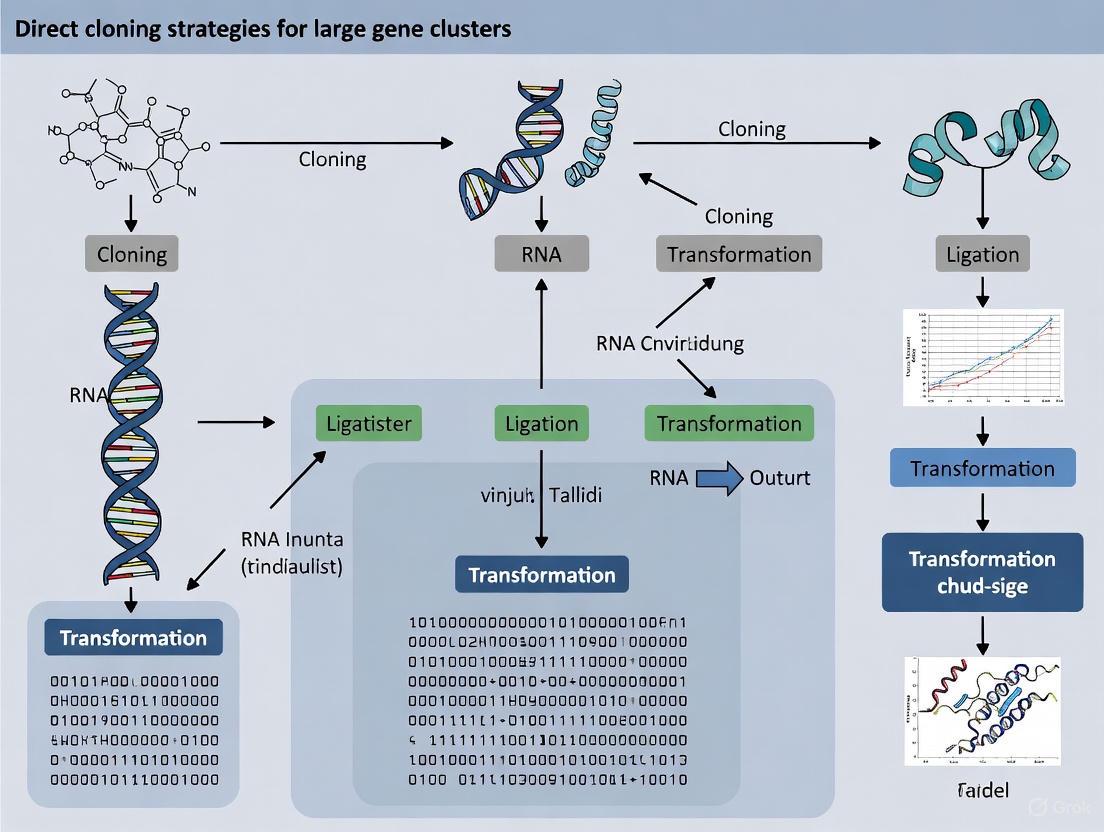
The microbial world represents a vast and largely untapped reservoir of natural products with immense potential for drug discovery. These compounds, often possessing complex structures and significant bioactivities, are synthesized by Biosynthetic Gene Clusters (BGCs)—groups of clustered genes in bacteria, fungi, and some plants that encode the machinery for secondary metabolite production [1] [2]. Large-scale genome-mining analyses have revealed that microbes potentially harbor a huge reservoir of uncharacterized BGCs [3]. However, a major challenge persists: the majority of these BGCs are silent (or cryptic) and are not expressed under standard laboratory conditions, making their corresponding natural products inaccessible for traditional discovery methods [4] [3]. This gap between genetic potential and characterized metabolites has driven the development of innovative strategies to access this hidden biosynthetic diversity.
Direct cloning has emerged as a pivotal genomic technique for the capture and heterologous expression of large, intact DNA fragments containing BGCs. It is defined as a methodology that enables the direct isolation of a specific, large DNA segment—often tens of kilobytes in size—from a source organism's genome and its subsequent assembly into a vector suitable for transfer and expression in a surrogate host [5] [3]. This strategy is particularly powerful because it bypasses the native host's regulatory constraints that often silence BGCs. By placing the cloned cluster in a genetically tractable, optimized heterologous host, researchers can activate the silent pathway and discover the novel compounds it produces [4]. Direct cloning is thus a cornerstone of modern heterologous host-based genome mining, providing a universal and enabling technology to prioritize the vast and ever-increasing number of uncharacterized BGCs identified in sequencing projects [3].
The Imperative for Direct Cloning in Natural Product Discovery
The drive to develop and refine direct cloning methodologies stems from the significant limitations of traditional approaches to BGC characterization and natural product discovery.
- The Problem of Silent BGCs: Genomic sequencing consistently reveals that Streptomyces and other prolific producers harbor a significantly larger number of BGCs than the number of identified natural products, suggesting that numerous clusters remain minimally expressed or completely inactive [4]. For instance, a study on Streptomyces thermolilacinus SPC6 uncovered 20 typical secondary metabolic BGCs, one of which,
dmx, was identified as completely silent until directly cloned and expressed in a heterologous host, leading to the discovery of a new lanthipeptide, dmxorosin [4]. - Limitations of In-Situ Activation Methods: While methods like promoter engineering, manipulation of global regulators, and the One Strain Many Compounds (OSMAC) strategy can sometimes activate silent BGCs in their native hosts, they are often hit-or-miss and not universally applicable [4]. When these approaches fail, isolating the biosynthetic gene cluster for heterologous expression becomes necessary [4].
- Advantages over Traditional Construction: Direct cloning is distinguished from earlier methods that relied on the painstaking, step-by-step in vitro assembly of a BGC from synthesized parts. By capturing the native cluster directly from genomic DNA (gDNA), direct cloning preserves the cluster's innate genetic architecture, including its native promoters, regulatory elements, and codon usage, which can be critical for successful expression. This makes it a faster and more faithful route to accessing biosynthetic diversity.
Core Principles and Methodological Framework of Direct Cloning
The execution of a direct cloning experiment, regardless of the specific technique, requires the resolution of three fundamental issues [3]. The table below outlines these core steps and their objectives.
Table 1: The Three Fundamental Issues in Direct Cloning of BGCs
| Step | Core Objective | Key Considerations |
|---|---|---|
| 1. Genomic DNA (gDNA) Preparation | To obtain high-quality, high-molecular-weight gDNA from the source organism. | DNA integrity is paramount; shearing must be minimized to preserve large DNA fragments containing the entire BGC. |
| 2. BGC Fragment Liberation | To precisely digest and release the intact target BGC from the genomic context. | Requires precise digestion at bilateral boundaries; methods range from restriction enzyme-based to homology-assisted. |
| 3. Vector Assembly | To ligate the liberated BGC fragment into a suitable capture vector. | The vector must be replicable in the heterologous host and often contains selectable markers and elements for genetic manipulation. |
The following diagram illustrates the logical workflow and decision points in a generalized direct cloning strategy.
Key Methodologies for Direct Cloning
Several highly effective methods have been established to address the core challenges of direct cloning.
- Red/ET Recombination (Recombination-Mediated Genetic Engineering): This is a powerful E. coli-based method that uses bacteriophage-derived proteins to mediate highly efficient homologous recombination between linear DNA fragments. In the context of direct cloning, short homology arms (50 base pairs) are added to the ends of the target BGC via PCR. These arms correspond to sequences in the linearized capture vector. When co-introduced into an E. coli strain expressing the Red/ET proteins, the BGC fragment recombines into the vector, seamlessly capturing the cluster [4]. This method was successfully used to directly clone the 23.5 kilobase pair (kbp)
dmxBGC from Streptomyces thermolilacinus [4]. - Transformation-Associated Recombination (TAR): TAR is a yeast-based cloning system that exploits the innate homologous recombination machinery of Saccharomyces cerevisiae. The method involves co-transforming gDNA alongside a linearized capture vector into yeast cells. The capture vector contains "hooks"—homology arms that target the regions flanking the desired BGC. Inside the yeast nucleus, the endogenous recombination systems assemble the circular plasmid by linking the vector to the target genomic fragment, effectively cloning the BGC directly from the gDNA mixture.
- CRISPR-Cas Assisted Cloning: The precision of CRISPR-Cas systems has been harnessed for direct cloning. For example, an in vitro protocol using CRISPR-Cas12a has been developed for the direct cloning of large DNA fragments [5]. In this approach, Cas12a ribonucleoproteins (RNPs) are programmed to make double-strand cuts at specific sites flanking the BGC, precisely liberating the fragment from the genome for subsequent capture. This method offers high specificity and flexibility.
Application Notes: A Protocol for Direct Cloning and Heterologous Expression
This protocol outlines the key steps for direct cloning of a BGC using Red/ET recombination, based on a successful case study [4].
Protocol: Direct Cloning of a BGC via Red/ET Recombination
Objective: To isolate a silent ~23 kbp lanthipeptide BGC (e.g., dmx) from Streptomyces thermolilacinus SPC6 and express it heterologously in a suitable Streptomyces host.
Materials: Table 2: Research Reagent Solutions for Direct Cloning
| Reagent / Material | Function / Explanation |
|---|---|
| Source Organism gDNA | High-quality, high-molecular-weight genomic DNA from the organism harboring the target BGC (e.g., S. thermolilacinus SPC6). Serves as the template for cloning. |
| Linearized Capture Vector | A bacterial Artificial Chromosome (BAC) or other suitable vector, linearized to contain homology arms for Red/ET recombination. Provides the backbone for BGC propagation and selection. |
| E. coli GBdir-red Strain | An engineered E. coli strain that inducibly expresses the Red/ET recombination proteins. Essential for facilitating the homologous recombination event. |
| PCR Reagents | For amplifying homology arms and potentially the entire BGC if a "capture" approach is used. |
| Electrocompetent E. coli Cells | For high-efficiency transformation of the recombined plasmid after the Red/ET reaction. |
| Heterologous Host Strains | Genetically tractable surrogate hosts (e.g., S. coelicolor, S. lividans). Must be easily transformable and provide a compatible biosynthetic background for BGC expression. |
Experimental Workflow:
Bioinformatic Analysis and Primer Design:
- Identify the exact boundaries of the target BGC (e.g.,
dmx) using genome mining tools like antiSMASH [1]. - Design PCR primers to amplify approximately 50 base pair homology arms corresponding to the very beginning and end of the target BGC. These arms must also match the ends of the linearized capture vector.
- Identify the exact boundaries of the target BGC (e.g.,
Preparation of the Targeting Cassette:
- Amplify the homology arms via PCR.
- If using a "capture" approach, assemble these arms onto a selectable marker cassette to create a linear "targeting cassette."
Red/ET Recombination in E. coli:
- Introduce the linearized capture vector and the targeting cassette (or the PCR-amplified BGC with homology arms) into the Red/ET-proficient E. coli strain (e.g., GBdir-red) via electroporation.
- Allow homologous recombination to occur, which results in the insertion of the target BGC into the capture vector, forming a circular, replicable plasmid.
Selection and Validation:
- Plate the transformation mixture on selective media to isolate clones containing the putative BGC plasmid.
- Screen colonies by colony PCR to confirm the presence of the BGC.
- Validate positive clones by restriction enzyme digestion and full-length sequencing of the plasmid insert to ensure fidelity and completeness.
Heterologous Expression:
- Introduce the validated plasmid containing the cloned BGC (e.g., pCAP-dmx) into a suitable Streptomyces heterologous host via protoplast transformation or conjugation.
- Culture the recombinant host under appropriate fermentation conditions.
Metabolite Analysis and Compound Discovery:
- Extract metabolites from the culture broth and mycelia.
- Analyze the extracts using Liquid Chromatography–High-Resolution Mass Spectrometry (LC-HRMS).
- Compare the metabolic profiles of the heterologous host containing the cloned BGC with a control strain (empty vector). New peaks present only in the experimental sample indicate compounds produced by the silent BGC, as demonstrated by the discovery of dmxorosin [4].
The experimental workflow from genome mining to novel compound discovery is summarized in the following diagram.
Integration with Computational and Analytical Workflows
Direct cloning does not operate in a vacuum; its success is heavily dependent on integrated bioinformatic and analytical pipelines.
- Genome Mining for BGC Prioritization: The initial identification of a target BGC relies on computational tools. Tools like antiSMASH (antibiotics & Secondary Metabolite Analysis Shell) are fundamental for the automated identification and annotation of BGCs in genomic data [1]. This allows researchers to survey an organism's biosynthetic potential and prioritize silent clusters for cloning based on novelty or predicted interesting features.
- The Role of Advanced Analytics: After heterologous expression, advanced metabolomics is critical. In the dmxorosin discovery, the Global Natural Products Social molecular networking platform and the COCONUT database were used to compare the MS data of the heterologous expresser against known compounds, allowing the researchers to confidently identify the produced compound as new [4]. This highlights the essential partnership between cloning and analytical chemistry.
Direct cloning has firmly established itself as an indispensable strategy in the modern natural product discovery pipeline. By providing a direct and robust route to capture, shuttle, and express silent biosynthetic gene clusters in tractable heterologous hosts, it effectively bypasses the regulatory constraints of native producers. This methodology is a key enabler for translating the immense genetic potential revealed by genome sequencing into tangible chemical entities. The continued refinement of direct cloning protocols—making them faster, more efficient, and applicable to ever-larger gene clusters—will undoubtedly accelerate the discovery of novel natural products. These compounds serve as crucial lead structures for the development of new pharmaceuticals, particularly in an era of rising antibiotic resistance. As such, direct cloning stands as a cornerstone technique for accessing the hidden biosynthetic diversity encoded within the microbial world.
The direct cloning of large biosynthetic gene clusters (BGCs) is a fundamental strategy in modern natural product research and drug discovery, enabling the heterologous expression and characterization of compounds from difficult-to-culture microorganisms [3]. However, this field faces three persistent technical challenges: the substantial size of many BGCs (often exceeding 100 kb), their frequent residence in GC-rich genomic regions, and their association with complex repetitive sequences [6] [7]. These characteristics complicate high-fidelity DNA extraction, precise enzymatic manipulation, and stable vector assembly. This Application Note details current, practical methodologies to overcome these hurdles, providing researchers with structured protocols and resource guides to accelerate the cloning of large, architecturally complex gene clusters.
Technical Hurdles and Strategic Solutions
The successful direct cloning of a BGC is contingent upon addressing its specific physicochemical and structural properties. The table below summarizes the primary challenges and the corresponding advanced strategies developed to counter them.
Table 1: Key Technical Hurdles and Corresponding Cloning Strategies
| Technical Hurdle | Underlying Cause of Difficulty | Recommended Strategy | Key Experimental Tools |
|---|---|---|---|
| Large Size (>50 kb) | Mechanical shearing during DNA isolation; low cloning efficiency in standard vectors [6]. | Preparation of High-Molecular-Weight (HMW) DNA; use of high-capacity vectors [6] [8]. | Cell embedding in agarose plugs; Cas9-guided excision (CISMR); Bacterial Artificial Chromosomes (BACs); Transformation-Associated Recombination (TAR) in yeast [6] [8]. |
| GC-Richness | Formation of stable secondary structures; impedes polymerase progression and restricts enzyme access [6]. | Optimization of PCR and enzymatic reaction buffers; use of specialized polymerases. | High-fidelity, GC-enhanced DNA polymerases (e.g., KAPA HiFi HotStart); additives like DMSO or betaine; optimized thermal cycling conditions [9] [10]. |
| Repetitive Sequences | Homologous recombination between repeats; misassembly during in vivo methods; incorrect sequence alignment [7]. | In vitro assembly methods; careful design of homology arms. | Gibson assembly; Golden Gate assembly; Red/ET Recombineering; designing unique homology arms for TAR cloning that flank, rather than reside within, repetitive regions [6] [7]. |
The Scientist's Toolkit: Essential Research Reagents
The successful implementation of the strategies outlined in Table 1 relies on a suite of specialized reagents and materials. The following table catalogues key solutions for direct cloning workflows.
Table 2: Research Reagent Solutions for Direct Cloning of Large Gene Clusters
| Research Reagent | Function / Application | Specific Example(s) / Notes |
|---|---|---|
| High-Capacity Vectors | Carrying large DNA inserts (50-200 kb) without instability [6]. | Bacterial Artificial Chromosomes (BACs), Cosmids (e.g., pWEB). |
| High-Fidelity DNA Polymerases | Accurate amplification of long, GC-rich templates; critical for epPCR library construction with low bias [9] [10]. | KAPA HiFi HotStart, Platinum SuperFi II, Hot-Start Pfu DNA Polymerase. |
| CRISPR-Cas9 System | Programmable excision of specific, large DNA fragments from a genome [6]. | In vitro Cas9 nuclease with sgRNAs designed to flank a target BGC. |
| Homology Assembly Enzymes | In vitro assembly of multiple DNA fragments via homologous recombination. | Gibson Assembly Master Mix. |
| TAR "Capture" Vectors | In vivo homologous recombination in S. cerevisiae to assemble or capture large DNA regions [8]. | Yeast shuttle vectors with short homology arms targeting the gene cluster flanking sequences. |
| Viscoelastic Liquids & Capillary Systems | Highly sensitive separation and concentration of HMW DNA fragments [6]. | Used post-Cas9 cleavage to isolate specific fragments from a complex background. |
Detailed Experimental Protocols
Protocol 1: TAR-Mediated Reassembly of a Large BGC from Overlapping Cosmids
Transformation-Associated Recombination (TAR) is a powerful method for assembling a single, large DNA construct from multiple overlapping clones, such as those obtained from a cosmid library [8].
Workflow Overview:
Diagram 1: TAR cloning workflow from cosmids.
Materials:
- Overlapping cosmid clones spanning the target BGC.
- TAR "capture" vector (a yeast-E. coli shuttle vector with a selection marker).
- Saccharomyces cerevisiae host strain (e.g., VL6-48).
- Standard reagents for yeast transformation (PEG/LiAc, single-stranded carrier DNA).
- Selective media plates (e.g., lacking uracil for selection).
Method:
- Capture Vector Design: Linearize the TAR capture vector. The ends of the linearized vector must contain short homology arms (approximately 50-150 bp) that are identical to the 5' and 3' ends of the target BGC you wish to assemble. Crucially, these arms must be designed from unique, non-repetitive sequences that flank the gene cluster to avoid aberrant recombination within repetitive internal regions [8].
- Prepare DNA Mixture: Combine the linearized capture vector with a mixture of the overlapping cosmid clones. The cosmids should provide full coverage of the entire BGC with sufficient overlap between adjacent clones (typically >5 kb).
- Yeast Transformation: Co-transform the DNA mixture into competent S. cerevisiae cells using a high-efficiency protocol, such as the PEG/LiAc method.
- Selection and Growth: Plate the transformed yeast cells onto selective medium. The selection should be for the marker on the successfully recombined TAR vector. Incubate at 30°C for 2-3 days until colonies form.
- Validation: Isolate yeast plasmid DNA and transform it into a suitable E. coli host for amplification and downstream analysis. Confirm the integrity and correct assembly of the captured BGC using restriction fragment length polymorphism (RFLP) analysis and long-read sequencing (e.g., PacBio or Nanopore).
Protocol 2: High-Throughput Construction of Mutagenesis Libraries for BGC Optimization
Directed evolution through mutagenesis libraries is a key strategy for optimizing the expression and function of cloned BGCs in heterologous hosts. This protocol outlines a high-throughput, chip-based oligonucleotide synthesis approach for creating precise, high-coverage libraries [10].
Workflow Overview:
Diagram 2: Mutagenesis library construction workflow.
Materials:
- Target gene or BGC sequence.
- High-fidelity, low-bias DNA polymerase (e.g., KAPA HiFi HotStart, Platinum SuperFi II).
- Gibson Assembly Master Mix.
- Chip-synthesized oligonucleotide pool.
- Appropriate expression vector.
Method:
- Library Design: Divide the target gene sequence into tiled sub-libraries. For each amino acid position to be mutated, design oligonucleotides that contain the desired mutation (e.g., an amber codon, TAG) flanked by 16-19 bp homology arms for subsequent assembly [10].
- Oligonucleotide Synthesis: The designed oligonucleotide library is commercially synthesized using high-throughput, chip-based parallel synthesis technology.
- Sub-library Amplification: Use a high-fidelity DNA polymerase to PCR-amplify each sub-library from the pooled oligonucleotides. The use of a high-fidelity polymerase is critical to minimize the introduction of secondary, unintended mutations and to reduce the formation of chimeric sequences caused by incomplete extension [10].
- Assembly and Cloning: Assemble the full-length mutated gene from the amplified sub-libraries using Gibson assembly and clone it into the destination vector.
- Library Validation: Transform the assembled library into E. coli and use next-generation sequencing (NGS) to assess the mutation coverage and uniformity of the library. Aim for high coverage (>90%) and analyze unmapped reads to identify and troubleshoot issues like synthesis errors or PCR chimeras [10].
Concluding Remarks
The direct cloning of large, complex BGCs is no longer an insurmountable challenge. By leveraging a toolkit of specialized strategies—including TAR for assembly, CRISPR for precise excision, and optimized reagents for handling difficult sequences—researchers can systematically overcome the hurdles of size, GC-richness, and repetitive elements. The protocols detailed herein provide a actionable framework for accessing the vast untapped reservoir of natural products encoded in microbial genomes, thereby accelerating the discovery and development of novel therapeutic agents.
The field of molecular cloning has undergone a revolutionary transformation, shifting from traditional library-based approaches to sophisticated direct capture and editing technologies. This evolution is particularly crucial for research on large gene clusters, such as natural product biosynthetic gene clusters (BGCs), where conventional methods often proved slow and inefficient [3]. The limitations of native host-based approaches, including poor expression of silent BGCs under laboratory conditions, have driven the development of heterologous host-based genome mining strategies [3]. This application note examines the trajectory of cloning technology within the context of direct cloning strategies for large gene clusters, providing researchers with current methodologies and practical frameworks for implementation.
The Paradigm Shift: From Library-Based Cloning to Direct Capture
Historical Limitations of Library Approaches
Traditional genomic library construction and screening presented significant bottlenecks for cloning large gene clusters. These methods involved fragmenting genomic DNA, cloning into vectors, transforming hosts, and laboriously screening thousands to millions of clones—a process that could require weeks to months [11]. The fundamental challenge lay in the random nature of library construction, which often resulted in incomplete coverage or fragmentation of large gene clusters essential for natural product synthesis.
The Direct Capture Revolution
Direct cloning strategies emerged to address these limitations by enabling targeted isolation of specific genomic regions of interest without library construction and screening. As summarized in recent advances, each direct cloning method must resolve three critical issues: (1) genomic DNA preparation, (2) bilateral boundary digestion for target BGC release, and (3) BGC and capture vector assembly [3]. This paradigm shift has dramatically accelerated the cloning of large gene clusters, reducing the process from months to days in some cases [11].
Table 1: Comparison of Cloning Technology Generations
| Technology Generation | Key Methodology | Maximum Insert Size | Key Applications |
|---|---|---|---|
| Genomic Libraries | Random fragmentation, library construction, and screening | 40-50 kb (cosmid); >100 kb (BAC) | Gene discovery, sequencing projects |
| Target Sequence Capture | Hybridization with RNA baits to genomic DNA | Dependent on bait design | Phylogenomics, variant discovery |
| Direct Cloning & Capture | PCR-independent capture using specific enzymes | >50 kb | Natural product BGC cloning |
| Programmable Chromosome Engineering | CRISPR-based and recombinase-based systems | Megabase scale | Chromosome engineering, crop improvement |
Modern Direct Capture Methodologies
CRISPR-Enhanced Retrieval Systems
The CRISPR Counter-Selection Interruption Circuit (CCIC) represents a significant advancement in clone retrieval from complex metagenomic libraries. This system utilizes nuclease-deficient Cas9 (dCas9) programmed with guide RNAs to target unique barcode sequences adjacent to cloned inserts, enabling selective survival under counter-selection conditions [11].
Experimental Protocol: CCIC-Based Clone Retrieval
Vector Design: Engineer a cloning vector (e.g., pCCIC cosmid) containing:
- A counter-selection marker (e.g., sacB gene for sucrose sensitivity)
- A promoter driving counter-selection gene expression
- A degenerate 24-bp barcode sequence between promoter and counter-selection gene
- Multiple cloning site for insert DNA
Library Construction:
- Fragment high-molecular-weight metagenomic DNA
- Ligate into barcoded pCCIC vectors
- Package using lambda phage and transform into E. coli
- Pool transformants to create library
Library Sequencing & Indexing:
- Sequence library pool using PacBio HiFi long-read technology
- Link barcode sequences to specific cloned inserts bioinformatically
- Identify target clones based on insert sequence
Target Retrieval:
- Design sgRNA targeting barcode of desired clone
- Introduce sgRNA plasmid into library pool
- Plate on sucrose-containing media
- Only clones with dCas9-silenced sacB expression survive
This method has demonstrated efficiency in retrieving target sequences from pools containing up to 50,000 non-target clones with positive hit rates exceeding 70% [11].
Programmable Chromosome Engineering
A groundbreaking advancement in large-scale DNA manipulation comes from Programmable Chromosome Engineering (PCE) systems, which enable precise editing of chromosomal segments ranging from kilobases to megabases [12] [13]. This technology overcomes historical limitations of the Cre-Lox system through three key innovations:
- Asymmetric Lox Site Design: Reduces reversible recombination activity by over 10-fold while maintaining high-efficiency forward recombination [12]
- AiCErec Recombinase Engineering: Uses AI-informed protein engineering to optimize Cre's multimerization interface, yielding a variant with 3.5 times higher recombination efficiency than wild-type [12]
- Scarless Editing Strategy: Employs prime editors with specifically designed Re-pegRNAs to replace residual Lox sites with original genomic sequence [12]
Experimental Protocol: PCE for Large-Scale Genome Engineering
Target Selection: Identify target genomic region (demonstrated for manipulations up to 12 Mb inversions and 4 Mb deletions) [13]
gRNA Design:
- Design asymmetric Lox sites flanking target region
- Create Re-pegRNAs for precise editing outcomes
System Delivery:
- Deliver PCE components (engineered Cre recombinase, asymmetric Lox sites, Re-pegRNAs) to plant or animal cells
- Use appropriate transformation method for target cell type
Selection & Validation:
- Apply appropriate selection for edited cells
- Validate edits via PCR, sequencing, and phenotypic assessment
- Confirm precise boundaries and absence of residual sequences
In proof-of-concept applications, researchers used PCE technology to create herbicide-resistant rice germplasm through a precise 315-kb chromosomal inversion [12].
Table 2: Capabilities of Programmable Chromosome Engineering Systems
| Edit Type | Demonstrated Scale | Experimental System | Key Application |
|---|---|---|---|
| Targeted Insertion | 18.8 kb | Plant and animal cells | Gene stacking |
| Sequence Replacement | 5 kb | Plant and animal cells | Allele swapping |
| Chromosomal Inversion | 12 Mb | Plant and animal cells | Gene regulation studies |
| Chromosomal Deletion | 4 Mb | Plant and animal cells | Functional genomics |
| Whole Chromosome Translocation | Entire chromosomes | Plant and animal cells | Chromosome engineering |
| Precise Inversion | 315 kb | Rice | Herbicide-resistant crops |
Application-Focused Workflows
Optimized Gene Cloning in Complex Genomes
Recent work on cloning disease resistance genes in wheat demonstrates an optimized workflow that combines multiple advanced techniques for rapid gene identification [14]. This approach successfully cloned the stem rust resistance gene Sr6 in just 179 days using only three square meters of plant growth space [14].
Experimental Protocol: High-Throughput Gene Cloning in Wheat
Mutagenesis Population:
- Treat wheat seeds with ethyl methanesulfonate (EMS)
- Sow M1 generation at high density (15 grains per 64 cm²)
- Harvest individual M2 spikes as separate families
Phenotypic Screening:
- Inoculate 3-week-old M2 seedlings with pathogen (Puccinia graminis)
- Identify loss-of-resistance mutants based on sporulation
- Transfer putative mutants to single pots and re-inoculate
Genomic Analysis:
- Harvest leaf tissue from confirmed mutants for RNA sequencing
- Generate isoform sequencing (Iso-Seq) data for wild-type parent
- Perform MutIsoSeq analysis to identify transcripts with EMS-type mutations
Validation:
- Amplify and sequence candidate gene from all mutants
- Develop KASP marker for genotyping
- Confirm gene identity via VIGS and CRISPR/Cas9 knockout
This workflow successfully identified Sr6 as encoding a CC-BED-domain-containing NLR immune receptor, with mutations found in 97 of 98 loss-of-function mutants [14].
Target Sequence Capture for Phylogenomics
For evolutionary studies, target sequence capture remains a powerful method when combined with appropriate bait design strategies [15]. This approach uses custom RNA baits to hybridize with and enrich complementary DNA regions before sequencing.
Experimental Protocol: Phylogenomic Target Capture
Bait Selection:
- Identify conserved loci across taxonomic group of interest
- Use existing bait sets (e.g., UCEs, AHE) or design custom baits
- Consider genetic divergence between bait source and study organisms
Library Preparation & Capture:
- Extract high-quality genomic DNA
- Fragment DNA and prepare sequencing libraries
- Hybridize with biotinylated RNA baits
- Capture bait-bound fragments using streptavidin beads
Sequencing & Analysis:
- Sequence captured libraries on appropriate platform
- Process data with phylogenetically-informed bioinformatic pipelines
This method is particularly valuable for degraded DNA samples from museum specimens, as the enrichment provides greater coverage of target loci [15].
The Scientist's Toolkit: Essential Research Reagents
Table 3: Key Research Reagents for Direct Cloning Applications
| Reagent/Resource | Function | Application Examples |
|---|---|---|
| dCas9 (nuclease-deficient) | Sequence-specific binding without cleavage | CCIC clone retrieval [11] |
| Asymmetric Lox Sites | Directional recombination with reduced reversibility | PCE systems [12] |
| Engineered Cre Recombinase (AiCErec) | High-efficiency recombination | Large DNA fragment manipulation [12] |
| Re-pegRNAs | Guide prime editors for scarless editing | Residual site removal in PCE [12] |
| Barcoded CCIC Vectors | Clone-specific identification and retrieval | Metagenomic library screening [11] |
| EMS (Ethyl Methanesulfonate) | Chemical mutagenesis to induce point mutations | Forward genetic screens [14] |
| Target Capture Baits | Hybridization-based enrichment of genomic regions | Phylogenomic studies [15] |
| Tapestri Technology | Single-cell DNA-RNA sequencing platform | Functional phenotyping of variants [16] |
The trajectory from genomic libraries to direct capture technologies represents a fundamental shift in how researchers approach gene cloning and manipulation. Modern methods now enable precise programming of cellular machinery to target, capture, and engineer genetic elements with unprecedented efficiency and scale. These advances are particularly transformative for the study of large gene clusters, where traditional methods often failed. As direct cloning strategies continue to evolve, they promise to further accelerate natural product discovery, crop improvement, and our fundamental understanding of genetic regulation.
The Critical Need for Large-Insert Cloning in Natural Product Discovery and Functional Genomics
The rapid accumulation of genomic data has revealed a vast reservoir of uncharacterized biosynthetic potential, particularly in the genomes of uncultured microorganisms. Large-insert cloning technologies serve as the critical bridge connecting this genetic potential to discoverable natural products and functional insights. These methods enable researchers to directly capture, manipulate, and express large genomic fragments that are often beyond the capacity of conventional cloning vectors. In the context of functional genomics, large-insert cloning facilitates the systematic study of gene function by allowing researchers to transfer entire gene clusters into model organisms for phenotypic analysis [17]. Similarly, in natural product discovery, these techniques provide access to extensive biosynthetic gene clusters (BGCs) encoding novel compounds with potential therapeutic value [8] [3]. This application note details the experimental frameworks and reagent solutions essential for successful large-insert cloning, emphasizing its transformative role in direct cloning strategies for large gene cluster research.
The Scientific Challenge: Why Large-Insert Cloning is Necessary
The Scale of Biological Complexity
Many biological functions are encoded not by single genes but by large, coordinated genetic elements. Biosynthetic gene clusters for natural products often span 30-150 kilobases, encompassing genes for biosynthesis, regulation, and transport [8] [3]. Similarly, functional studies of gene regulatory elements or multi-gene complexes require capturing large genomic contexts to preserve biological activity. Conventional plasmid vectors typically accommodate only 2-3kb inserts, creating a fundamental technical gap in studying these complex genetic systems [18].
The Problem of Uncultured Microbes
Single gram of soil can contain thousands of unique bacterial species, most of which have never been cultured in laboratory settings [8]. This represents one of biology's largest reservoirs of unexplored genetic diversity and natural product potential. Large-insert cloning from environmental DNA (eDNA) allows researchers to access this uncultured majority by capturing their genetic material directly from environmental samples and expressing it in tractable host organisms [8].
Table 1: Comparison of Cloning Vectors by Insert Capacity
| Vector Type | Typical Insert Size | Primary Applications | Key Advantages |
|---|---|---|---|
| Plasmid | 2-3 kb | Gene expression, protein production | High copy number; easy manipulation |
| Cosmid | 30-45 kb | Small gene clusters, metagenomic libraries | Efficient packaging and transduction |
| Bacterial Artificial Chromosome (BAC) | Up to 350 kb | Large gene clusters, genomic libraries | Very large insert capacity; stable maintenance |
| Yeast Artificial Chromosome (YAC) | Up to 1,000 kb | Extremely large gene clusters, synthetic biology | Largest capacity; eukaryotic features |
Quantitative Foundations: Understanding Library Scale Requirements
The probability of capturing any specific large gene cluster from a complex environmental sample depends on both the insert size of the cloning vector and the overall size of the library. Research with soil-derived eDNA cosmid libraries has empirically demonstrated that library complexity must be substantial to ensure complete coverage of large biosynthetic pathways [8].
Table 2: Empirical Library Size Requirements for Soil eDNA Studies
| Library Source | Library Size | Insert Size (Cosmid) | Theoretical Coverage | Practical Outcome |
|---|---|---|---|---|
| Utah Soil Library | ~10 million clones | 30-40 kb | Extensive but incomplete for large BGCs | Enabled identification of overlapping clones for reassembly |
| California Soil Library | ~15 million clones | 30-40 kb | More comprehensive coverage | Facilitated recovery of complete pathways via overlapping clones |
These studies revealed that while constructing 30-40kb insert cosmid libraries from environmental samples is now routine, capturing BGCs larger than a single cosmid insert requires either extremely large libraries or strategies to reassemble complete pathways from multiple overlapping clones [8]. This fundamental limitation has driven the development of specialized methods for cloning large genomic fragments in the 50-150kb range [19].
Experimental Framework: TAR-Mediated Reassembly of Large Gene Clusters
Transformation-associated recombination (TAR) in Saccharomyces cerevisiae provides a powerful method for reassembling large natural product biosynthetic gene clusters from collections of overlapping eDNA cosmid clones. This approach leverages the highly efficient homologous recombination system of yeast to assemble large DNA constructs that are challenging to manipulate using traditional restriction enzyme-based methods [8].
Protocol: TAR Reassembly of Large BGCs from Overlapping Cosmid Clones
Step 1: Library Construction and Screening
- High Molecular Weight DNA Extraction: Extract DNA directly from environmental samples (e.g., soil) using gentle lysis buffer (2% SDS, 100 mM Tris-HCl, 100 mM EDTA, 1.5 M NaCl, 1% CTAB). Incubate at 70°C for 2 hours, then remove particulates by centrifugation at 4,000 × g for 30 minutes [8].
- DNA Purification and Size Selection: Precipitate DNA with isopropyl alcohol, then perform gel electrophoresis (1% agarose, 16h, 20V) to isolate high molecular weight DNA (>40 kb) [8].
- Cosmid Library Construction: Blunt-end the purified DNA, ligate into cosmid vectors (e.g., pWEB or pWEB-TNC), package into lambda phage, and transduce into E. coli host cells (e.g., EC100) [8].
- Library Formatting and Screening: Create glycerol stocks and DNA minipreps arrayed in 8×8 grids representing 250,000-320,000 clones. Pool rows and columns for efficient PCR-based screening using degenerate primers targeting conserved biosynthetic genes (e.g., β-ketoacyl synthase sequences for Type II PKS systems) [8].
Step 2: Identification of Overlapping Clones
- PCR Screening: Use degenerate primers to identify cosmids containing portions of the target BGC:
- For β-ketoacyl synthase domains: dp:KSβ (5′-TTCGGSGGNTTCCAGWSNGCSATG-3′) and dp:ACP (5′-TCSAKSAGSGCSANSGASTCGTANCC-3′) [8].
- PCR conditions: 50 ng eDNA template, 2.5 μM each primer, 2 mM dNTPs, ThermoPol Buffer, 0.5 U Taq polymerase, 5% DMSO.
- Touchdown protocol: Initial denaturation (95°C, 2 min); 8 touchdown cycles (95°C/45s, 65°C→58°C/1min, 72°C/2min); 35 standard cycles (95°C/45s, 58°C/1min, 72°C/2min); final extension (72°C, 2 min) [8].
- Clone Verification: Sequence amplicons of correct size (~1.5 kb for Type II PKS) to confirm identity and identify overlapping cosmids for reassembly [8].
Step 3: TAR Assembly
- Capture Vector Design: Construct a TAR "capture" vector containing:
- Yeast replication origin (ARS/CEN)
- Yeast selectable marker (e.g., URA3)
- Homology arms (500-1000 bp) matching the 5' and 3' ends of the target BGC
- Co-transformation: Combine the capture vector with overlapping cosmid clones (typically 2-4 clones spanning the entire BGC) and transform into S. cerevisiae using standard yeast transformation protocols [8].
- Selection and Verification: Select for yeast transformants containing the reassembled BGC using appropriate selection media. Verify correct assembly by PCR and restriction mapping [8].
Figure 1: TAR-mediated reassembly workflow for large biosynthetic gene clusters from overlapping cosmid clones.
Direct Cloning Strategies for Large Genomic Fragments
Recent advances in direct cloning methods have expanded capabilities for capturing large genomic fragments (50-150 kb) without the need for library construction and screening. These approaches address the three fundamental challenges of large fragment cloning: genomic DNA preparation, precise bilateral boundary digestion for target release, and efficient assembly with capture vectors [3].
Methodological Approaches for Direct Cloning
Vector Systems for Large-Insert Cloning
- Bacterial Artificial Chromosomes (BACs): Capable of maintaining inserts up to 350 kb, though typically yielding metagenomic libraries 2-3 orders of magnitude smaller than cosmid-based libraries [8].
- Transformation-Associated Recombination (TAR): Enables targeted capture of specific large genomic regions using homology arms, bypassing library construction [8].
- Advanced Direct Cloning Methods: Newer techniques including:
- Cas9-assisted targeting of chromosome segments (CATS)
- Metagenomic direct cloning approaches [19]
Critical Considerations for Direct Cloning
- DNA Quality and Integrity: Isolate high molecular weight DNA with minimal shearing using gentle extraction methods [8] [3].
- Boundary Determination: Precisely identify the 5' and 3' boundaries of target gene clusters through bioinformatic analysis prior to cloning [3].
- Vector-Host Compatibility: Select appropriate vector-host systems that support stable maintenance of large inserts without rearrangement [8] [18].
Figure 2: Decision framework for selecting appropriate large-insert cloning strategies based on project requirements.
The Scientist's Toolkit: Essential Research Reagent Solutions
Successful implementation of large-insert cloning strategies requires specialized reagents and systems optimized for handling high molecular weight DNA and maintaining large constructs.
Table 3: Essential Research Reagents for Large-Insert Cloning
| Reagent Category | Specific Examples | Function in Large-Insert Cloning | Key Considerations |
|---|---|---|---|
| Cloning Vectors | pWEB, pWEB-TNC cosmids; BAC vectors; TAR capture vectors | Provide backbone for insert propagation and selection | Stability with large inserts; appropriate origin of replication; selectable markers |
| Host Systems | E. coli EC100 (cosmid); S. cerevisiae (TAR); BAC-compatible E. coli | Enable replication and maintenance of large inserts | Recombination deficiency (recA-); restriction modification systems; transformation efficiency |
| Enzymes for DNA Manipulation | End-It Blunt-Ending Kit; T4 DNA Ligase; High-Fidelity Restriction Enzymes | Modify DNA ends for cloning; ligate fragments | Minimal shearing activity; high efficiency with large fragments; methylation sensitivity |
| DNA Purification Systems | Gel electrophoresis; Silica column purification; SPRI beads | Size selection and purification of large DNA fragments | Gentle handling to prevent shearing; efficient recovery of high molecular weight DNA |
| Screening Tools | Degenerate primers for BGC detection; antibiotic resistance markers; blue/white screening | Identification of correct clones and assemblies | Specificity for target sequences; minimal background; visual differentiation |
Applications and Future Perspectives
Large-insert cloning technologies are driving advances in two primary domains: functional genomics and natural product discovery. In functional genomics, CRISPR-based functional genomics tools enable high-throughput screening of gene function in vertebrate models, with large-insert cloning providing the means to transfer entire gene regulatory elements or multi-gene complexes into model organisms [17]. For natural product discovery, these methods facilitate the cloning and heterologous expression of large biosynthetic gene clusters from uncultured microorganisms, providing access to novel chemical entities [8] [3].
The future of large-insert cloning will likely see continued development of more efficient direct cloning methods, improved vector systems for even larger inserts, and integration with synthetic biology approaches for refactoring and optimizing cloned gene clusters. As these technologies mature, they will further accelerate the connection between genomic sequence and biological function, enabling researchers to fully explore the functional potential of complex genomes.
A Methodological Deep Dive: From RecE/RecT to CRISPR-Cas12a for Capturing BGCs
The expanding field of natural product discovery and synthetic biology necessitates the precise cloning and manipulation of large biosynthetic gene clusters (BGCs), which often range from 10 kb to over 200 kb in size [20]. These clusters encode the machinery for producing diverse compounds with biological activities, including antibiotics and anticancer agents. Traditional cloning methods, which rely on restriction enzymes and ligation, are often inadequate for capturing these large DNA sequences due to their limited cloning capacity, dependency on suitable restriction sites, and low efficiency [21]. Functional analysis of the genome sequences being delivered by massively parallel sequencing requires more efficient cloning methods [22].
Transformation-associated recombination (TAR) in Saccharomyces cerevisiae represents one alternative, but its efficiency is relatively low (0.1–2%) due to vector recircularisation by non-homologous end joining (NHEJ), necessitating intensive screening [20]. Escherichia coli–Streptomyces shuttle bacterial artificial chromosomal (BAC) vectors can carry large-sized BGCs, but the construction of BAC libraries is laborious, expensive, and results in cloning of random genome parts rather than a specific BGC of interest [20]. Against this backdrop, recombineering (recombination-mediated genetic engineering) has emerged as a powerful approach, with the RecE/RecT system from the Rac prophage demonstrating particular efficacy for the direct cloning of large genomic sequences [22] [23].
Mechanistic Basis of the RecE/RecT System
System Components and Functional Relationship
The RecE/RecT system constitutes a dedicated homologous recombination pathway encoded by the Rac prophage in E. coli. This system functions independently of the native RecA/RecBCD pathway and comprises two essential proteins that operate as an orthologous pair, meaning recombination proceeds efficiently only when both components from the same origin are co-expressed [24].
- RecE: A potent 5'→3' exonuclease that processes double-stranded DNA (dsDNA) ends. It resects one strand of the DNA, generating 3'-ended single-stranded DNA (ssDNA) overhangs [24] [23].
- RecT: A single-stranded DNA annealing protein (SSAP) that binds to the ssDNA overhangs generated by RecE. RecT facilitates the annealing of this single-stranded DNA to complementary sequences, promoting strand invasion or annealing [24] [23].
The functional synergy between these proteins is critical. Neither RecE nor RecT alone can mediate efficient recombination, and they cannot be functionally substituted by their lambda phage orthologs (Redα/Redβ) or by the host RecA protein [24]. This specificity is attributed to a required protein-protein interaction between the two components of an orthologous pair [24].
The following diagram illustrates the functional relationship and process of double-stranded break repair mediated by the RecE/RecT system:
Comparative Analysis of Recombineering Systems
Different recombineering systems exhibit distinct functional characteristics and operational requirements. The table below provides a comparative overview of the RecE/RecT system alongside other prominent systems:
Table 1: Comparison of Key Recombineering Systems
| System | Origin | Core Components | Key Mechanism | Notable Features |
|---|---|---|---|---|
| RecE/RecT | Rac prophage | RecE (exonuclease), RecT (SSAP) | Linear-linear homologous recombination | Highly efficient for large fragment cloning (>50 kb); requires full-length RecE for optimal activity [22] [23] |
| Redα/Redβ (Lambda Red) | Lambda phage | Redα (exonuclease), Redβ (SSAP) | Homologous recombination initiated at dsDNA breaks | Functionally similar but mechanistically distinct from RecE/RecT; orthologous pairing required [24] |
| SSAP-only strategies | Various phages | Beta protein (from Lambda Red) | Annealing of ssDNA to replication fork | Can function without exonuclease partner; used to enhance CRISPR editing efficiency [25] |
| CRISPR-Cas9/Beta | Hybrid system | Cas9 (nuclease), Beta (SSAP) | DSB creation followed by SSAP-mediated repair | Provides selective pressure via DSB lethality; improved editing efficiency with Beta [25] |
A key advancement in this field was the discovery that the full-length RecE protein significantly enhances the efficiency of linear-linear homologous recombination compared to the truncated versions previously studied [22]. This full-length RecE facilitates the direct cloning of large genomic sequences, including megasynthetase gene clusters ranging from 10–52 kb, enabling bioprospecting for natural products [22].
Application Notes: Experimental Protocol for Direct Cloning
This section provides a detailed methodology for implementing RecE/RecT recombineering for direct cloning of large genomic fragments, such as biosynthetic gene clusters.
The complete experimental process, from target identification to heterologous expression, involves multiple stages as illustrated below:
Detailed Stepwise Protocol
Step 1: Target Identification and Hook Design
- Bioinformatic Analysis: Identify the target BGC and its flanking sequences from genomic data.
- Homology Hook Design: Design 50-base pair homology arms (hooks) complementary to the regions immediately flanking the target BGC. These hooks will be incorporated into the linear vector backbone [20].
Step 2: Vector Preparation
- Vector Linearization: Prepare a linearized vector backbone (e.g., a yeast-E. coli-Streptomyces shuttle vector) containing the necessary elements for selection and maintenance in various hosts.
- Homology Arm Incorporation: Incorporate the designed homology hooks at both ends of the linearized vector using PCR amplification or enzymatic assembly [20].
Step 3: Genomic DNA Isolation
- High-Molecular-Weight DNA Extraction: Isolate intact, high-quality genomic DNA from the source organism using methods that preserve large DNA fragments (e.g., agarose plug embedding or gentle phenol-chloroform extraction).
Step 4: Co-transformation with RecE/RecT
- RecE/RecT Expression: Use a host strain (e.g., E. coli GB2005) that expresses the full-length RecE and RecT proteins. Alternatively, co-transform with a plasmid expressing these genes under inducible promoters [22].
- Transformation Mixture: Co-transform the linear vector (50-100 ng) and genomic DNA (200-500 ng) into the recombinase-expressing host via electroporation [20] [22].
Step 5: Recombinant Selection
- Selective Plating: Plate transformation mixtures onto selective media containing appropriate antibiotics.
- Screening: Screen resulting colonies for correct recombinants using:
- PCR screening across cloning junctions
- Restriction analysis
- Comparative genome hybridization
- Counter-Selection: Implement counterselection markers (e.g., yeast killer toxin K1 cassette, URA3 with 5-FOA) to reduce background from empty vectors [20].
Step 6: Heterologous Expression
- Vector Isolation: Isate the recombinant vector containing the captured BGC.
- Host Transformation: Introduce the construct into a suitable heterologous host (e.g., Streptomyces albus Del14) for expression.
- Product Analysis: Screen for the production of expected compounds using metabolomic approaches (e.g., LC-MS) [20].
Critical Parameters for Success
- Homology Arm Length: Optimal homology arm length is approximately 50 base pairs for efficient recombination [24]. While shorter arms (35-40 bp) can function, efficiency decreases significantly.
- Protein Expression Balance: Recombination efficiency correlates with the stoichiometric balance between RecE and RecT, with a slight excess of the annealing protein (RecT) being beneficial [24].
- DNA Substrate Quality: Use high-quality, undegraded genomic DNA as substrate to maximize the yield of full-length clones.
- Host Strain Selection: Use recombination-proficient strains such as E. coli GB2005 or WM6026 (for conjugation), or engineering strains to express the RecE/RecT system [20] [22].
Performance Data and Applications
Quantitative Performance Metrics
The RecE/RecT system has demonstrated remarkable efficiency in cloning large genomic fragments, as summarized in the table below:
Table 2: Performance Metrics of RecE/RecT-Mediated Direct Cloning
| Application | Insert Size | Efficiency | Key Outcome |
|---|---|---|---|
| Megasynthetase clusters from Photorhabdus luminescens | 10-52 kb | Highly efficient | Successful cloning of all 10 targeted clusters; heterologous expression yielded luminmycin A and luminmide A/B [22] |
| Chelocardin BGC from Amycolatopsis sulphurea | 35 kb | Functional cloning | Successful heterologous expression in S. albus Del14 resulted in antibiotic production [20] |
| Daptomycin BGC from Streptomyces filamentosus | 67 kb | Functional cloning | Heterologous expression generated the corresponding antibiotic [20] |
| cDNA and BAC segment cloning | Variable | High efficiency | Precise cloning of exactly defined DNA segments [22] |
The Scientist's Toolkit: Essential Research Reagents
Table 3: Key Research Reagents for RecE/RecT Recombineering
| Reagent / Material | Function / Purpose | Examples / Specifications |
|---|---|---|
| Full-length RecE/RecT expression system | Mediates homologous recombination between linear DNA fragments | Codon-optimized versions for enhanced expression in desired hosts [22] |
| Shuttle vectors | Cloning and maintenance of large inserts in multiple hosts | Yeast-E. coli-Streptomyces shuttle vectors (e.g., pCAP01, pTARa) [20] |
| High-efficiency competent cells | Transformation of large constructs | E. coli GB2005, E. coli WM6026 (for conjugation), S. cerevisiae BY4742 ΔKu80 (for TAR) [20] |
| Counterselectable markers | Elimination of empty vectors | Yeast killer toxin K1 cassette, URA3 with 5-FOA, sacB for negative selection in bacteria [20] |
| Homology hooks | Target-specific sequence recognition | 50 bp sequences homologous to regions flanking the target BGC [20] |
| High-fidelity DNA polymerase | Accurate amplification of vector backbones and homology arms | Phusion Hot Start II High-Fidelity DNA Polymerase [21] |
The RecE/RecT recombineering system represents a powerful and efficient methodology for the direct cloning of large genomic sequences, particularly biosynthetic gene clusters. Its ability to facilitate linear-linear homologous recombination enables the capture of gene clusters exceeding 50 kb with high fidelity, overcoming the limitations of traditional restriction enzyme-based cloning. The discovery that full-length RecE dramatically enhances this process has been instrumental in advancing bioprospecting efforts [22].
Future developments in this field are likely to focus on several key areas. First, the systematic discovery of novel recombinases from microbial sequencing data will expand the toolbox available for genome engineering [26]. Second, the integration of recombineering with other advanced technologies, such as CRISPR-Cas systems, will further enhance editing efficiency and specificity [25]. Finally, the continued optimization of delivery systems and host strains will broaden the application of these techniques across diverse bacterial species, including non-model organisms and pathogens.
As the field of synthetic biology continues to advance, the RecE/RecT system and related recombineering technologies will play an increasingly vital role in accessing and harnessing the vast genetic diversity encoded in microbial genomes for drug discovery, bioengineering, and fundamental biological research.
Direct cloning of large biosynthetic gene clusters (BGCs) is a fundamental challenge in natural product discovery and synthetic biology. Microorganisms, particularly actinobacteria, represent an unrivalled source of bioactive small molecules, with many clinically used compounds deriving directly from these natural products [27]. However, genome sequencing has revealed that the vast majority of BGCs are cryptic, meaning they are not expressed under standard laboratory conditions [27]. Furthermore, more than 10% of characterized BGCs exceed 80 kb in size, with 40% having GC content greater than 70% [27]. This combination of large size and high GC content makes these clusters particularly difficult to clone and express using conventional methods.
The emergence of CRISPR-based technologies has revolutionized large-fragment cloning by enabling precise targeting and efficient capture of specific genomic regions. Within this context, this Application Note focuses on CRISPR-enhanced capture methods, specifically CAT-FISHING (CRISPR/Cas12a-mediated fast direct biosynthetic gene cluster cloning) and related approaches, for isolating superlarge BGCs exceeding 100 kb. These technologies address critical limitations of earlier methods by combining programmable nucleases with advanced recombination systems, opening new avenues for natural product-based drug discovery [27] [28].
Core Principles of CRISPR-Enhanced Capture
CRISPR-enhanced BGC cloning methods leverage the programmability of CRISPR nucleases to create precise double-strand breaks at flanking regions of target clusters. The fundamental process involves two indispensable steps: targeted release of the large genomic fragment and its subsequent capture into an appropriate vector system [28]. CAT-FISHING specifically utilizes Cas12a (Cpf1), which offers distinct advantages over other CRISPR nucleases, including recognition of T-rich PAM sites and generation of staggered ends with 4-5 nt overhangs that facilitate subsequent assembly steps [27].
The technology combines Cas12a cleavage with advanced features of bacterial artificial chromosome (BAC) library construction, creating a robust platform for capturing large BGCs with high GC content [27]. This synergy addresses a significant challenge in the field, as traditional BAC library construction, while suitable for large DNA fragments with high GC content, is notoriously time-consuming, labor-intensive, and technically demanding [27].
Comparative Analysis of Large-Fragment Cloning Methods
Table 1: Comparison of Major Large-Fragment Cloning Methods
| Method | Key Features | Maximum Clone Size | Advantages | Limitations |
|---|---|---|---|---|
| CAT-FISHING | Combines Cas12a cleavage with BAC features | 145 kb (demonstrated) [27] | Handles high GC content (>70%); high efficiency | Specialized vector construction required |
| CRISPR-Cas9/Gibson Assembly | Uses Cas9 for cleavage with Gibson Assembly | 77 kb (demonstrated) [29] | Simpler vector construction; high fidelity for <50 kb fragments | Requires agarose gel embedding technique [29] |
| TAR | Based on homologous recombination in yeast | Varies | Efficient for some fragment types | Challenging plasmid extraction from yeast; complex restriction analysis [29] |
| ExoCET | Integrates in vitro annealing with in vivo recombination in E. coli | >150 kb (reported) [28] | Does not require restriction sites | Limited by restriction sites for BGC acquisition [29] |
| CATCH | Cas9-assisted targeting of chromosome segments | Varies | Not restricted by restriction sites | Uses agarose gel embedding technique [29] |
Experimental Protocols
CAT-FISHING Workflow for Large BGC Capture
3.1.1 Capture Plasmid Construction The capture plasmid is constructed by introducing the lacZ gene and two PCR-amplified homology arms (each ≥30 bp containing at least one PAM site) corresponding to the flanking regions of the target BGC into the pBAC2015 vector [27].
- Procedure:
- Mix 100 ng of amplified pBAC2015 backbone, 50 ng of each homology arm DNA, and 50 ng of lacZ cassette DNA
- Add 2 μL buffer and 2 μL recombinase (EZmax one-step seamless cloning kit)
- Incubate at 37°C for 30 minutes
- Use mixture directly for transformation [27]
Alternatively, if homology arms contain only one PAM site, two 30-bp arms (4-nt PAM site + 26-nt target recognition sequence) can be used. The linearized capture plasmid can be obtained by one-step PCR with homology arm-incorporated primers using pBAC2015 as template [27].
3.1.2 Genomic DNA Preparation High-quality, high-molecular-weight genomic DNA is essential for success. For actinomycetes:
- Culture strain in TSB medium supplemented with glycine (5 g/L) or sucrose (10.3%) at 30°C for 24-48 hours
- Collect mycelium by centrifugation (4°C, 4000 × g, 5 minutes)
- Resuspend in TE25S buffer (25 mM Tris-HCl, 25 mM EDTA, 0.3 M sucrose, pH 8)
- Adjust mycelium density to OD₆₀₀ of 1.9-2.0 with TE25S
- Mix with equal volume of 1.0% LMP agarose at 50°C
- Pour into plug mold and solidify
- Incubate blocks at 37°C for 1 hour in lysozyme solution (2 mg/mL in TE25S)
- Replace with proteinase K solution (1 mg/mL in NDS) and incubate at 50°C for 2 hours
- Wash blocks with TE buffer (10 mM Tris-HCl, 1 mM EDTA, pH 8) [27]
3.1.3 Cas12a Cleavage and Fragment Capture The critical step involves Cas12a-mediated release of the target fragment and its homologous recombination with the capture plasmid.
Figure 1: CAT-FISHING Workflow for BGC Capture
CRISPR-Cas9/Gibson Assembly Method
3.2.1 sgRNA Synthesis and Purification
- Design 20 bp oligonucleotides using available resources (e.g., Zhang lab design tool)
- Obtain double-stranded transcription template DNA by PCR annealing
- Perform in vitro transcription using T7 High Yield RNA Transcription Kit
- Purify RNA using VAHTS RNA Clean Beads [29]
3.2.2 Cas9 Expression and Purification
- Clone Cas9 gene fragments into pET28a vector with SalI and NcoI restriction sites
- Transform into E. coli BL21(DE3)
- Grow in LB medium at 37°C until OD₆₀₀ reaches 0.6-0.8
- Induce with 0.4 mM IPTG at 16°C for 20 hours
- Purify recombinant protein using AKTA system [29]
3.2.3 In Vitro Cas9 Cleavage of Genomic DNA
- Reaction Setup:
- 800 nM Cas9
- 400 nM sgRNA1 + 400 nM sgRNA2
- 30 μL 10× NEB buffer 3.1
- 1 μL recombinant ribonuclease inhibitor
- Total volume: 300 μL (without genomic DNA)
- Incubate at 37°C for 20 minutes
- Add 0.02-0.04 nM genomic DNA
- Incubate at 37°C for 2 hours [29]
3.2.4 DNA Purification and Gibson Assembly
- Add equal volume phenol-chloroform-isoamyl alcohol to cleavage reaction
- Centrifuge and collect supernatant
- Add 3M sodium acetate to supernatant
- Precipitate with ethanol
- Perform Gibson assembly with purified fragments and vector [29]
The Scientist's Toolkit: Essential Research Reagents
Table 2: Key Research Reagents for CRISPR-Enhanced BGC Capture
| Reagent Category | Specific Examples | Function and Application Notes |
|---|---|---|
| CRISPR Nucleases | Cas12a (Cpf1), Cas9 | Programmable DNA cleavage; Cas12a preferred for staggered ends and T-rich PAM sites [27] |
| Vector Systems | pBAC2015, Bacterial Artificial Chromosomes | Stable maintenance of large inserts; essential for >100 kb fragments [27] |
| Host Strains | E. coli derivatives, Streptomyces albus J1074 (heterologous expression) | E. coli for cloning; specialized Streptomyces strains for expression of actinobacterial BGCs [27] |
| Enzyme Kits | EZmax one-step seamless cloning kit, T7 High Yield RNA Transcription Kit, Gibson Assembly mix | Streamline key steps including assembly, in vitro transcription, and recombination [27] [29] |
| Selection Markers | lacZ (blue/white screening), antibiotic resistance genes | Enable selection of successful recombinants and maintain vector stability [27] |
| Culture Media | Luria-Bertani, Soybean flour-mannitol, TSB with glycine/sucrose | Optimized growth conditions for source organisms and heterologous hosts [27] |
Application Case Study: Marinolactam A Discovery
The power of CAT-FISHING is exemplified by the discovery of marinolactam A, a novel macrolactam compound with promising anticancer activity. Researchers successfully captured a 110 kb cryptic polyketide encoding BGC from Micromonospora sp. 181 and heterologously expressed it in a Streptomyces albus J1074-derived cluster-free chassis strain [27]. This breakthrough demonstrates the practical utility of CRISPR-enhanced capture for unlocking silent biosynthetic potential.
The process involved:
- Identification of the cryptic BGC through bioinformatic analysis
- Precise targeting and capture using CAT-FISHING methodology
- Heterologous expression in a optimized Streptomyces chassis
- Compound isolation and structure elucidation
- Bioactivity testing revealing anticancer properties [27]
This case study validates CAT-FISHING as a powerful method for complicated BGC cloning and highlights its importance to the entire community of natural product-based drug discovery [27].
Technical Considerations and Optimization
Critical Parameters for Success
Homology Arm Design: Arms should be ≥30 bp with at least one PAM site. Optimal GC content and length improve recombination efficiency. For difficult regions, consider extending arm length to 50-100 bp [27].
GC-Rich Content Challenges: BGCs from actinobacteria often have >70% GC content. Optimize hybridization temperatures and use betaine or similar additives in PCR and recombination reactions to mitigate challenges.
Fragment Size Limitations: While CAT-FISHING has captured fragments up to 145 kb, efficiency decreases with increasing size. For fragments >100 kb, optimize Cas12a cleavage time and use high-efficiency electrocompetent cells for transformation.
Troubleshooting Common Issues
- Low Capture Efficiency: Verify Cas12a activity with control substrates, ensure high-quality genomic DNA preparation, and optimize homology arm length
- Vector Self-Ligation: Implement robust counter-selection (e.g., lacZ blue/white screening) and consider additional purification steps after cleavage
- Heterologous Expression Failure: Optimize codon usage, promoter elements, and consider different chassis organisms for expression of captured BGCs
The vast majority of biosynthetic gene clusters (BGCs) in microorganisms remain silent or "cryptic" under standard laboratory conditions, presenting a significant challenge and opportunity for natural product discovery [30] [31]. Activating these cryptic pathways is essential for accessing novel chemical compounds with potential pharmaceutical applications. This application note details a case study utilizing the CAT-FISHING (CRISPR/Cas12a-mediated fast direct biosynthetic gene cluster cloning) method to directly clone and heterologously express a cryptic gene cluster, leading to the discovery of marinolactam A, a novel macrolactam with promising anticancer activity [32]. The methodology and principles described herein are framed within the broader context of direct cloning strategies for large genomic fragments, a field rapidly advancing through innovations in molecular biology [28].
Key Principles of Direct Cloning for Cryptic Gene Clusters
Direct cloning strategies bypass the need for traditional library construction and screening, enabling targeted capture and heterologous expression of large BGCs. These methods generally address three critical steps: (1) preparation of high-quality genomic DNA, (2) precise release of the target BGC from the genome, and (3) efficient assembly of the fragment into a suitable capture vector [3].
The transition from analyzing life to rewriting it necessitates technologies capable of manipulating large DNA segments. While restriction enzymes were foundational, the programmability of CRISPR/Cas systems has driven their extensive adoption for fragment release due to their flexibility and precision [28]. For capturing the released fragments, techniques such as Homologous Recombination (HR), single-strand annealing (SSA), and site-specific recombination (SSR) have been optimized to meet diverse cloning needs [28].
The CAT-FISHING Methodology
The CAT-FISHING platform combines the programmability of the CRISPR/Cas12a system with refined aspects of bacterial artificial chromosome (BAC) library construction, creating an efficient in vitro platform for capturing large BGCs [32].
Experimental Workflow and Protocol
The following diagram illustrates the key stages of the CAT-FISHING protocol for direct cloning and activation of a cryptic biosynthetic gene cluster.
Step 1: Target Identification and gDNA Preparation
- Bioinformatic Analysis: Identify the cryptic BGC of interest (e.g., a polyketide-derived macrolactam cluster) using tools like antiSMASH [33] [34].
- gDNA Isolation: Extract high-molecular-weight genomic DNA from the source actinomycete, Micromonospora sp. 181, using a standard phenol-chloroform protocol. Assess DNA integrity and purity via agarose gel electrophoresis and spectrophotometry.
Step 2: Cas12a-Mediated Fragment Release
- crRNA Design: Design and synthesize two crRNAs targeting sequences immediately upstream and downstream of the ~110 kb target BGC.
- In Vitro Digestion: Set up a 50 µL reaction containing:
- 1-5 µg of genomic DNA
- 200 nM of each crRNA
- 50 nM Lachnospiraceae bacterium Cas12a (LbCas12a) enzyme
- 1X Cas12a reaction buffer
- Incubate at 37°C for 2 hours. Heat-inactivate the enzyme at 65°C for 20 minutes.
Step 3: Vector Preparation and Capture
- Linearize the Capture Vector (e.g., a BAC vector) using restriction enzymes or PCR.
- Generate Homology Arms: Amplify by PCR two ~800 bp homology arms that correspond to the ends of the target BGC. Clone these into the linearized vector.
- Assemble the Construct: Use an in vitro recombination system (e.g., Gibson Assembly) to combine the Cas12a-released genomic fragment with the prepared capture vector.
- Transform the assembled product into competent E. coli cells and select on appropriate antibiotic plates.
Step 4: Heterologous Expression
- Isolate the Plasmid containing the captured BGC from E. coli.
- Transform the plasmid into a suitable Streptomyces expression chassis (e.g., S. coelicolor or S. lividans) via protoplast transformation or conjugation.
- Culture the recombinant strain in appropriate liquid media for 5-7 days to allow for compound production.
Step 5: Compound Isolation and Characterization
- Extract metabolites from the culture broth using organic solvents (e.g., ethyl acetate).
- Purify the compound of interest using chromatographic methods (HPLC, silica gel).
- Characterize the structure using spectroscopic techniques (LC-MS, NMR).
Performance Metrics of CAT-FISHING
Table 1: Performance Metrics of the CAT-FISHING Method in the Marinolactam A Study
| Parameter | Performance | Experimental Details |
|---|---|---|
| Maximum BGC Size Captured | 145 kb | Successfully cloned from actinomycetal genomic DNA [32]. |
| GC Content Tolerance | Up to 75% | Demonstrated with high-GC content BGCs [32]. |
| Target BGC Size | 110 kb | The specific marinolactam A cluster from Micromonospora sp. 181 [32]. |
| Key Outcome | Discovery of Marinolactam A | A novel macrolactam with promising anticancer activity [32]. |
The Scientist's Toolkit: Essential Research Reagents
Table 2: Key Research Reagents and Solutions for CAT-FISHING
| Reagent/Solution | Function | Specific Example/Note |
|---|---|---|
| CRISPR/Cas12a System | Programmable enzymatic release of the target BGC from gDNA. | Lachnospiraceae bacterium Cas12a (LbCas12a) was used for its precision and efficiency [32]. |
| Homology Arms | Facilitate precise recombination between the vector and target DNA ends. | ~800 bp arms designed based on BGC flanking sequences; critical for capture success [32]. |
| BAC (Bacterial Artificial Chromosome) Vector | Stable maintenance and propagation of large DNA inserts in E. coli. | Essential for cloning fragments >100 kb without instability [35]. |
| Heterologous Chassis | Provides a tractable background for expressing silent BGCs. | A Streptomyces species (e.g., S. coelicolor) is often optimal for actinomycete BGCs [32] [31]. |
| In Vitro Recombination Kit | Seamlessly assembles the released fragment and linearized vector. | Gibson Assembly is a common choice, though other methods exist [36] [31]. |
Discussion and Comparative Analysis
The success of CAT-FISHING in discovering marinolactam A underscores the power of direct cloning approaches in functional genomics and natural product discovery. This method effectively addresses several challenges: it is independent of the host's genetic tractability, avoids the time-consuming process of library screening, and minimizes the introduction of mutations that can occur during PCR-based assembly [32] [36].
CAT-FISHING is one of several advanced direct cloning methods. Other notable techniques include CAPTURE, which uses Cas12a and in vivo Cre-lox recombination and has demonstrated success in cloning 47 BGCs with nearly 100% efficiency, leading to the discovery of 15 novel natural products [37]. Similarly, RecET-mediated linear-plus-linear homologous recombination (LLHR) has been used to clone gene clusters up to 52 kb from Photorhabdus luminescens [36] [35].
A primary advantage of CRISPR-based methods like CAT-FISHING is their programmability, which allows for precise targeting without reliance on rare restriction enzyme sites [28] [3]. Furthermore, direct cloning preserves the native genetic context of the BGC, which can be crucial for successful expression, though it does not guarantee it. The heterologous host may still lack necessary activators or contain incompatible regulatory elements [36].
The CAT-FISHING method represents a significant asset in the growing toolkit for direct cloning of large genomic fragments. By enabling the efficient capture and heterologous expression of a 110 kb cryptic gene cluster, it facilitated the discovery of marinolactam A, demonstrating a clear path from genomic sequence to novel bioactive compound. As direct cloning technologies continue to evolve, they will undoubtedly accelerate the systematic mining of microbial genomes, fueling the discovery of new drugs and biomaterials from nature's vast, untapped genetic reservoir.
The rapidly expanding repository of microbial genomic data has revealed a vast, untapped reservoir of biosynthetic gene clusters (BGCs) that encode for potentially novel bioactive natural products [38]. This discovery is particularly significant for drug development, as natural products and their derivatives constitute a substantial proportion of clinical drugs, including 61% of anticancer drugs and 49% of anti-infection medicines [39]. However, a significant challenge persists: the majority of these BGCs are "silent" or "cryptic," meaning they are poorly expressed or not expressed at all under standard laboratory conditions [38] [3]. This limitation impedes the discovery and characterization of novel compounds, necessitating robust strategies to access this chemical diversity.
Within this context, heterologous expression has emerged as a powerful and versatile strategy for natural product discovery [40]. This approach involves the transfer of BGCs from their native hosts into genetically tractable surrogate hosts, thereby facilitating the activation, production, and engineering of encoded compounds. Its utility is especially pronounced for BGCs derived from unculturable microorganisms or those that are genetically intractable [38]. The seamless execution of this strategy, however, relies on the integration of several core disciplines: genome mining for the identification of BGCs, direct cloning for their physical capture, and pathway engineering for the optimization of production titers and structural diversification. This application note details the protocols and methodologies that underpin this integrated workflow, providing a structured guide for researchers and drug development professionals aiming to leverage direct cloning strategies for large gene cluster research.
Research Reagent Solutions and Essential Materials
The successful execution of heterologous expression and genome mining projects is contingent upon the availability of specialized reagents and tools. The table below catalogs key resources essential for experiments in this field.
Table 1: Essential Research Reagents and Tools for Heterologous Expression and Genome Mining
| Item Name | Type | Function/Application | Examples & Notes |
|---|---|---|---|
| antiSMASH | Bioinformatics Software | Prediction and annotation of BGCs in genomic sequences [41]. | Critical for initial genome mining; integrates with genomic databases [38] [42]. |
| CMNPD | Database | Comprehensive Marine Natural Products Database for structural determination [38]. | Assists in dereplication and pharmacokinetic property calculation [38]. |
| TAR Cloning Vector (e.g., pCAP01) | Molecular Biology Vector | Direct capture and manipulation of large BGCs (>50 kb) in yeast [39]. | Contains elements for shuttling between yeast, E. coli, and actinobacteria [39]. |
| pSBAC Vector | Bacterial Artificial Chromosome (BAC) | Cloning and stable maintenance of very large BGCs (up to ~200 kb) [39]. | An E. coli-Streptomyces shuttle vector; enables conjugal transfer and chromosomal integration [39]. |
| ΦBT1 Integrase System | Recombination System | Integrase-mediated site-specific recombination for BGC capture [39]. | Used in the IR (Integrase-Mediated Recombination) cloning system [39]. |
| RecE/RecT Proteins | Recombineering System | Facilitates direct cloning of genomic fragments into linearized vectors in E. coli [36]. | Enables homologous recombination between two linear DNA molecules [36]. |
| Streptomyces coelicolor | Heterologous Host | Model actinomycete for expression of actinobacterial BGCs [39]. | Well-characterized genetics and metabolism; frequently used [39]. |
| Streptomyces albus | Heterologous Host | Heterologous host with fast growth and efficient genetic system [39]. | Known for its relatively simple secondary metabolome, reducing background interference. |
Genome Mining andIn silicoBGC Identification
Application Notes
Genome mining serves as the critical first step in the discovery pipeline, allowing researchers to transition from raw genomic data to candidate BGCs for experimental characterization. This bioinformatics-driven process leverages publicly available genomic databases and sophisticated algorithms to identify and prioritize silent BGCs encoded within sequenced genomes [41] [42]. The power of this approach is magnified when applied to metagenomic sequences derived from environmental samples (metagenome-assembled genomes), offering access to the biosynthetic potential of uncultured marine and soil microorganisms [38] [42]. Effective genome mining connects BGC sequences to their encoded natural products, significantly accelerating the identification of metabolites with therapeutic relevance [42].
Protocol: BGC Prediction using antiSMASH
This protocol outlines the use of the antiSMASH (antibiotics & Secondary Metabolite Analysis Shell) pipeline to identify BGCs in a novel bacterial strain, such as Streptomyces [41].
- Input Preparation:
- Obtain the complete genome sequence of the target organism in FASTA format. This can be a newly sequenced, assembled genome.
- Software Access:
- Navigate to the antiSMASH web server (https://antismash.secondarymetabolites.org/) or install the standalone version (antiSMASH 7.0 or newer is recommended) [41].
- Job Submission & Analysis:
- Upload your genome FASTA file to the server.
- Specify the organism type (e.g., "bacteria") and select appropriate analysis parameters. It is advisable to enable all available analysis modules, including those for detecting core biosynthetic enzymes, regulatory elements, and resistance genes.
- Initiate the analysis. The processing time will depend on the genome size and server load.
- Output Interpretation:
- antiSMASH will generate an interactive results page. Each predicted BGC will be displayed within its genomic context.
- Analyze the "Cluster Type" annotation for each BGC, which identifies the primary biosynthetic machinery (e.g., Type I PKS, NRPS, lantipeptide).
- Examine the detailed annotations for individual genes within the cluster, including functional predictions for biosynthetic, regulatory, and transport-related genes.
- Use the "KnownClusterBlast" and "MIBiG" comparison features to assess the similarity of your candidate BGC to clusters encoding known natural products. This helps in prioritizing novel clusters and predicting chemical scaffolds [38] [41].
The following diagram illustrates the core computational workflow for genome mining and subsequent experimental prioritization of BGCs.
Direct Cloning Strategies for Large Gene Clusters
Application Notes
Direct cloning is the pivotal, rate-limiting step that physically captures the BGC identified through genome mining for heterologous expression [3]. Traditional methods like cosmid library construction are laborious and often limited to clusters under 40 kb, which is insufficient for many large BGCs [39]. Recent advances have yielded several highly efficient systems designed to capture large genomic segments directly from the source DNA without the need for complex library construction and screening [36] [3]. These methods generally address three key issues: genomic DNA preparation, precise release of the target BGC from the chromosome, and its efficient assembly into a capture vector [3]. The choice of system depends on factors such as BGC size, source organism, and available genetic tools.
Protocol A: Direct Cloning using RecET Recombineering
This method exploits the high efficiency of full-length RecE and RecT proteins in E. coli to recombine a linearized vector with a large, restriction-enzyme-digested genomic fragment [36].
- Vector and DNA Preparation:
- Design Homology Arms: Identify unique restriction enzyme sites flanking the target BGC. PCR-amplify ~500 bp homology arms corresponding to the sequences immediately outside these restriction sites.
- Linearize Vector: Amplify your expression vector (e.g., a standard E. coli expression vector) with primers that incorporate the homology arms at the ends, producing a linear vector molecule.
- Prepare Genomic DNA: Isolate high-quality, high-molecular-weight genomic DNA from the source organism. Digest the genomic DNA with the restriction enzymes identified in step 1 to release the intact BGC.
- Co-transformation:
- Co-transform the linearized vector and the digested genomic DNA into an E. coli strain (e.g., GB05-dir) that is induced to express the full-length RecE and RecT proteins.
- Selection and Screening:
- Plate the transformation mixture on selective media. Screen resulting colonies by colony PCR or restriction digest to identify clones containing the correct, full-length BGC insert.
- Limitations and Adaptation:
- For very large BGCs (>50 kb), a second recombineering step with an additional antibiotic marker may be required to eliminate background from recircularized empty vectors [36].
Protocol B: Transformation-Associated Recombination (TAR) in Yeast
The TAR system utilizes the highly efficient innate homologous recombination machinery of Saccharomyces cerevisiae to capture large BGCs [39].
- Vector Construction:
- Use a yeast capture vector (e.g., pCAP01) that contains yeast, E. coli, and actinobacterial elements. Clone the two homology arms (corresponding to the 5' and 3' ends of the target BGC) into this vector.
- Linearize the capturing vector within the sequence between the two homology arms.
- Yeast Transformation:
- Co-transform the linearized capturing vector and restriction-enzyme-digested genomic DNA (containing the target BGC) into competent yeast cells.
- Selection and Validation:
- Select for yeast transformants on appropriate dropout media. Isolate yeast plasmid DNA and transform it into E. coli for recovery and amplification.
- Validate the captured BGC by restriction analysis and sequencing. The final construct can then be transferred into a Streptomyces heterologous host via conjugation [39].
Table 2: Comparison of Direct Cloning Methods for Large BGCs
| Method | Principle | Typical Insert Size | Key Advantage | Key Limitation |
|---|---|---|---|---|
| RecET Direct Cloning [36] | Homologous recombination between two linear DNA molecules in E. coli. | ~10 - 52 kb (demonstrated) | Bypasses PCR, minimizing mutations; cloning and expression in E. coli. | Requires specific flanking restriction sites; efficiency drops with larger sizes. |
| TAR Cloning [39] | Natural in vivo homologous recombination in yeast. | >50 kb (e.g., 67 kb taromycin cluster) | Very high capacity; useful for capturing BGCs from metagenomic DNA. | Requires yeast handling; can be less efficient for some genomic regions. |
| pSBAC System [39] | Restriction digestion and self-ligation of large fragments into a BAC vector. | Up to ~200 kb | Extremely high capacity; stable maintenance in E. coli and Streptomyces. | Requires unique restriction sites or prior engineering of the genome. |
| Integrase-Mediated Recombination (IR) [39] | ΦBT1 integrase-mediated site-specific recombination. | Large BGCs (e.g., daptomycin cluster) | Precise excision of the BGC from the native chromosome. | Requires genetic manipulation of the original producer strain. |
The following workflow summarizes the major experimental steps from BGC capture to heterologous expression analysis.
Heterologous Expression and Pathway Engineering
Application Notes
Once a BGC is successfully cloned into a suitable vector, it must be transferred and expressed in a heterologous host. This step is crucial for activating cryptic pathways and producing sufficient quantities of the target compound for characterization [38] [40]. The selection of an appropriate heterologous host is critical; the closer the host is phylogenetically to the native producer, the more likely it is to possess the necessary substrates, cofactors, and transcriptional machinery for successful expression [38]. Streptomyces coelicolor, S. lividans, and S. albus are among the most frequently used hosts for expressing actinomycete BGCs due to their well-understood biology and genetic tractability [39]. Heterologous expression not only facilitates the discovery of novel compounds but also provides a platform for yield improvement and biosynthetic pathway engineering through genetic manipulation [40] [39].
Protocol: Heterologous Expression inStreptomyces
This protocol outlines the key steps for introducing a captured BGC into a Streptomyces host and analyzing its expression.
- Host Selection and Preparation:
- Select an appropriate heterologous host (e.g., S. coelicolor M1152 or S. albus J1074). Prepare spores or mycelium of the receptor Streptomyces strain as per standard procedures [41].
- Conjugal Transfer from E. coli:
- Donor Preparation: Transform the BGC-containing vector (e.g., pCAP01 or pSBAC, which contain an oriT for conjugation) into a donor E. coli strain, such as ET12567/pUZ8002, which carries the conjugation machinery.
- Conjugation: Mix the donor E. coli cells with the prepared Streptomyces spores or mycelium. Plate the mixture on appropriate solid media and incubate to allow conjugation.
- Selection and Overlay: After a suitable period for conjugation, overlay the plates with antibiotics that select for the Streptomyces exconjugants (containing the integrated BGC) and counter-select against the E. coli donor. Common antibiotics include apramycin or thiostrepton, depending on the vector's resistance marker [41] [39].
- Fermentation and Metabolite Analysis:
- Screen Streptomyces exconjugants for successful integration of the BGC by PCR.
- Inoculate positive clones into liquid production media. The OSMAC (One Strain Many Compounds) approach can be applied here by varying culture parameters (media, aeration) to optimize production [38].
- After fermentation, extract the culture broth and mycelia with organic solvents (e.g., ethyl acetate or methanol).
- Compound Detection and Characterization:
- Analyze crude extracts using analytical techniques such as Liquid Chromatography-Mass Spectrometry (LC-MS) or High-Performance Liquid Chromatography (HPLC).
- Compare the metabolic profiles of the BGC-containing strain with a negative control (the host containing an empty vector). Look for new, specific peaks that indicate heterologous production of the target compound.
- Scale up fermentation for the productive strain, and use guided isolation (e.g., preparative HPLC) to purify the compound for structural elucidation via Nuclear Magnetic Resonance (NMR) spectroscopy.
Pathway Engineering via DNA Assembler
For the engineering of BGCs (e.g., site-directed mutagenesis, promoter swapping, or hybrid pathway creation), the DNA assembler method is a powerful tool. This method uses in vivo homologous recombination in S. cerevisiae to assemble and engineer large biochemical pathways in a one-step fashion [43]. The process involves:
- Designing overlapping fragments for the desired genetic modifications.
- Co-transforming these fragments with a linearized vector into yeast.
- Screening yeast clones for the correct assembled construct, which can then be shuttled back into the production host for functional analysis [43].
Analytical Techniques for Validation and Characterization
The final phase of the workflow involves rigorous analytical validation of the heterologously produced natural product. As detailed in the protocol, LC-MS and HPLC are fundamental for initial detection and comparative metabolomics. Further purification via techniques like preparative HPLC or flash chromatography is necessary to obtain pure compound for definitive structural characterization, primarily using NMR spectroscopy. For complex datasets, such as those generated in multi-omics studies, quantitative quality control measures—like those implemented in the software CITESeQC for transcriptomic and proteomic data—emphasize the importance of standardized, quantitative metrics to ensure data reliability [44]. Applying this principle to natural product discovery, robust analytical validation ensures that the observed biological activity can be correctly assigned to the characterized compound produced by the heterologously expressed BGC.
Navigating Experimental Pitfalls: A Troubleshooting Guide for Complex Cloning Projects
Addressing No-Transformation and High-Background Scenarios
Within the broader research on direct cloning strategies for large gene clusters, achieving successful heterologous expression hinges on efficiently generating correct recombinant constructs. Two of the most pervasive technical challenges in this process are no-transformation (few or no colonies after transformation) and high-background (excessive numbers of colonies lacking the desired insert) scenarios. These issues are particularly pronounced when cloning large biosynthetic gene clusters (BGCs), which are essential for discovering novel natural products with potential pharmacological applications [6] [3]. This application note details the underlying causes of these problems and provides standardized, actionable protocols to overcome them, enabling more reliable and efficient cloning of large DNA fragments.
Troubleshooting Common Cloning Scenarios
A systematic approach to troubleshooting begins with understanding the specific symptoms and their most probable causes. The tables below summarize these scenarios and link them to the solutions detailed in subsequent sections.
Table 1: Addressing No-Transformation Scenarios
| Symptom & Description | Primary Associated Causes | Recommended Solution Pathways |
|---|---|---|
| No Colonies: Transformation plates show little to no growth after incubation. | • Cell viability or competency is low.• DNA fragment is toxic to host cells.• Ligation reaction failed or was inefficient.• Cloned construct is too large for standard systems. | • Verify cell competency and transformation efficiency (4.1).• Use specialized host strains for toxic genes (4.2).• Optimize ligation conditions and DNA quality (4.3).• Employ high-capacity vectors and direct cloning methods (4.4). |
Table 2: Addressing High-Background Scenarios
| Symptom & Description | Primary Associated Causes | Recommended Solution Pathways |
|---|---|---|
| Empty Vectors: Excessive colonies that contain recircularized vector without the insert. | • Incomplete digestion of the vector.• Inefficient dephosphorylation of vector ends.• Inadequate removal of phosphatase prior to ligation. | • Validate restriction enzyme digestion (5.1).• Implement robust dephosphorylation protocols (5.2).• Use advanced counterselection strategies (e.g., CcdB) (5.3). |
| Incorrect Constructs: Colonies contain plasmids with unwanted mutations, multiple inserts, or incorrect sequences. | • Recombination in the host (e.g., RecA+ strains).• Internal restriction sites within the insert.• Methylated DNA (e.g., from mammalian/plant sources) is degraded. | • Use recombination-deficient strains ( recA-) (5.4).• Analyze insert sequence for internal sites.• Use mcrA-, mcrBC-, mrr- deficient strains (5.4). |
The Scientist's Toolkit: Research Reagent Solutions
Selecting the appropriate reagents and host systems is critical for the success of direct cloning projects, especially for large and complex gene clusters.
Table 3: Essential Reagents and Host Strains for Direct Cloning
| Item | Function/Application | Example Products / Strains |
|---|---|---|
| Recombineering Systems | Mediates homologous recombination between linear DNA fragments, bypassing traditional restriction-ligation. | RecET (Rac prophage), Redαβ (lambda phage) [45] [36]. |
| CRISPR-Based Cloning | Enables precise in vitro excision of large BGCs from genomic DNA using targeted cleavage. | CRISPR-Cas12a crRNAs and enzyme [46]. |
| High-Capacity Vectors | Carries large DNA inserts (up to hundreds of kb) for heterologous expression. | Bacterial Artificial Chromosomes (BACs) [6]. |
| Specialized E. coli Strains | Provides a suitable host for large, unstable, or toxic DNA fragments; prevents degradation of methylated DNA. | NEB 10-beta, NEB Stable ( recA-, deficient in McrA, McrBC, Mrr) [47]. |
| T4 DNA Ligase | Joins blunt-ended or cohesive-ended DNA fragments. Requires optimization for blunt-end ligation [48]. | Concentrated T4 DNA Ligase, Quick Ligation Kit [47]. |
| Alkaline Phosphatase | Removes 5' phosphate groups from linearized vectors to prevent self-ligation and reduce background. | Calf Intestinal Phosphatase (CIP), Shrimp Alkaline Phosphatase (SAP) [47] [48]. |
Experimental Protocols for Key Scenarios
Protocol: Verification of Cell Competency and Transformation Efficiency
Purpose: To diagnose and rule out host-cell-related issues as a cause of no-transformation.
- Step 1: Transform competent cells with 100 pg–1 ng of an uncut, validated control plasmid (e.g., pUC19).
- Step 2: Plate the transformation on LB agar containing the appropriate antibiotic. Include a negative control (water) to confirm sterility.
- Step 3: Incubate overnight at 37°C and count colonies.
- Step 4: Calculate transformation efficiency (CFU/μg DNA). For high-efficiency cloning, this should be >1 x 10^7 CFU/μg for chemically competent cells. If efficiency is low (<10^4 CFU/μg), the competent cells should be remade or a commercial high-efficiency strain should be used [47].
Protocol: RecET Direct Cloning of Large Gene Clusters
Purpose: To directly clone large BGCs (10-150 kb) from genomic DNA into an expression vector, bypassing traditional library construction [45] [36].
- Step 1: Prepare Genomic DNA. Isolate high-molecular-weight (HMW) genomic DNA from the source organism. Integrity is critical; methods involving embedding cells in agarose plugs or grinding in liquid nitrogen can yield fragments >80 kb [6].
- Step 2: Digest Genomic DNA and Linearize Vector. Use restriction enzymes or CRISPR-Cas12a [46] to release the target BGC. In parallel, amplify the expression vector with primers that introduce 40-50 bp homology arms matching the ends of the target BGC.
- Step 3: Co-transform Linear DNA. Co-transform the digested genomic DNA and the linearized, homologous vector into an E. coli host strain (e.g., NEB 10-beta) expressing the full-length RecET recombineering system.
- Step 4: Select and Screen. Select for transformants on antibiotic plates. For difficult-to-clone large fragments (>50 kb), a second recombineering step with Redαβ and a second antibiotic marker can be used to counterselect against empty vectors [45] [36].
Protocol: Optimizing Blunt-End Ligation to Overcome Low Efficiency
Purpose: To enhance the efficiency of blunt-end ligations, which are inherently less efficient than sticky-end ligations.
- Step 1: Prepare DNA. Ensure the insert is phosphorylated. If using synthetic fragments or PCR products, treat with T4 Polynucleotide Kinase if necessary [48]. Dephosphorylate the linearized vector with SAP to prevent re-ligation.
- Step 2: Set Up Ligation. Use a higher concentration of T4 DNA Ligase (e.g., 3 units/20 μL reaction) than for cohesive-end ligation. Optimize the insert:vector molar ratio, typically between 3:1 and 10:1, using a calculator like NEBioCalculator.
- Step 3: Incubate. Extend the ligation time to overnight at room temperature (or 16-25°C). Some protocols recommend an initial 1-hour incubation with high DNA concentrations, followed by dilution and further incubation to promote circularization over concatemer formation [48].
- Step 4: Clean Up. Purify the ligation mixture (e.g., with a spin column) before transformation, especially for electroporation, as salts and PEG can cause arcing [47].
Advanced Methodologies for Intractable Problems
Counterselection with CcdB to Minimize Background
Purpose: To dramatically reduce background from empty vectors by using a toxic gene.
- Procedure: Clone the target BGC into a vector containing the
ccdBtoxin gene within the cloning site. Successful recombination and circularization result in the displacement and loss of theccdBgene. Transformation into accdB-sensitive strain will kill any cells that harbor an empty vector or incorrectly assembled plasmid, allowing only cells with the desired construct to survive [45].
CRISPR-Cas12a-Mediated Direct Cloning
Purpose: For high-precision cloning of BGCs from complex genomes, particularly those with high GC content.
- Procedure: Design crRNAs to target the boundaries of the BGC. Incubate HMW genomic DNA with Cas12a and the crRNAs in vitro to excise the target fragment precisely. Simultaneously, linearize the capture plasmid. Ligate the purified, excised BGC into the linearized plasmid and transform into a suitable E. coli host [46]. This method offers superior specificity compared to restriction enzyme-based approaches.
Strategic Host Strain Selection
Purpose: To address DNA toxicity, recombination, and methylation sensitivity.
- For Toxic DNA: Use strains with tighter transcriptional control, such as NEB 5-alpha F' Iq, or lower incubation temperatures (25–30°C) [47].
- To Prevent Recombination: Always use
recA-strains (e.g., NEB 5-alpha, NEB 10-beta) to maintain plasmid and insert stability [47]. - For Methylated DNA: When cloning from mammalian or plant genomes, use strains deficient in the methylation-dependent restriction systems McrA, McrBC, and Mrr (e.g., NEB 10-beta) to prevent degradation of the incoming DNA [47].
The cloning of large genomic sequences, particularly biosynthetic gene clusters (BGCs) for natural product discovery, represents a frontier in modern molecular biology and drug development [28] [49]. These sequences often present significant technical challenges due to their GC-rich composition, repetitive elements, and extended lengths, which can exceed 150 kb [28]. Traditional cloning methods, such as PCR-based amplification and restriction enzyme cloning, frequently struggle with these complex templates due to introduced mutations, inefficient amplification, and the absence of unique restriction sites [36] [49]. Consequently, optimizing strategies for direct cloning of these difficult templates is paramount for advancing research in functional genomics, synthetic biology, and pharmaceutical development. This application note details current methodologies and protocols to successfully navigate these challenges, framed within the broader context of direct cloning strategies for large gene clusters.
Understanding the Challenge: Complex Sequence Architectures
Complex DNA sequences impede standard cloning workflows through several distinct mechanisms. GC-rich regions (typically >65% GC content) form stable secondary structures that hinder polymerase processivity during PCR and can reduce the efficiency of restriction enzyme digestion and ligation [28]. Repetitive sequences are prone to homologous recombination in bacterial hosts, leading to vector instability and rearrangements that compromise clone integrity [50]. The challenge of long sequences is multifaceted; shearing forces can damage large DNA during isolation, and transformation efficiency into host cells decreases significantly as insert size increases [28] [49]. Furthermore, the preparation of high-quality, high-molecular-weight (HMW) DNA is a critical first step, as integrity is threatened by both mechanical shearing and endogenous DNases [49].
Strategic Approaches and Optimized Protocols
A range of in vitro and in vivo techniques has been developed to circumvent the limitations of traditional cloning. The choice of strategy depends on the specific nature of the template and the desired application.
Preparation of High-Molecular-Weight DNA
The foundation of successful large fragment cloning is the isolation of intact genomic DNA.
- Standard Protocol for HMW DNA Extraction:
- Cell Lysis: Use a combination of enzymatic (e.g., lysozyme, proteinase K) and chemical (e.g., SDS) disruption in a buffer containing EDTA to chelate metal ions and suppress DNase activity [49].
- Purification: Remove contaminants using CTAB and/or phenol-chloroform extraction [49].
- Recovery: Precipitate DNA gently with isopropanol or ethanol. Avoid vortexing; instead, use wide-bore pipette tips for all transfers to minimize mechanical shearing.
- Advanced Method: Agarose Plug Embedding: For maximum DNA integrity, embed microbial protoplasts directly in low-melting-point agarose plugs. This immobilizes the DNA, allowing for in-situ lysis and washing, which produces megabase-sized DNA fragments ideal for subsequent steps like CRISPR-Cas9-assisted liberation of target clusters [49].
Direct Cloning and In Vivo Assembly Methods
These methods avoid the pitfalls of in vitro PCR amplification by leveraging cellular machinery.
- Transformation-Associated Recombination (TAR) Cloning: This in vivo method uses the innate homologous recombination capability of Saccharomyces cerevisiae. Linearized vector and genomic DNA fragments are co-transformed into yeast. The vector contains homology arms (typically 200-1000 bp) targeting regions flanking the gene cluster of interest, guiding the precise assembly of the entire cluster within the yeast cell [28] [49].
- Direct Cloning Using Full-Length RecE/T (e.g., ExoCET): This E. coli-based system utilizes the full-length RecE and RecT proteins from the Rac phage, which are exceptionally efficient at catalyzing homologous recombination between two linear DNA molecules [36]. The workflow involves:
- Digesting genomic DNA with restriction enzymes that flank the target cluster.
- Amplifying a linear expression vector with primers that add 5' homology arms matching the ends of the target genomic fragment.
- Co-transforming the digested genomic DNA and the linearized vector into an E. coli strain expressing full-length RecE and RecT.
- Selecting for clones where the target fragment has recombined with the vector [36]. This method has been used to clone fragments up to 52 kb in a single step [36].
Advanced In Vitro Assembly Techniques
For sequences that are difficult to clone directly, or for the assembly of multiple fragments, sophisticated in vitro methods are available.
- Gibson Assembly: This is an isothermal, single-reaction method that assembles multiple overlapping DNA fragments. The method uses three enzymatic activities in a single master mix: a 5' exonuclease to create single-stranded 3' overhangs, a DNA polymerase to fill in the gaps, and a DNA ligase to seal the nicks [51] [50]. For sequences with high GC content, a modified Gibson Assembly protocol has been developed to improve efficiency [28].
- Golden Gate Assembly: This method uses Type IIS restriction enzymes (e.g., BsaI, BsmBI), which cleave DNA outside of their recognition sites. This allows for the creation of custom, non-palindromic overhangs. The original recognition sites are absent from the final assembled product, enabling seamless, scarless assembly in a single pot [51] [50]. It is particularly effective for assembling repetitive sequences because the predefined overhangs prevent misassembly.
Table 1: Comparison of Key Cloning Strategies for Difficult Templates
| Strategy | Mechanism | Max Insert Size (approx.) | Best Suited For | Key Advantage |
|---|---|---|---|---|
| TAR Cloning [28] [49] | In vivo homologous recombination in yeast | > 150 kb | Very large clusters, repetitive DNA | Avoids PCR; high fidelity for complex repeats |
| RecE/T Direct Cloning [36] | Linear-linear recombination in E. coli | ~50-100 kb | Large fragments from genomic DNA | Bypasses library construction; PCR-free |
| Gibson Assembly [51] [50] | In vitro exonuclease, polymerase, and ligase activity | 1-15 fragments | Multi-fragment assemblies; GC-rich (with optimization) | Isothermal and rapid; highly flexible |
| Golden Gate Assembly [51] [50] | Type IIS restriction enzyme digestion and ligation | 1-20 fragments | Repetitive sequences; modular construction | Scarless and seamless; high precision |
The Scientist's Toolkit: Essential Reagents and Materials
Table 2: Key Research Reagent Solutions for Difficult Cloning
| Reagent / Material | Function & Importance | Example Use Case |
|---|---|---|
| High-Fidelity DNA Polymerase [50] | Reduces errors during PCR amplification of homology arms or sub-fragments. Critical for maintaining sequence fidelity. | Amplifying vector homology arms for RecE/T cloning. |
| Type IIS Restriction Enzymes (e.g., BsaI-HFv2) [51] [50] | Enable Golden Gate Assembly by creating unique, user-defined overhangs not present in the final construct. | Assembling a repetitive gene cluster from multiple synthetic fragments. |
| T4 DNA Ligase [50] | Catalyzes phosphodiester bond formation during ligation-based cloning. Essential for traditional and Golden Gate methods. | Joining DNA fragments with compatible ends in restriction enzyme cloning. |
| T4 Polynucleotide Kinase (PNK) [52] | Phosphorylates 5' ends of DNA fragments, a prerequisite for ligation. Crucial when using synthetic oligonucleotides or PCR products. | Preparing PCR-amplified inserts for ligation into a vector. |
| RecE/RecT Proteins [36] | Catalyze homologous recombination between linear DNA molecules in E. coli, enabling direct cloning from genomic DNA. | Direct cloning of a ~40 kb BGC from Photorhabdus luminescens [36]. |
| BAC Vectors [49] | Bacterial Artificial Chromosome vectors stablely maintain very large DNA inserts (100-300 kb) in E. coli. | Constructing genomic libraries to capture large gene clusters. |
| Electrocompetent Cells [51] [50] | E. coli or yeast cells prepared for transformation via electroporation, which offers higher efficiency for large DNA constructs. | Transforming large plasmid assemblies (>100 kb) from Gibson or TAR cloning. |
Visualizing Key Workflows
The following diagrams illustrate the logical flow of two primary protocols for handling difficult templates.
Diagram 1: Core workflows for direct cloning and DNA assembly.
The efficient cloning of GC-rich, repetitive, and long DNA sequences is no longer an insurmountable barrier. By selecting the appropriate strategy—whether it is direct in vivo cloning via TAR or RecE/T systems, or sophisticated in vitro assembly like Gibson and Golden Gate—researchers can reliably access the vast functional information encoded in large gene clusters. The continued refinement of these protocols, coupled with a deep understanding of the underlying challenges, will undoubtedly accelerate discovery in natural product mining, therapeutic development, and synthetic biology.
Mitigating Instability and Toxicity in Heterologous Hosts
The direct cloning and heterologous expression of large biosynthetic gene clusters (BGCs) is a powerful strategy for discovering novel natural products, including potential pharmaceuticals [53]. However, a significant challenge in this process is the frequent occurrence of host instability and toxicity, which can severely reduce titers or lead to complete failure of production [54] [55]. These issues often stem from the inherent incompatibility between the foreign genetic material and the host's cellular machinery, the redirection of crucial cellular resources (metabolic burden), or the production of proteins or metabolites that are toxic to the production organism [55] [56]. Success in heterologous expression therefore depends not only on the efficient cloning of large DNA fragments but also on the implementation of robust strategies to mitigate these destabilizing effects. This application note outlines the primary sources of instability and toxicity and provides detailed, practical protocols to overcome them, framed within the context of modern direct cloning strategies for large genomic segments.
Understanding the Challenges: Instability and Toxicity
Metabolic Burden
The production of heterologous proteins or secondary metabolites consumes cellular resources—including nucleotides, amino acids, energy (ATP), and co-factors—that would otherwise be allocated to native cellular processes like growth and maintenance [55] [56]. This competition for resources, termed metabolic burden, forces the host cell to reallocate its metabolic fluxes, which can negatively impact cell fitness, trigger stress responses, and ultimately lead to a reduction in the final yield of the desired product [55]. In severe cases, high burden can select for mutant populations that have lost or inactivated the expression construct, leading to a non-productive culture.
Product and Intermediate Toxicity
The expression of a heterologous gene cluster may result in the production of an enzyme or a final metabolic product that is toxic to the host cell [54]. This is a common challenge in the production of recombinant toxins for therapeutic purposes, such as immunotoxins for cancer therapy, where even minimal expression can lead to host cell intoxication [54]. Similarly, in natural product discovery, the synthesis of novel antibiotics or other bioactive compounds can poison the heterologous host, preventing the accumulation of high yields.
Table 1: Common Sources of Instability in Heterologous Hosts
| Source of Instability | Impact on Host Cell | Potential Consequence |
|---|---|---|
| Metabolic Burden [55] [56] | Depletion of cellular resources (energy, precursors), activation of stress responses | Reduced growth, low product titer, genetic instability |
| Protein Overexpression [54] | Saturation of protein folding/secretion machinery, formation of inclusion bodies | Cell death, protein aggregation, unfolded protein response |
| Toxic Product/Intermediate [54] | Inhibition of essential host enzymes, membrane disruption | Cell lysis, selection for non-producing mutants |
| Genetic Incompatibility | Disruption of native gene regulation, incompatible GC content/codon usage | Poor transcription/translation, silencing of the cluster |
Direct Cloning Strategies for Large Gene Clusters
The first step in heterologous expression is the efficient and faithful cloning of the target BGC. Recent advances have moved beyond traditional library-based methods to direct cloning techniques that allow for the targeted capture of large genomic fragments (>50 kb) in a single step [28] [53] [36]. These methods generally involve three key steps: the release of the target fragment from the source genome, its capture into a suitable vector, and assembly [28].
Table 2: Comparison of Direct Cloning Methods for Large Gene Clusters
| Cloning Method | Principle | Fragment Capacity | Key Advantage | Key Disadvantage |
|---|---|---|---|---|
| TAR (Transformation-Associated Recombination) [53] | In vivo homologous recombination in S. cerevisiae | <100 kb | Cas9-facilitated high efficiency; suitable for large regions | Technically challenging; requires yeast spheroplasts |
| LLHR (Linear-Linear Homologous Recombination) [36] | RecET-mediated recombination of two linear DNA molecules in E. coli | < ~52 kb | Technically easier; uses short homologous arms | Lower efficiency for very large BGCs |
| CATCH (Cas9-Assisted Targeting of Chromosome segments) [53] | Cas9-mediated digestion and Gibson assembly | < ~150 kb | Highly specific; suitable for very large genomic regions | Requires careful DNA preparation in gel |
| ExoCET [53] | CRISPR/Cas9 digestion & RecET-mediated recombination | < ~102 kb | Combines specific digestion with efficient recombination | Can have low efficiency for the largest BGCs |
The following workflow diagram illustrates the general process for direct cloning and subsequent mitigation of instability and toxicity:
Direct Cloning and Mitigation Workflow
Experimental Protocols
Protocol 1: Mitigating Metabolic Burden via Inducible Expression
This protocol uses a tightly regulated inducible promoter system to separate the growth phase from the production phase, minimizing burden during high-density cultivation [54] [55].
Key Research Reagent Solutions:
| Reagent/Equipment | Function/Description |
|---|---|
| pET Series Vectors (Novagen) | Expression vectors featuring the T7/lac system for tight, IPTG-inducible control in E. coli. |
| pAOX1-based Vectors | Vectors utilizing the alcohol oxidase 1 promoter for strong, methanol-inducible expression in Komagataella phaffii. |
| L-Rhamnose (Catalog # R3875) | Inducer for rhaBAD promoter; provides fine-tuned, graded expression levels to balance burden and yield. |
| Tetracycline (Catalog # T7660) | Antibiotic for plasmid selection; also used in some systems (e.g., ExoCET vectors) for inducible expression [36]. |
Methodology:
- Cloning and Transformation: Clone the target BGC into an expression vector containing an inducible promoter (e.g., T7, pAOX1, rhaBAD) using a direct cloning method such as ExoCET or CATCH [53] [36]. Transform the construct into the selected heterologous host (E. coli, S. cerevisiae, or K. phaffii).
- Biomass Growth Phase: Inoculate the production strain into a defined, rich medium containing the necessary antibiotics but no inducer. Incubate with vigorous shaking (e.g., 220 rpm for E. coli) at the optimal growth temperature (e.g., 37°C for E. coli) until the culture reaches the mid- to late-exponential phase (OD600 ~0.6-0.8).
- Production Induction Phase: Add the specific inducer to the culture. For example:
- For T7/lac systems: Add IPTG to a final concentration of 0.1 - 1.0 mM.
- For pAOX1 systems: Centrifuge cells and resuspend in methanol-containing medium (e.g., 0.5% v/v).
- Lower the incubation temperature (e.g., to 18-25°C) to slow down growth and reduce burden, favoring proper protein folding and stability.
- Monitoring and Harvest: Continue incubation post-induction for the desired period (typically 4-24 hours). Monitor cell density and viability. Harvest cells by centrifugation (e.g., 4,000 x g, 20 min, 4°C) for intracellular products, or collect the supernatant for secreted products.
Protocol 2: Circumventing Toxicity via Fusion Tags and Export Systems
This protocol employs fusion partners and secretion signals to neutralize the activity of toxic proteins during production and to direct them out of the cytoplasm [54] [55].
Key Research Reagent Solutions:
| Reagent/Equipment | Function/Description |
|---|---|
| GST-Tag Vector (e.g., pGEX) | Fusion tag that aids solubility; allows purification via glutathione-sepharose and can mask toxicity of passenger protein. |
| MBP-Tag Vector (e.g., pMAL) | Maltose-binding protein fusion tag; highly effective at improving solubility and reducing proteolysis of toxic proteins. |
| SUMO-Tag Vectors | Small Ubiquitin-like Modifier tag; enhances solubility and can be cleaved with high specificity by SUMO proteases. |
| Signal Peptides (e.g., PelB, OmpA) | Leader sequences that direct recombinant proteins to the periplasm (E. coli) or extracellular medium (yeast), isolating them from cytosolic targets. |
Methodology:
- Construct Design: Design a fusion construct where the toxic gene (e.g., Diphtheria Toxin Fragment A) is cloned downstream of a solubilizing tag (e.g., GST, MBP) and/or a secretion signal sequence. Ensure the cloning strategy maintains the correct reading frame. For toxin domains, consider cloning only the catalytic subunit if the goal is an immunotoxin [54].
- Expression and Secretion:
- Follow Protocol 4.1 for transformation and growth.
- Upon induction, the fusion protein will be synthesized. If a secretion signal is used, the protein will be translocated to the periplasm (in bacteria) or secreted into the culture medium (in yeast).
- For intracellular fusions, the solubilizing tag will keep the toxic protein in a soluble, less active state.
- Purification and Cleavage:
- Harvest cells and lyse using a method appropriate for the host (e.g., lysozyme for E. coli, glass beads for yeast).
- Purify the fusion protein using affinity chromatography specific to the tag (e.g., Glutathione Sepharose for GST).
- For toxins requiring activation, incubate the purified fusion protein with the specific protease (e.g., Thrombin, TEV, or SUMO protease) to release the active toxic subunit. Remove the cleaved tag and protease via a second chromatography step.
The Scientist's Toolkit: Essential Reagents for Direct Cloning and Mitigation
Table 3: Key Research Reagent Solutions
| Category | Item | Function/Description |
|---|---|---|
| Cloning Systems | RecET / Redαβ Recombineering System [36] | Enzyme pairs for homologous recombination in E. coli, essential for methods like LLHR and ExoCET. |
| CRISPR/Cas9 System [28] [53] | For programmable digestion of genomic DNA to release target BGCs in methods like CATCH. | |
| Expression Hosts | E. coli BL21(DE3)-pLysS [54] | Engineered for toxic protein expression; carries T7 lysozyme to suppress basal expression. |
| Komagataella phaffii [55] [56] | Methylotrophic yeast; high secretory capacity, GRAS status, strong inducible AOX1 promoter. | |
| Saccharomyces cerevisiae [54] [55] | Conventional yeast; GRAS status, well-characterized genetics, eukaryotic protein processing. | |
| Mitigation Tools | Inducible Promoter Systems (T7, pAOX1, rhaBAD) [54] [55] | Enable temporal control over gene expression to decouple growth and production. |
| Solubility Enhancement Tags (GST, MBP, SUMO) | Fusion partners that improve solubility and can mask the activity of toxic proteins during production. | |
| Secretion Signals (PelB, α-factor) [55] | Direct recombinant proteins to the extracellular space, minimizing intracellular toxicity. |
The strategic relationships between the sources of instability, the mitigation strategies, and the molecular tools involved can be visualized as follows:
Mitigation Strategy Relationships
Best Practices in Fragment Preparation, Purification, and Transformation
Within the field of natural product discovery and synthetic biology, direct cloning of large biosynthetic gene clusters (BGCs) presents a powerful strategy for the heterologous expression and characterization of novel compounds [49] [36]. Success in these endeavors is critically dependent on the initial steps of fragment preparation, purification, and transformation. Efficient cloning of large genomic sequences, often ranging from 10 to over 150 kb, requires high-quality, high-molecular-weight DNA, precise assembly methods, and optimized transformation protocols [49] [36]. This application note details established and emerging best practices to support research in the direct cloning of large gene clusters, providing a framework for reproducible and efficient experimentation.
Fragment Preparation: Generating High-Quality DNA
DNA Extraction and Fragmentation
The initial quality of genomic DNA (gDNA) is the most critical factor for successfully cloning large fragments. The goal is to obtain high-molecular-weight DNA with minimal shearing and nuclease contamination [49].
Key Methods for gDNA Preparation:
- Lysis and Purification: Cell lysis can be achieved through chemical (e.g., SDS), enzymatic (e.g., lysozyme, proteinase K), or physical (e.g., grinding, sonication) methods. Subsequent purification involving CTAB and/or phenol-chloroform effectively removes proteins and other cellular debris [49]. To inhibit endogenous DNases, the use of proteinase K, phenol, and EDTA is recommended [49].
- Agarose Plug Method: For megabase-sized DNA, microbial cells can be embedded in low-melting-point agarose plugs and treated with lysozyme and proteinase K. This method minimizes mechanical shearing but is complex and time-consuming (approximately 3 days) [49].
- Rapid Magnetic Bead-Based Method: A faster alternative (∼1 hour) involves cell grinding in liquid nitrogen, lysis with an SDS-based buffer, and purification using carboxylated magnetic beads. By fine-tuning grinding duration, vibrational frequency, and lysis conditions, DNA fragments ranging from 79 to 145 kb can be obtained [49].
Targeted Fragmentation for Specific BGCs: When the sequence of the target BGC is known, precise excision can be achieved using the CRISPR-Cas9 system. Genomic DNA embedded in an agarose plug is treated with Cas9 and sgRNA pairs designed to create double-strand breaks at the boundaries of the gene cluster. This allows for the isolation of specific DNA segments from 50 kb up to megabase sizes, as verified by pulsed-field gel electrophoresis (PFGE) [49].
Physical and Enzymatic Fragmentation for Library Construction
For the construction of genomic DNA libraries, fragmentation is a key step. While traditional restriction enzyme digestion (e.g., with frequent four-base cutters like Sau3AI) is widely used, physical fragmentation methods can reduce bias [57].
- Physical Shearing: Devices like the g-TUBE provide a reproducible method for physical DNA fragmentation, avoiding the sequence bias associated with enzymatic digestion [57].
- Blunt-End Generation: After fragmentation, DNA ends are often repaired to create blunt ends compatible with blunt-end cloning strategies [57].
Table 1: DNA Fragmentation Methods for Library Construction
| Method | Principle | Advantages | Limitations |
|---|---|---|---|
| Restriction Enzyme (e.g., Sau3AI) | Sequence-specific cleavage | Generates cohesive ends for specific vector ligation | Introduces sequence bias; may shear intact BGCs |
| Physical Shearing (g-TUBE) | Hydrodynamic shearing force | Random fragmentation; reduces sequence bias | Requires subsequent end-repair step |
| CRISPR-Cas9 | RNA-guided endonuclease cleavage | Enables precise excision of target BGCs | Requires known sequence and complex preparation |
Fragment Purification and Size Selection
Purification is essential to remove enzymes, salts, and other impurities that can inhibit downstream assembly and transformation reactions. The chosen method significantly impacts DNA quality and yield.
- PCR Product Purification: For PCR-amplified fragments, if the reaction produces a single, strong band of the correct size, purification may be optional. However, if multiple products or a smear are present, purification is necessary [58]. Limiting unpurified PCR products to 20% of the final assembly reaction volume is advised [58].
- Gel Extraction vs. Magnetic Beads: While gel extraction can purify specific fragments, it can introduce contaminants like guanidine thiocyanate from the gel-dissolving buffer, which reduces assembly efficiency [58]. As an alternative, magnetic bead-based purification offers a faster, solution-based method that minimizes DNA loss and avoids harmful chemicals [57]. This is particularly beneficial for library construction, where agarose gel separation can lead to significant DNA loss, reducing genome representation in the final library [57].
The following workflow illustrates the key decision points in fragment preparation and purification for different cloning strategies:
DNA Assembly Methods for Multi-Fragment Cloning
Assembling multiple DNA fragments, whether for constructing entire BGCs or complex vectors, requires highly efficient and accurate molecular techniques. Seamless cloning methods have largely surpassed traditional restriction-enzyme based approaches.
Seamless Assembly Method Comparison
Table 2: Comparison of DNA Assembly Methods for Multi-Fragment Cloning
| Method | Principle | Optimal Overlap Length | Recommended Molar Ratio (Insert:Vector) | Incubation Time | Key Advantages |
|---|---|---|---|---|---|
| NEBuilder HiFi DNA Assembly | Exonuclease creates single-stranded overhangs; polymerase fills gaps; ligase seals nicks | 15-20 bp (2-3 fragments)\n20-30 bp (4-6 fragments) | 2:1 (2-3 fragments)\n1:1 (4-6 fragments) | 60 minutes | High accuracy and efficiency; optimized system |
| Gibson Assembly | Similar exonuclease-based mechanism | 15-25 bp (2-3 fragments)\n20-80 bp (4-6 fragments) | 2-3:1 (2-3 fragments)\n1:1 (4-6 fragments) | Up to 60 minutes (increases with fragment number) | One-step isothermal assembly |
| In-Fusion Snap Assembly | Proprietary enzyme mix creates single-stranded overhangs for annealing | 15 bp (single fragment)\n20 bp (multiple fragments) | 2:1 per insert | 15 minutes (regardless of fragment number) | Ligase- and polymerase-independent; fastest incubation |
Performance Notes: A comparative test assembling five inserts showed that In-Fusion Snap Assembly yielded approximately ten times more colonies than Gibson Assembly, with accuracy ≥90% compared to 20% for Gibson [59]. For NEBuilder HiFi Assembly, the total amount of DNA in the reaction should be 0.03-0.2 pmol for 2-3 fragments and 0.2-0.5 pmol for 4-6 fragments [58].
Direct Cloning Using RecET Linear-Linear Recombination
A direct cloning method utilizes the full-length RecE and RecT proteins from the Rac phage in E. coli, which efficiently catalyze homologous recombination between two linear DNA molecules [36]. This approach allows the direct transfer of large genomic regions (10-52 kb demonstrated) into a linearized expression vector without requiring PCR amplification, thus minimizing mutations [36].
Workflow:
- Genomic DNA is digested with restriction enzymes that cleave upstream and downstream of the target BGC.
- A standard expression vector is amplified with primers introducing ends homologous to the BGC boundaries.
- The linearized vector and digested genomic DNA are co-transformed into E. coli expressing full-length RecET.
- Successful recombination yields circular plasmids containing the large genomic insert [36].
Limitation: This method requires unique restriction enzyme sites flanking the target cluster and efficiency decreases with larger inserts (>50 kb) [36].
Transformation and Validation
Transformation Best Practices
Transformation is the final critical step in delivering the assembled construct into a host cell for propagation and expression.
- Competent Cells: Always use high-efficiency competent cells with a transformation efficiency of 10⁸ - 10⁹ cfu/µg. E. coli strains like NEB 5-alpha or NEB 10-beta are recommended [58]. For challenging multi-fragment assemblies, using competent cells optimized for a specific assembly system (e.g., Stellar competent cells for In-Fusion Snap Assembly) can provide a synergistic effect and improve results [59].
- Heat-Shock Transformation: A standard protocol involves incubating the assembly reaction with competent cells on ice for 20-30 minutes, a heat shock at 42°C for 45 seconds, and recovery in LB medium at 37°C for 45-60 minutes before plating [60].
- Plating Volume: For multi-fragment cloning, which typically yields fewer colonies, plating a larger volume (e.g., 1/5 to 1/3 of the transformation reaction) may be necessary to obtain a sufficient number of colonies for analysis [59].
Validation of Successful Assembly and Transformation
After transformation, it is crucial to verify that the assembly was successful and the correct construct was recovered.
- PCR Assay: A rapid in vitro method to check assembly success is to use the assembly reaction itself as a PCR template. Dilute 1 µl of the assembly reaction with 3 µl water, then use 1 µl of the dilution as a template in a 50 µl PCR with primers that anneal to the vector and amplify across the insert. Note: Do not use primers that anneal across the assembly junction, as this can produce false positives [58].
- Colony PCR and Sequencing: Picking individual colonies for colony PCR and subsequent sequencing of the insert remains the gold standard for confirming the presence and correctness of the assembled fragment.
- Functional Complementation: If available, a functional assay that restores a detectable phenotype (e.g., antibiotic resistance, fluorescence) in the host can be a powerful indicator of successful cloning.
The Scientist's Toolkit: Essential Reagents and Kits
Table 3: Key Research Reagent Solutions for Fragment-Based Cloning
| Reagent/Kits | Function | Example Use Case |
|---|---|---|
| NEBuilder HiFi DNA Assembly Master Mix | All-in-one mix for seamless assembly of multiple fragments | Assembling 2-6 PCR fragments into a vector in a single reaction [58] |
| In-Fusion Snap Assembly Master Mix | Proprietary enzyme mix for rapid, seamless cloning | Challenging multi-fragment cloning with short incubation time [59] |
| Magnetic Beads (Carboxylated) | Solution-based DNA purification and size selection | Rapid purification of fragmented gDNA for library construction [57] |
| High-Efficiency Competent E. coli | Host cells for plasmid transformation and propagation | NEB 5-alpha or NEB 10-beta for high transformation efficiency [58] |
| CRISPR-Cas9 System | Targeted excision of specific genomic regions | Isolating a specific BGC from native genomic DNA [49] |
| g-TUBE | Physical fragmentation of genomic DNA | Preparing randomly sheared DNA for library construction [57] |
| RecET Recombineering System | Direct cloning of large genomic fragments | Capturing large BGCs (10-150 kb) directly from genomic DNA [36] |
The successful cloning of large gene clusters hinges on meticulous attention to each step of the process: from obtaining high-quality, high-molecular-weight DNA, selecting the appropriate purification method to maintain integrity, choosing an assembly strategy that matches the project's scale and complexity, and finally, employing optimized transformation and validation techniques. By adhering to these best practices and leveraging the advanced tools now available, researchers can significantly enhance the efficiency and success rate of their direct cloning endeavors, accelerating the discovery and characterization of novel natural products.
Ensuring Fidelity: Validation, Sequencing, and Comparative Analysis of Cloning Methods
The Imperative of Sequence Verification and Automated Tools like ACE
The successful heterologous expression of large biosynthetic gene clusters is a cornerstone of modern natural product discovery, enabling the identification of novel compounds with potential pharmaceutical applications such as antibiotics and chemotherapeutics [36]. However, a significant technical challenge lies in the direct cloning of these large genomic segments, which can range from 10 kb to over 100 kb, and the subsequent imperative for complete sequence verification of the constructed plasmids [36] [35]. This application note details the critical role of sequence verification within the context of direct cloning strategies, framing it as an non-negotiable step to ensure the fidelity of cloned constructs and the success of downstream functional expression experiments. The integration of automated, high-throughput verification tools, analogous to the efficiency principles of automated commercial environments, provides a framework for managing the scale and complexity of this essential process.
The Direct Cloning Workflow and Its Verification Checkpoints
The process of cloning large gene clusters directly from genomic DNA presents specific challenges that make sequence verification a critical control point. The following workflow delineates the primary strategies and the stages where verification is paramount.
Two prominent methods for direct cloning are RecET-mediated linear-plus-linear homologous recombination (LLHR) and the Cre/loxP plus BAC strategy [36] [35].
Quantitative Comparison of Direct Cloning Techniques
The choice of cloning strategy depends on the project's goals, with key performance characteristics outlined below.
Table 1: Comparison of Direct Cloning Strategies for Large DNA Fragments
| Strategy | Mechanism | Typical Insert Size | Key Advantages | Key Limitations | Reported Success Rate |
|---|---|---|---|---|---|
| RecET-mediated LLHR [36] | Homologous recombination between two linear DNA molecules using RecE exonuclease and RecT annealing protein. | 10 - 52 kb | Circumvents library generation; no PCR amplification needed, minimizing mutations. | Requires unique flanking restriction sites; efficiency drops with larger fragments. | ~90% (9/10 clusters cloned; 2 with 5' end mutations) [36] |
| Cre/loxP plus BAC [35] | Site-specific recombination between two loxP sites integrated flanking the target cluster via Cre recombinase. | Up to ~78 kb (demonstrated with siderophore cluster) | Effective for very large fragments; uses stable BAC backbone. | Requires multi-step chromosomal integration; more complex initial setup. | Successfully cloned 32 kb T3SS and 78 kb siderophore clusters [35] |
The Critical Role of Sequence Verification in Direct Cloning
Sequence verification is not merely a final confirmatory step but an integral part of troubleshooting and quality control in direct cloning workflows.
Despite the advantages of direct cloning, several steps in the process can introduce errors that compromise the integrity of the cloned insert:
- Imperfect Homology: In RecET cloning, imperfect homology arms can lead to truncated or incorrectly recombined fragments, as evidenced by consistent mutations at the 5' end of some cloned clusters [36].
- Restriction Enzyme Inefficiency: The requirement for unique restriction sites can force the use of enzymes that do not cleave with 100% efficiency, potentially resulting in incomplete fragment extraction [36].
- Host Cell Repair Mechanisms: E. coli cellular machinery may introduce mutations during the recombination or plasmid propagation stages, particularly with large, complex inserts [36].
Consequences of Unverified Sequences
Proceeding to heterologous expression with an unverified construct can lead to:
- Failed Expression: Missing or mutated core genes can prevent the pathway from being expressed.
- Production of Unexpected Metabolites: Errors in regulatory or enzyme-encoding regions can lead to the production of incorrect compounds, misguiding discovery efforts.
- Resource Depletion: Wasting significant time and resources on fermentation and chemical analysis of non-functional strains.
Modern Sequence Verification Methodologies
The gold standard for sequence verification has evolved from traditional Sanger sequencing to more comprehensive and efficient long-read sequencing technologies.
Verified Protocol: High-Accuracy Plasmid Sequencing using Oxford Nanopore Technology
This protocol is designed for complete sequence verification of pure plasmid populations, such as those generated from direct cloning, and is capable of achieving 100% accuracy [61].
1. Library Preparation and Sequencing
- Linearization: Linearize the plasmid (e.g., BAC construct) using a restriction enzyme that generates 5' overhangs. This is critical to maintain the full plasmid sequence during the library preparation end-repair step [61].
- Library Construction: Use the ONT SQK-LSK110 Ligation Sequencing kit, following the Genomic DNA by Ligation protocol.
- Sequencing: Load the library onto a MinION flow cell (e.g., R10.3) and sequence for up to 72 hours to generate a deep dataset (~3-4 million raw reads) [61].
2. Data Processing and Consensus Generation
- Base Calling and Filtering: Perform base calling (e.g., using Guppy) and retain only reads where all bases have a quality score (Q-score) of 12 or higher. Filter reads to those within 250 bp of the expected plasmid length [61].
- Pseudopairing for Accuracy: To correct for strand-specific base-calling biases, pseudopair sense and antisense reads. Use this list to perform duplex consensus base calling (e.g., with Bonito base caller). This step reduces the error rate from approximately 5.3% to 0.53% [61].
- Generate High-Quality Consensus: Use a pileup approach on the duplex base-called reads to generate a final consensus sequence, taking the most-evidenced base at each position. This provides per-base counts and confidence scores [61].
Comparison of Verification Methods
Selecting the appropriate verification method depends on the requirements for accuracy, throughput, and cost.
Table 2: Comparison of Sequence Verification Methods for Cloned Constructs
| Method | Key Principle | Max Read Length | Throughput | Key Advantage | Primary Limitation |
|---|---|---|---|---|---|
| Sanger Sequencing [61] | Dideoxy chain termination with capillary electrophoresis. | ~800 bp per read | Low | Low per-read cost; established gold standard. | Requires primer walking for large inserts; costly for full plasmid Q/C. |
| ONT MinION (This Protocol) [61] | Nanopore-based detection of DNA strands. | Entire plasmid in a single read | Medium (1 plasmid/flow cell) | Single-read captures entire plasmid; detects structural variants. | Higher raw error rate requires deep coverage and duplex calling. |
| Short-Read NGS (e.g., Illumina) [62] | Sequencing by synthesis of short fragments. | 150-300 bp | Very High | Extremely high accuracy and base-level resolution. | Poor for repetitive regions; requires complex assembly for large inserts. |
The Scientist's Toolkit: Research Reagent Solutions
A successful direct cloning and verification pipeline relies on a suite of specialized reagents and tools.
Table 3: Essential Research Reagents for Direct Cloning and Verification
| Reagent / Tool | Category | Function in Workflow | Example Use Case |
|---|---|---|---|
| pBeloBAC11 Vector [35] | Cloning Vector | Provides a bacterial artificial chromosome (BAC) backbone for stable propagation of large DNA inserts in E. coli. | Used in Cre/loxP strategy to clone a 78 kb siderophore gene cluster [35]. |
| RecE & RecT Proteins [36] | Recombineering Enzyme | Facilitate homologous recombination between two linear DNA molecules (genomic fragment and linearized vector). | Direct cloning of 10-52 kb megasynthetase clusters from P. luminescens [36]. |
| Cre Recombinase [35] | Site-Specific Recombinase | Catalyzes recombination between two loxP sites, excising the intervening DNA segment as a circular plasmid. | Excision of the T3SS gene cluster from the P. luminescens chromosome into a BAC vector [35]. |
| Restriction Enzymes [36] [61] | Molecular Biology Enzyme | Used for genomic DNA digestion (cloning) and plasmid linearization (verification). | Creating unique ends for recombination or preparing plasmid for ONT library prep [36] [61]. |
| ONT MinION R10.3 Flow Cell [61] | Sequencing Platform | Generates long reads spanning entire plasmid inserts for comprehensive sequence verification. | Achieving 100% accuracy consensus sequence for clinical-grade plasmid verification [61]. |
The integration of robust sequence verification protocols is a non-negotiable component of the direct cloning pipeline for large gene clusters. Methodologies like the high-accuracy ONT MinION protocol provide the comprehensive data needed to confirm the fidelity of large and complex constructs, ensuring that downstream resources are invested in functionally intact pathways. As cloning strategies advance to capture even larger genomic segments, the role of verification will only grow in importance. Embracing automated, high-throughput verification tools—operating on principles of efficiency and reliability analogous to automated systems like ACE—will be imperative for scaling up discovery efforts and reliably unlocking the vast potential of microbial natural products.
Within the context of a broader thesis on direct cloning strategies for large gene clusters, this application note provides a critical evaluation of the key performance metrics for modern cloning techniques. The ability to clone large biosynthetic gene clusters (BGCs)—which can range from 10 to over 150 kb—is fundamental to accessing the vast reservoir of uncharacterized natural products encoded in microbial genomes [6] [28]. While bioinformatics tools can readily identify these clusters, connecting them to their chemical products requires physical cloning and heterologous expression [63] [3]. Traditional methods, reliant on the construction and screening of genomic libraries, are often time-consuming, labor-intensive, and limited by insert size [8]. This document systematically assesses contemporary direct cloning methods, providing researchers with standardized metrics and detailed protocols to guide experimental design in natural product discovery and development.
Comparative Analysis of Direct Cloning Methods
The following table summarizes the core performance characteristics of several established direct cloning methods, providing a basis for strategic selection.
Table 1: Key Metrics of Prominent Direct Cloning Methods
| Method | Key Technology | Maximum Cloned Size (kb) | Reported Efficiency | Fidelity/Key Challenges |
|---|---|---|---|---|
| TAR Cloning | In vivo homologous recombination in S. cerevisiae | ~67 [63] (theoretically higher) | High efficiency in yeast [8] | Can be hampered by repetitive sequences [64] |
| CAPTURE | Cas12a digestion + in vivo Cre-lox recombination | 113 [64] | ~100% for 47 tested BGCs [64] | Robust for high-GC and repetitive sequences [64] |
| CAT-FISHING | Cas12a digestion + in vitro ligation | 145 [65] | High, PFGE-free option available [65] | Optimized for high-GC actinobacteria [65] |
| CATCH | Cas9 digestion + Gibson Assembly | ~100 [28] | Varies | Lower efficiency with high GC-content and large fragments [64] |
| ExoCET | Cas9 + RecET recombination | >150 [28] | Highly efficient [28] | Combines CRISPR targeting with homologous recombination [28] |
| HR Cloning (e.g., pRMT) | RecET/Redαβ in E. coli | 10 [66] | 4.3 x 10⁴ CFU/μg [66] | Requires specific receiver plasmids; potential for empty vectors [66] |
Detailed Experimental Protocols
Protocol 1: CAPTURE (Cas12a-Assisted Precise Targeted Cloning Using In Vivo Cre-loxRecombination)
The CAPTURE method is highly efficient for cloning large BGCs, including those with high GC-content and repetitive sequences [64].
3.1.1 Workflow Overview
The CAPTURE method involves targeted release of the gene cluster and sophisticated in vivo circularization. The following diagram illustrates this multi-stage process:
3.1.2 Step-by-Step Procedure
Targeted Release of BGC Fragment:
- Design a pair of crRNAs targeting sequences immediately flanking the BGC of interest.
- Digest 1-5 µg of high-quality, high-molecular-weight genomic DNA with Cas12a enzyme and the designed crRNAs in an appropriate buffer. Incubate at 37°C for 1-2 hours [64].
Preparation of DNA Receivers:
- Amplify two DNA receiver fragments via PCR from universal receiver plasmids (e.g., designed for Streptomyces or B. subtilis expression). Each receiver must contain a loxP site at its terminus but lacks either a resistance marker or an E. coli origin of replication on its own [64].
- Purify PCR products using a gel extraction or PCR cleanup kit.
T4 Polymerase Exo + Fill-in DNA Assembly:
- Combine the Cas12a-digested genomic DNA (without prior purification of the target fragment) with the two purified DNA receivers.
- Assemble using a T4 polymerase-based assembly system. The T4 DNA polymerase exhibits exonuclease activity to create overhangs, followed by a fill-in synthesis to seamlessly join the fragments [64].
- Incubate at 25°C for 30 minutes, followed by a 5-minute inactivation step at 80°C.
In Vivo Circularization and Transformation:
- Transform the entire assembly reaction into competent E. coli cells (e.g., JM109) harboring the helper plasmid pBE14. This plasmid expresses Cre recombinase and the phage lambda Red Gam protein.
- Plate the transformation on LB agar with the appropriate antibiotic to select for the assembled plasmid. The Red Gam protein inhibits the E. coli RecBCD complex, protecting the linear DNA, while Cre recombinase mediates efficient circularization at the loxP sites in vivo [64].
Clone Verification:
- Screen resulting colonies by colony PCR or restriction digest to verify the correct clone.
- Sanger sequence the boundaries to ensure precise assembly. The helper plasmid can be cured from the E. coli host due to its temperature-sensitive origin of replication [64].
Protocol 2: CAT-FISHING (CRISPR/Cas12a-Mediated Fast Direct Biosynthetic Gene Cluster Cloning)
CAT-FISHING is optimized for cloning high-GC content BGCs from actinomycetes and offers a PFGE-free option for rapid processing [65].
3.2.1 Workflow Overview
This method uses Cas12a for precise excision and direct in vitro ligation, streamlining the cloning process as shown below:
3.2.2 Step-by-Step Procedure
Preparation of High-Molecular-Weight Genomic DNA:
- To minimize mechanical shearing, embed actinomycete cells in low-melting-point agarose plugs.
- Lyse cells within the plugs using lysozyme and proteinase K. Wash plugs thoroughly to remove cellular debris and inhibitors [65].
Cas12a-Mediated Gene Cluster Excision:
- Incubate a slice of the DNA-containing agarose plug in Cas12a cleavage buffer with designed crRNA pairs. Cas12a generates DNA fragments with 4- or 5-nt sticky ends, facilitating subsequent ligation [65].
- The resulting mixture, containing the digested genomic DNA and the released BGC fragment, can be used directly for ligation in a PFGE-free workflow. For improved efficiency, the target fragment can be purified by PFGE.
Vector Ligation and Transformation:
- Ligate the Cas12a-digested product directly to a BAC plasmid that has been digested with a compatible enzyme to create complementary cohesive ends. Use a high-efficiency DNA ligase.
- Transform the ligation product into competent E. coli cells and plate on selective media [65].
Heterologous Expression:
- Isiply the correct BAC clone and introduce it into a suitable heterologous host, such as Streptomyces coelicolor, for expression of the cryptic gene cluster [65].
The Scientist's Toolkit: Essential Research Reagents
Successful execution of these protocols relies on key reagents and genetic elements.
Table 2: Essential Research Reagents for Direct Cloning
| Reagent / Component | Function | Example & Notes |
|---|---|---|
| Cas12a (Cpf1) Nuclease | Programmable endonuclease for targeted DNA cleavage. Preferable to Cas9 for generating sticky ends. | From Francisella novicida; used in CAPTURE [64] and CAT-FISHING [65]. |
| crRNA Guides | Short RNA guides that direct Cas12a to specific genomic loci. | Two guides are designed to flank the target BGC precisely. |
| Helper Plasmid | Provides proteins for in vivo recombination and circularization in E. coli. | pBE14 (for CAPTURE): Expresses Cre recombinase and λ Red Gam protein [64]. |
| Receiver / Capture Vector | Plasmid backbone for incorporating and maintaining the cloned BGC. | pCAP01 (for TAR): A shuttle vector for yeast, E. coli, and actinobacteria [63]. BAC vectors (for CAT-FISHING) [65]. |
| T4 DNA Polymerase | Enzyme for in vitro assembly of DNA fragments via exonuclease and fill-in activities. | Used in the CAPTURE method's assembly step as an alternative to Gibson Assembly [64]. |
| High-Fidelity DNA Polymerase | Amplification of receiver vectors and other constructs with minimal error rates. | KOD-FX polymerase is recommended for large DNA fragments (>10 kb) [66]. |
| Universal Receiver Plasmids | Pre-designed vectors containing host-specific elements for heterologous expression. | Can include origins of replication, conjugation elements, and integration sites for specific hosts like Streptomyces or B. subtilis [64]. |
The direct cloning methodologies detailed herein, particularly those leveraging CRISPR nucleases like Cas12a combined with advanced in vivo or in vitro assembly, have dramatically increased the throughput and success rate of capturing large BGCs. Methods such as CAPTURE and CAT-FISHING demonstrate that challenges like high GC-content and repetitive sequences are no longer insurmountable barriers. By providing comparative metrics, standardized protocols, and a catalog of essential reagents, this application note equips researchers to select and implement the optimal strategy for their specific gene cluster of interest. The continued refinement of these techniques promises to accelerate the discovery of novel bioactive compounds from the vast untapped genomic reservoir.
{#topic}
Cloning vs. Direct Sequencing: Evaluating the Consensus for Damaged Templates
Within the broader research on direct cloning strategies for large gene clusters, the accurate analysis of damaged DNA templates presents a significant methodological challenge. Template damage, such as that induced by ionizing radiation, can manifest as single-strand breaks (SSBs), double-strand breaks (DSBs), and oxidized bases, which complicate subsequent sequencing efforts [67]. Researchers are thus often faced with a critical choice: to employ a traditional clone-based sequencing (CBS) approach or to move directly to direct next-generation sequencing (NGS). Each method possesses distinct advantages and drawbacks, particularly in its handling of lesions and its capacity to reveal the true complexity of a nucleic acid population. This application note provides a structured comparison of these two paradigms, summarizes quantitative data on their performance, and offers detailed protocols to guide researchers in selecting the optimal path for their work on large genomic fragments, such as biosynthetic gene clusters (BGCs) [28].
Comparative Analysis: Clone-Based Sequencing vs. Direct Next-Generation Sequencing
The core difference between the two methods lies in their workflow and underlying architecture. CBS involves a physical separation of template molecules via bacterial cloning before Sanger sequencing of individual clones, creating a discrete dataset. In contrast, direct NGS, such as ultradeep pyrosequencing (UDPS), sequences a library of template molecules en masse in a parallelized fashion, generating a continuous, high-depth dataset [68]. This fundamental distinction dictates their respective abilities to detect damage-induced artifacts and resolve population heterogeneity.
Table 1: Quantitative Comparison of Clone-Based Sequencing and Direct Next-Generation Sequencing
| Feature | Clone-Based Sequencing (CBS) | Direct Next-Generation Sequencing (NGS) |
|---|---|---|
| Underlying Principle | Physical separation of molecules via cloning before Sanger sequencing [68]. | Massively parallel sequencing of a library of templates [68]. |
| Throughput | Low (typically tens to hundreds of clones) [68]. | Very high (hundreds of thousands of sequences) [68]. |
| Sensitivity to Low-Abundance Variants | Limited, as low-frequency variants can be missed due to undersampling [68]. | High; can detect variants present at frequencies <1% [68]. |
| Quantification of Heterogeneity (Quasispecies Complexity) | Lower, often underestimates true diversity [68]. | Higher, provides a more nuanced and accurate picture of population structure [68]. |
| Handling of Damaged Templates | "Jumping PCR" can create chimeric sequences during amplification, which are then ascribed to individual clones, leading to false positives [69]. | Damage can cause base-calling errors, but its high coverage allows for statistical confidence in consensus building; errors are often "averaged out" [69]. |
| Key Artifact from Damage | Chimeric sequences from template switching are cloned and treated as real sequences [69]. | Incorrect base incorporation, which may appear as low-frequency noise rather than discrete false haplotypes [69]. |
| Typical Output (e.g., Amino Acid Substitutions) | 9.7 ± 1.1 per sample [68]. | 16.2 ± 1.4 per sample [68]. |
A critical consideration for damaged DNA is the phenomenon known as the "jumping polymerase chain reaction." When a DNA polymerase encounters a lesion such as a break or an apurinic site, it may terminate synthesis, then jump to another template molecule and continue, resulting in an in vitro recombination product [69]. In CBS, these chimeric molecules are cloned and sequenced as though they are genuine, leading to the misinterpretation of the template population. In direct sequencing, however, while these artifacts occur, they are generally averaged out across the vast number of sequences and do not manifest as discrete, clonable entities [69].
Table 2: Performance in the Context of Specific DNA Lesions (Data from Ionizing Radiation Studies)
| Technique | Primary Lesion Detected | Identified Consensus Sequence Preference | Key Finding |
|---|---|---|---|
| Linear Amplification/Polymerase Stop Assay | Base damage (e.g., oxidized guanine) [67]. | 5'-GG* [67] | Detects damage predominantly at guanine (G) nucleotides. |
| End-Labelling Procedure | Single-Strand Breaks (SSBs) [67]. | 5'-AGGC*C [67] | Detects cleavage predominantly at cytosine (C) nucleotides. |
| Illumina Genome-Wide Sequencing | Double-Strand Breaks (DSBs) [67]. | 5'-GGC*MH (H is not G) [67] | Detects cleavage predominantly at cytosine (C) nucleotides. |
Experimental Protocols
Protocol 1: Clone-Based Sequencing (CBS) for a Target Region
This protocol is adapted from methods used to sequence the hepatitis B virus reverse transcriptase (RT) region from serum samples [68].
1. Nucleic Acid Extraction and Target Amplification:
- Extract genomic DNA using a commercial kit (e.g., QIAamp DNA Blood Mini Kit).
- Perform high-fidelity PCR to amplify the target region (e.g., ~1.1 kb RT region). Use primers designed for your specific target and a high-fidelity polymerase (e.g., AccuPrime Pfx SuperMix). Cycling conditions: initial denaturation at 95°C for 5 min; 35 cycles of 95°C for 15 s, 55°C for 30 s, and 68°C for 90 s; final extension at 68°C for 10 min.
- Purify the PCR product using a gel extraction kit (e.g., QIAquick Gel Extraction Kit).
2. Molecular Cloning:
- Ligate the purified PCR product into a cloning vector (e.g., pGEM-T Easy Vector System) following the manufacturer's instructions.
- Transform the ligation product into competent E. coli cells (e.g., TOP10 strain) and plate onto ampicillin-containing agar plates with appropriate selection for positive clones (e.g., blue-white screening).
- Incubate overnight at 37°C.
3. Clone Selection and Sequencing:
- Pick individual bacterial colonies and culture them in a small volume of LB broth with ampicillin.
- Identify positive clones by colony PCR.
- Submit positive clones for Sanger sequencing using vector-specific primers.
4. Data Analysis:
- Manually curate sequences to identify and remove any obvious chimeric reads.
- Align sequences using a multiple sequence alignment tool (e.g., MUSCLE, ClustalW).
- Analyze the aligned sequences for genetic variants and calculate population complexity.
Protocol 2: Direct Sequencing via Ultra-Deep Pyrosequencing (UDPS)
This protocol outlines a method for direct NGS of a target region, using a barcoded, amplicon-based approach to enable multiplexing [68].
1. Barcoded Library Preparation:
- Design three to four pairs of primers to generate overlapping amplicons (e.g., ~400 bp each) that cover your entire target region.
- Incorporate sample-specific barcode sequences into the primers to allow for multiplexing of multiple samples in a single sequencing run.
- Perform PCR amplification for each segment using high-fidelity polymerase. Cycling conditions: 1 cycle at 95°C for 5 min; 35 cycles of 95°C for 30 s, 55°C for 30 s, and 72°C for 1 min; final extension at 72°C for 10 min.
- Purify the PCR amplicons and quantify them using a fluorometric method.
- Pool equimolar amounts of each barcoded amplicon from all samples to create the final sequencing library.
2. Sequencing and Primary Bioinformatic Analysis:
- Subject the pooled library to ultra-deep pyrosequencing on a platform such as the Roche 454 GS-FLX, or an equivalent Illumina platform with a similar long-read capability.
- Quality Control: Use a tool like Trimmomatic to remove low-quality bases and adapter sequences from the raw sequencing reads [70].
- Demultiplexing: Assign reads to their respective samples based on the unique barcode sequences.
- Alignment: Map the quality-filtered reads to a reference sequence using an aligner like BWA (Burrows-Wheeler Aligner) [70].
3. Variant Calling and Population Genetics Analysis:
- Identify variants (single nucleotide polymorphisms, insertions/deletions) using a variant caller like GATK (Genome Analysis Toolkit) [70].
- Calculate quasispecies complexity using diversity indices (e.g., Shannon entropy, Sn = -Σi(pilnpi)/lnN) [68].
Diagram Title: Workflow Comparison for Damaged DNA Analysis
Diagram Title: How DNA Damage Leads to Chimeric Sequences
The Scientist's Toolkit: Research Reagent Solutions
Table 3: Essential Reagents for Sequencing Damaged Templates and Large Fragments
| Reagent / Tool | Function / Application | Example Product / Note |
|---|---|---|
| High-Fidelity DNA Polymerase | Reduces PCR errors during amplification of target regions, which is critical for distinguishing true variants from polymerase errors [68]. | AccuPrime Pfx SuperMix [68] |
| Cloning & Vector System | Provides the backbone for inserting and propagating individual DNA fragments in a bacterial host for CBS. | pGEM-T Vector Systems [68] |
| Direct Cloning Systems | Designed to capture and clone very large genomic fragments (e.g., >50 kb) directly from complex genomes, bypassing library construction [28]. | TAPE, CAT-FISHING, CATCH [28] |
| CRISPR-dCas9 Systems | Used for programmable retrieval of specific large-fragment clones from complex metagenomic libraries without laborious screening [11]. | CCIC (CRISPR counter-selection interruption circuit) [11] |
| Barcoded Primers | Enable multiplexing of numerous samples in a single NGS run by tagging each sample's DNA with a unique nucleotide sequence [68]. | Custom-designed primers |
| NGS Library Prep Kit | Facilitates the conversion of purified DNA fragments into a library compatible with a specific NGS platform. | TruSeq Nano DNA Library Prep Kit [67] |
The choice between cloning and direct sequencing for damaged templates is not a matter of which is universally superior, but which is more appropriate for the specific research question. Clone-based sequencing remains a robust, low-throughput method that provides discrete, physically separated sequences, but it is highly susceptible to artifacts from "jumping PCR" when templates are damaged. Direct next-generation sequencing, with its massive throughput and deep sampling, offers a more resilient and quantitative view of a heterogeneous population, as technical errors and chimeras are diluted and can be handled statistically. For contemporary research focused on large genomic fragments, such as biosynthetic gene clusters, the integration of direct cloning strategies [28] with powerful NGS workflows [68] represents the most powerful path forward, enabling researchers to accurately capture and interpret complex genetic information from even challenging, damaged samples.
A Critical Look at Method Limitations and Inherent Biases
Direct cloning strategies for large biosynthetic gene clusters (BGCs) have emerged as powerful tools for accessing the vast reservoir of uncharacterized natural products encoded in microbial genomes. While these methods bypass the need for tedious library construction and minimize PCR-introduced mutations, a critical examination reveals significant methodological limitations and inherent biases that can impact research outcomes and drug discovery efforts. This analysis, framed within the context of a broader thesis on direct cloning strategies, details these constraints and provides standardized protocols to help researchers navigate these challenges.
Methodological Limitations in Direct Cloning
Direct cloning techniques, despite their advantages, present several specific technical hurdles that can limit their universal application and efficiency.
DNA Digestion and Size Constraints
The initial step of digesting genomic DNA to release an intact target BGC is a primary bottleneck. The requirement for unique restriction enzyme sites flanking the cluster is often difficult to fulfill, and the 5' site must be particularly close to the start of the first gene to achieve high recombination efficiency [36]. Furthermore, cloning efficiency decreases significantly as the size of the target cluster increases. For instance, while clusters up to 52 kb have been successfully captured, this required a two-step cloning procedure with an additional selection marker, and even then, only 29% of the resulting clones were correct [36]. This size limitation inherently biases discovery toward small and medium-sized BGCs, potentially overlooking large clusters encoding complex molecules.
Host-Centric Biases in Heterologous Expression
A fundamental assumption of heterologous expression is that the chosen host can successfully express the cloned pathway. However, the genetic context of the native host is often not preserved, leading to failures in expression. Key issues include:
- Promoter Recognition: The heterologous host may not recognize the native promoters of the BGC, leading to incomplete or unbalanced expression of the biosynthetic genes [36].
- Regulatory Elements: The presence of native repressors or the absence of necessary activators within the cloned fragment can lead to silent clusters in the new host [36].
- Precursor Supply and Toxicity: The heterologous host may lack the necessary metabolic precursors for biosynthesis, or the expressed natural product may be toxic to the host, preventing its accumulation [71] [36].
Table 1: Key Limitations of Direct Cloning Methods
| Limitation Category | Specific Challenge | Impact on Research |
|---|---|---|
| Technical Constraints | Dependence on unique flanking restriction sites [36] | Limits the number of BGCs that can be targeted from a given genome. |
| Low efficiency with large fragments (>50 kb) [36] | Biases discovery towards smaller clusters, potentially missing complex molecules. | |
| Biological Biases | Unsuitable genetic context for heterologous hosts [36] | Can lead to silent clusters despite successful cloning. |
| Inability to process eukaryotic gene clusters with introns [36] | Renders the method largely unsuitable for fungal BGCs without additional steps. | |
| Functional Biases | Unbalanced gene expression and precursor supply [71] [36] | May result in no product or low yields of the target natural product. |
| Toxicity of expressed compounds to the host [36] | Prevents accumulation and detection of the final product. |
Experimental Protocols for Direct Cloning
The following protocols outline the core methodology for direct cloning, highlighting steps where biases can be introduced.
Protocol: Direct Cloning Using Linear-Linear Recombineering
This protocol is adapted from the RecET-based method, which facilitates recombination between two linear DNA molecules [36].
1. Preparation of Genomic DNA:
- Isolate high-quality, high-molecular-weight genomic DNA from the microbial strain of interest. Gently handled DNA is critical to avoid shearing of large BGCs [71].
2. Vector Preparation:
- Design a pair of oligonucleotides to amplify your expression vector, introducing 5' ends that are homologous to the boundaries of your target BGC.
- Amplify the linear vector backbone using a high-fidelity DNA polymerase.
3. Genomic DNA Digestion:
- Identify unique restriction enzyme sites that flank the target BGC and are as close as possible to its start and end.
- Digest the genomic DNA with the selected restriction enzymes to release the linear BGC fragment.
4. Co-transformation and Recombination:
- Co-transform the linearized vector and the digested genomic DNA into an E. coli host strain expressing the full-length RecE and RecT proteins.
- Plate the transformation on selective media to recover recombinant clones.
5. Screening and Validation:
- Screen colonies by colony PCR or restriction digest to identify clones containing the correct insert.
- For very large clones (>50 kb), a second recombineering step with an additional antibiotic marker may be necessary to eliminate background from recircularized empty vectors [36].
- Validate the final construct by sequencing.
Protocol: TAR Cloning inS. cerevisiae
The Transformation-Associated Recombination (TAR) method exploits the high homologous recombination efficiency of yeast [71].
1. Capture Vector Construction:
- Construct a BAC-based shuttle vector (e.g., pTARa) containing a selective marker for yeast and E. coli.
- Incorporate homology arms (e.g., 200-500 bp) into the vector; one arm homologous to a sequence upstream of the BGC, and the other to a sequence downstream.
2. Vector Linearization and Co-transformation:
- Linearize the capture vector by restriction enzyme digestion between the two homology arms.
- Co-transform the linearized vector and the source genomic DNA (gently isolated) into S. cerevisiae.
3. Yeast Culture and Plasmid Rescue:
- Culture the transformed yeast under selective conditions to maintain the circularized recombinant plasmid.
- Isolve the plasmid DNA from yeast and transform it into E. coli for amplification and subsequent analysis.
4. Heterologous Expression:
- The captured BGC can be shuttled into an appropriate heterologous host, such as Streptomyces, for expression and compound detection [71].
The Scientist's Toolkit: Research Reagent Solutions
Table 2: Essential Reagents for Direct Cloning and Heterologous Expression
| Reagent / Tool | Function / Application |
|---|---|
| RecE/RecT Protein Pair | Catalyzes homologous recombination between two linear DNA molecules, enabling the direct cloning method [36]. |
| pTARa Vector | A BAC-based shuttle vector designed for TAR cloning in S. cerevisiae, with maintenance in E. coli and conjugation into Streptomyces [71]. |
| High-Fidelity DNA Polymerase | Used for the accurate amplification of vector backbones with homology arms, minimizing introduction of mutations during PCR [36]. |
| S. cerevisiae Strain | A yeast strain with high recombination efficiency, serving as the in vivo factory for assembling the BGC and the capture vector via TAR [71]. |
| Heterologous Host (e.g., S. coelicolor) | A genetically tractable host organism used for expressing the cloned BGC, often chosen for its well-characterized metabolism and lack of competing pathways [71]. |
Visualizing Workflows and Logical Relationships
Direct Cloning Workflow
Cloning Method Comparison
Direct cloning represents a significant advancement in our ability to mine microbial genomes for novel natural products. However, its utility is bounded by technical and biological constraints that introduce clear biases into research outcomes. A critical understanding of these limitations—including size restrictions, host compatibility issues, and sequence dependencies—is essential for properly designing experiments and interpreting results. As the field progresses, overcoming these biases through methodological improvements and multi-faceted approaches will be crucial for fully unlocking the potential of silent biosynthetic gene clusters for drug discovery.
Conclusion
Direct cloning technologies have revolutionized our ability to access and exploit the vast functional potential encoded within large genomic fragments, particularly BGCs. The evolution from recombineering-based methods to advanced CRISPR-Cas12a systems like CAT-FISHING and ACQUIRE has dramatically expanded the size limit of clonable fragments, enabling the discovery of novel therapeutic compounds. However, challenges remain in maximizing efficiency for the largest clusters, ensuring perfect heterologous expression, and standardizing validation protocols. Future progress will hinge on developing even more efficient in vitro and in vivo recombination systems, engineering specialized host chassis, and fully automating the clone validation pipeline. These advancements promise to accelerate the translation of genomic information into tangible clinical and biotechnological breakthroughs, firmly establishing direct cloning as an indispensable pillar of synthetic biology and drug discovery.
References
- 1. Computational advances in biosynthetic gene cluster ... [https://www.sciencedirect.com/science/article/abs/pii/S0734975025000187]
- 2. Computational advances in biosynthetic gene cluster ... [https://pubmed.ncbi.nlm.nih.gov/39924008/]
- 3. Recent advances in the direct cloning of large natural ... [https://www.sciencedirect.com/science/article/pii/S2667370323000176]
- 4. Direct Cloning and Heterologous Expression of the Dmxorosin ... [https://pmc.ncbi.nlm.nih.gov/articles/PMC11855644/]
- 5. An in vitro CRISPR-Cas12a-mediated protocol for direct ... [https://www.sciencedirect.com/science/article/pii/S2666166723004021]
- 6. Recent Advances in Strategies for the Cloning of Natural ... [https://pmc.ncbi.nlm.nih.gov/articles/PMC8314000/]
- 7. Variation in surface protein expression leads to ... [https://www.nature.com/articles/s41467-025-64900-2]
- 8. Cloning large natural product gene clusters from the ... [https://pmc.ncbi.nlm.nih.gov/articles/PMC2895911/]
- 9. One-step generation of error-prone PCR libraries using ... [https://pmc.ncbi.nlm.nih.gov/articles/PMC3349575/]
- 10. A Rapid, High-Throughput Method for the Construction of ... [https://www.mdpi.com/2218-273X/15/11/1511]
- 11. High-throughput retrieval of target sequences from ... [https://pmc.ncbi.nlm.nih.gov/articles/PMC11042918/]
- 12. Scientists just cracked the code to editing entire ... [https://www.sciencedaily.com/releases/2025/08/250805041612.htm]
- 13. Chinese Scientists Unveil Major Breakthrough in Large ... [https://english.cas.cn/newsroom/cas_media/202508/t20250806_1049222.shtml]
- 14. An optimized disease resistance gene cloning workflow for ... [https://www.nature.com/articles/s41467-025-60033-8]
- 15. A Guide to Carrying Out a Phylogenomic Target Sequence ... [https://pmc.ncbi.nlm.nih.gov/articles/PMC7047930/]
- 16. Functional phenotyping of genomic variants using joint ... [https://www.nature.com/articles/s41592-025-02805-0]
- 17. CRISPR-based functional genomics tools in vertebrate ... [https://www.nature.com/articles/s12276-025-01514-0]
- 18. Molecular Cloning: A Detailed Introduction [https://www.goldbio.com/blogs/articles/molecular-cloning-detailed-intro?srsltid=AfmBOope8z6i-7SMbc0C_v8iEt4FZQSd3S43Yz34eWh1vXTYHtPYfKqi]
- 19. Direct cloning strategies for large genomic fragments [https://pubmed.ncbi.nlm.nih.gov/39637950/]
- 20. An Improved Transformation‐Associated Recombination ... [https://pmc.ncbi.nlm.nih.gov/articles/PMC11626649/]
- 21. A Practical Comparison of Ligation-Independent Cloning ... [https://pmc.ncbi.nlm.nih.gov/articles/PMC3871625/]
- 22. Full-length RecE enhances linear-linear homologous ... [https://pubmed.ncbi.nlm.nih.gov/22544021/]
- 23. Mechanisms and Application Research Progress of Bacterial ... [https://biotech.aiijournal.com/EN/10.13560/j.cnki.biotech.bull.1985.2024-0457]
- 24. RecE/RecT and Redα/Redβ initiate double-stranded break ... [https://genesdev.cshlp.org/content/14/15/1971.long]
- 25. Increasing the efficiency of gene editing with CRISPR ... [https://www.sciencedirect.com/science/article/abs/pii/S0141813024032367]
- 26. Systematic discovery of recombinases for efficient ... [https://www.nature.com/articles/s41587-022-01494-w]
- 27. Activating cryptic biosynthetic gene cluster through a CRISPR ... [https://pmc.ncbi.nlm.nih.gov/articles/PMC8989516/]
- 28. Direct cloning strategies for large genomic fragments [https://www.sciencedirect.com/science/article/abs/pii/S0734975024001885]
- 29. A CRISPR-Cas9-Mediated Large-Fragment Assembly ... [https://www.mdpi.com/2076-2607/12/7/1462]
- 30. Insights into microbial cryptic gene activation and strain ... [https://www.nature.com/articles/ja201682]
- 31. Recent Advances in Silent Gene Cluster Activation ... [https://www.frontiersin.org/journals/bioengineering-and-biotechnology/articles/10.3389/fbioe.2021.632230/full]
- 32. Activating cryptic biosynthetic gene cluster through a ... [https://pubmed.ncbi.nlm.nih.gov/35323947/]
- 33. Activation of Cryptic Antibiotic Biosynthetic Gene Clusters ... [https://pmc.ncbi.nlm.nih.gov/articles/PMC8465540/]
- 34. Genome mining for macrolactam-encoding gene clusters ... [https://www.nature.com/articles/s42004-023-01034-w]
- 35. “Cre/loxP plus BAC”: a strategy for direct cloning of large ... [https://www.nature.com/articles/srep29087]
- 36. Direct cloning of large genomic sequences - PMC [https://pmc.ncbi.nlm.nih.gov/articles/PMC4324733/]
- 37. Direct cloning method CAPTUREs novel microbial natural ... [https://chbe.illinois.edu/news/stories/direct-cloning-method-captures-novel-microbial-natural-products]
- 38. Recent Advances in the Heterologous Expression of ... [https://pmc.ncbi.nlm.nih.gov/articles/PMC9225448/]
- 39. Cloning and Heterologous Expression of a Large-sized ... [https://www.frontiersin.org/journals/microbiology/articles/10.3389/fmicb.2017.00394/full]
- 40. Heterologous expression facilitates the discovery and ... [https://www.sciencedirect.com/science/article/pii/S2667370323000693]
- 41. A guide to genome mining and genetic manipulation ... [https://www.jmicrobiol.or.kr/journal/view.php?number=2974]
- 42. A guide to genome mining and genetic manipulation of ... [https://pubmed.ncbi.nlm.nih.gov/40313146/]
- 43. Construction and engineering of large biochemical pathways via DNA... [https://experts.illinois.edu/en/publications/construction-and-engineering-of-large-biochemical-pathways-via-dn]
- 44. Quantitative measures to assess the quality of cellular ... [https://www.frontiersin.org/journals/bioinformatics/articles/10.3389/fbinf.2025.1630161/full]
- 45. RecET direct and Redαβ recombineering... | Nature cloning Protocols [https://www.nature.com/articles/nprot.2016.054?error=cookies_not_supported&code=5e241650-c6da-49fb-8993-ca59494d9556]
- 46. An in vitro CRISPR-Cas12a-mediated protocol for direct of... cloning [https://pubmed.ncbi.nlm.nih.gov/37432853/]
- 47. Troubleshooting Guide for Cloning [https://www.neb.com/en-us/tools-and-resources/troubleshooting-guides/troubleshooting-guide-for-cloning?srsltid=AfmBOopmWCnx3lGW4Gs3R5VT5DmL07lhHZBfZ_7uDNNN8rey5ywm88_C]
- 48. Expert Tips for Efficient Blunt-End Cloning Strategies | IDT [https://www.idtdna.com/page/support-and-education/decoded-plus/cloning-strategies-part-3-blunt-end-cloning/]
- 49. Recent Advances in Strategies for the Cloning of Natural ... [https://www.frontiersin.org/journals/bioengineering-and-biotechnology/articles/10.3389/fbioe.2021.692797/full]
- 50. Molecular Cloning Technology - Past, Present and Future [https://www.neb.com/en-us/tools-and-resources/feature-articles/foundations-of-molecular-cloning-past-present-and-future?srsltid=AfmBOooscVtvKdOflD60kayOkpI1qqXXGBEhAXSs30kWiNtOdyVxlei8]
- 51. Brief guide to gene cloning - PMC [https://pmc.ncbi.nlm.nih.gov/articles/PMC12302143/]
- 52. New to Cloning? [https://www.neb.com/en-us/new-to-cloning/new-to-cloning?srsltid=AfmBOorP_cxyySJLMlDD95Cf3C0wvswFQ0tzVYnzW4B_nO1SzNKuUXCk]
- 53. Bioprospecting Through Cloning of Whole Natural Product ... [https://pmc.ncbi.nlm.nih.gov/articles/PMC7290108/]
- 54. Hosts and Heterologous Expression Strategies of ... [https://pmc.ncbi.nlm.nih.gov/articles/PMC10748335/]
- 55. Burden Imposed by Heterologous Protein Production in ... [https://www.frontiersin.org/journals/fungal-biology/articles/10.3389/ffunb.2022.827704/full]
- 56. Burden Imposed by Heterologous Protein Production in ... [https://pmc.ncbi.nlm.nih.gov/articles/PMC10512257/]
- 57. Plasmid Library Construction From Genomic DNA - PMC [https://pmc.ncbi.nlm.nih.gov/articles/PMC11752411/]
- 58. Optimization Tips for NEBuilder® HiFi DNA Assembly and ... [https://www.neb.com/en-us/tools-and-resources/usage-guidelines/optimization-tips-for-nebuilder-hifi-dna-assembly-and-neb-gibson-assembly?srsltid=AfmBOopqWZ3TYvBuEFT4r2IdmDTuDIk8EWjSav8r6laiVtPA9ba2JNfn]
- 59. It's a snap! 9 considerations for easy multi-fragment cloning [https://www.takarabio.com/about/bioview-blog/tips-and-troubleshooting/its-a-snap-9-considerations-for-easy-multi-fragment-cloning?srsltid=AfmBOooCRIPAFLo5PTJHvVCKyl96F-brlX5L7DZhWukJYJUBtUOM6QNF]
- 60. Transformation and Golden Gate Assembly Training [https://2025.igem.wiki/ubc-vancouver/protocols/transformation-and-golden-gate-assembly-training]
- 61. Complete sequence verification of plasmid DNA using the ... [https://bmcbioinformatics.biomedcentral.com/articles/10.1186/s12859-023-05226-y]
- 62. Advancements in Whole-Genome Sequencing Protocols [https://www.jmaj.jp/detail.php?id=10.31662%2Fjmaj.2025-0159]
- 63. Direct cloning and refactoring of a silent lipopeptide biosynthetic gene cluster yields the antibiotic taromycin A - PMC [https://www.ncbi.nlm.nih.gov/pmc/articles/PMC3918841/]
- 64. Cas12a-assisted precise targeted cloning using in vivo Cre ... [https://www.nature.com/articles/s41467-021-21275-4]
- 65. Breakthrough in efficient cloning and activation of large cryptic biosynthetic gene clusters from high GC actinobacteria - PMC [https://www.ncbi.nlm.nih.gov/pmc/articles/PMC9289545/]
- 66. Generating In Vivo Cloning Vectors for Parallel Cloning of Large Gene Clusters by Homologous Recombination - PMC [https://pmc.ncbi.nlm.nih.gov/articles/PMC3823602/]
- 67. Comparison of Different Methods to Determine the DNA ... [https://pmc.ncbi.nlm.nih.gov/articles/PMC7016695/]
- 68. Comparison of Next-Generation Sequencing and Clone ... [https://pmc.ncbi.nlm.nih.gov/articles/PMC3838070/]
- 69. DNA damage promotes jumping between templates during ... [https://www.sciencedirect.com/science/article/pii/S0021925819396218]
- 70. Next generation DNA Sequencing data analysis and its ... [https://www.sciencedirect.com/science/article/abs/pii/S034403382500473X]
- 71. Recent advances in the direct cloning of large natural ... [https://pmc.ncbi.nlm.nih.gov/articles/PMC11611023/]