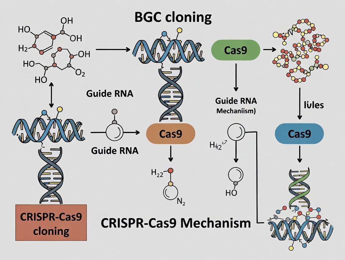
Harnessing CRISPR-Cas9 for BGC Cloning: A Complete Guide for Natural Product Discovery
This article provides a comprehensive overview of CRISPR-Cas9 applications for Biosynthetic Gene Cluster (BGC) cloning, targeting researchers and drug discovery professionals.
Harnessing CRISPR-Cas9 for BGC Cloning: A Complete Guide for Natural Product Discovery
Abstract
This article provides a comprehensive overview of CRISPR-Cas9 applications for Biosynthetic Gene Cluster (BGC) cloning, targeting researchers and drug discovery professionals. We explore the foundational principles of targeting BGCs in complex genomes, detail step-by-step methodologies from guide RNA design to heterologous expression, address common troubleshooting and optimization challenges, and validate the approach by comparing it to traditional methods like PCR and cosmids. The guide synthesizes current best practices to enable efficient mining of microbial genomes for novel therapeutics.
CRISPR-Cas9 and BGCs 101: From Genome Mining to Precise Targeting
This whitepaper explores the architecture, function, and pharmaceutical potential of Biosynthetic Gene Clusters (BGCs). Framed within a broader thesis on leveraging CRISPR-Cas9 for BGC cloning and engineering, this guide provides an in-depth technical overview for research and drug development professionals. The integration of advanced genome mining and precision genetic tools is revolutionizing the discovery and optimization of novel bioactive compounds.
Biosynthetic Gene Clusters are genomic loci containing co-localized genes encoding enzymes and regulatory proteins for a single specialized metabolic pathway. They produce secondary metabolites (natural products) with diverse biological activities. Historically discovered through activity-guided screening, modern genomics reveals that only a fraction of BGCs in sequenced microbes are expressed under laboratory conditions, representing a vast "hidden" reservoir of chemical diversity with immense pharmaceutical value.
Core Architecture and Key Classes
BGCs typically consist of core biosynthetic genes (e.g., for polyketide synthases [PKS], non-ribosomal peptide synthetases [NRPS]), tailoring enzymes (e.g., oxidases, methyltransferases), regulatory genes, and often resistance genes. The table below summarizes major BGC classes and their pharmaceutical significance.
Table 1: Major Classes of BGCs and Their Pharmaceutical Products
| BGC Class | Core Enzymes | Example Product | Pharmaceutical Application |
|---|---|---|---|
| Type I/II Polyketide (PKS) | Multi-domain Polyketide Synthases | Erythromycin (PKS I) | Antibiotic |
| Non-Ribosomal Peptide (NRPS) | Non-ribosomal Peptide Synthetases | Daptomycin | Antibiotic (anti-MRSA) |
| Ribosomally synthesized and post-translationally modified peptides (RiPPs) | Precursor peptide + modifying enzymes | Nisin | Food preservative, antimicrobial |
| Terpene | Terpene synthases/cyclases | Artemisinin | Antimalarial |
| Hybrid (e.g., PKS-NRPS) | Mixed PKS and NRPS assemblies | Bleomycin | Anticancer |
BGCs in the CRISPR-Cas9 Era: Cloning and Engineering
The precision and programmability of CRISPR-Cas9 have addressed key bottlenecks in BGC research: capturing silent clusters and engineering pathways for optimized production or novel analogs.
Experimental Protocol: CRISPR-Cas9-Mediated BGC Capture
This protocol outlines the capture of a target BGC from a microbial genome into a shuttle vector for heterologous expression.
- Target Identification & Guide RNA (gRNA) Design:
- Use genome mining tools (antiSMASH, PRISM) to identify BGC boundaries.
- Design two pairs of gRNAs targeting sequences flanking the BGC (approx. 20-40 kb). gRNAs should have high on-target scores and minimal off-targets in the host genome.
- Cas9 Cleavage in vitro or in vivo:
- In vitro Approach: Isolate genomic DNA. Perform a Cas9 ribonucleoprotein (RNP) cleavage reaction with the two gRNAs. This liberates the linear BGC fragment with defined ends.
- In vivo Approach: Transform the native host with a plasmid expressing Cas9 and the gRNAs. Induce double-strand breaks at the chromosomal locus.
- Vector Preparation & Assembly:
- Use a Bacterial Artificial Chromosome (BAC) or yeast-based vector linearized with ends homologous to the Cas9-cleaved BGC ends (via Gibson Assembly or yeast homologous recombination).
- Recombination & Capture:
- Co-transform/electroporate the cleaved BGC fragment and the linearized vector into an assembly host (e.g., S. cerevisiae). Select for clones containing the assembled construct.
- Heterologous Expression & Screening:
- Isolate the recombinant BAC and transform it into an optimized heterologous host (e.g., Streptomyces coelicolor, Pseudomonas putida).
- Induce expression and screen for metabolite production via LC-MS.
Diagram Title: CRISPR-Cas9 Workflow for BGC Capture and Expression
Experimental Protocol: CRISPR-Cas9-Mediated BGC Refactoring
This protocol describes the replacement of a native BGC promoter with a constitutive strong promoter for activation.
- Reporter Construction: Create a donor DNA cassette containing the desired strong promoter (e.g., ermEp*) flanked by ~500 bp homology arms matching sequences upstream and downstream of the native promoter region.
- gRNA Design: Design a gRNA targeting the native promoter sequence within the BGC.
- Delivery: Introduce a CRISPR-Cas9 plasmid (expressing Cas9 and the gRNA) and the donor DNA cassette into the host strain via conjugation or transformation.
- Selection & Screening: Select for double-crossover recombinants using a selectable marker on the donor cassette. Verify promoter swap via colony PCR and Sanger sequencing.
- Metabolite Analysis: Culture engineered and wild-type strains under identical conditions. Extract metabolites and compare profiles using HPLC or LC-MS.
The Scientist's Toolkit: Research Reagent Solutions
Table 2: Essential Materials for CRISPR-Cas9 BGC Engineering
| Item | Function in BGC Research | Example/Supplier (Illustrative) |
|---|---|---|
| antiSMASH Database | In silico identification & prediction of BGC boundaries in genomic data. | https://antismash.secondarymetabolites.org/ |
| Cas9 Nuclease (S. pyogenes) | Creates targeted double-strand breaks at BGC boundaries for excision or within clusters for editing. | Commercial recombinant protein (e.g., NEB). |
| Gibson Assembly Master Mix | Seamless assembly of large, homologous BGC fragments into cloning vectors. | New England Biolabs (NEB). |
| Yeast Transformation Kit | Facilitates homologous recombination-based capture of large BGCs into yeast vectors (e.g., pCAP01). | Commercial kits (e.g., Zymo Research). |
| BAC Vector (e.g., pBeloBAC11) | Stable maintenance of large (>100 kb) DNA inserts for BGC library construction and heterologous expression. | Addgene. |
| Heterologous Host Strains | Clean genetic background for expressing captured BGCs; optimized for secondary metabolism. | Streptomyces coelicolor M1152, P. putida KT2440. |
| HPLC-MS / LC-HRMS | Critical for detecting, quantifying, and structurally characterizing novel metabolites produced from activated BGCs. | Agilent, Thermo Fisher, Waters. |
Pharmaceutical Potential and Quantitative Impact
The systematic mining and engineering of BGCs directly feed the drug discovery pipeline. The table below highlights the quantitative scope and success of this approach.
Table 3: Quantitative Impact of BGC-Derived Natural Products
| Metric | Data | Context / Source |
|---|---|---|
| Approved Drugs from Natural Products | ~34% of all FDA-approved small-molecule drugs (1981-2019) are natural products or direct derivatives. | Newman & Cragg, 2020 |
| Microbial BGCs per Genome | Streptomyces spp. average 20-40 BGCs per genome; >90% are transcriptionally silent under lab conditions. | Zarins-Tutt et al., 2016 |
| Discovery Rate with Genomics | Genome mining increases the rate of novel metabolite discovery by 10-100x compared to traditional screening. | Research Review |
| Yield Improvement via Engineering | Promoter refactoring & gene editing in BGCs can improve titers by >100-fold for scaled production. | Case studies (e.g., avermectin) |
Diagram Title: From BGC to Drug Candidate via CRISPR Engineering
CRISPR-Cas9 technology has fundamentally transformed BGC research from a discovery-centric field into an engineering discipline. By enabling precise cloning, refactoring, and manipulation of these complex genetic loci, it unlocks the vast silent metabolome for systematic exploration. This integration of genomics, synthetic biology, and analytics creates a powerful, accelerated pipeline for discovering and developing the next generation of pharmaceutical leads, including novel antibiotics, anticancer agents, and immunosuppressants.
Clustered Regularly Interspaced Short Palindromic Repeats (CRISPR) and CRISPR-associated protein 9 (Cas9) constitute a bacterial adaptive immune system repurposed as a revolutionary genome-editing tool. In the context of Biosynthetic Gene Cluster (BGC) cloning research—aimed at harnessing microbial pathways for novel drug discovery—CRISPR-Cas9 provides unparalleled precision for the targeted capture, refactoring, and heterologous expression of large, complex genetic loci. This whitepaper details the core molecular mechanism of the Type II CRISPR-Cas9 system, emphasizing its application as a programmable "molecular scissor and guide."
Core Molecular Mechanism
The CRISPR-Cas9 system functions through a coordinated two-component complex: the Cas9 endonuclease and a single guide RNA (sgRNA).
The Cas9 Endonuclease
Cas9 is a multi-domain protein with two distinct nuclease lobes: the HNH domain and the RuvC-like domain. The HNH domain cleaves the DNA strand complementary to the guide RNA (target strand), while the RuvC domain cleaves the non-complementary strand (non-target strand). This results in a blunt-ended or near-blunt-ended double-strand break (DSB).
The Guide RNA (sgRNA)
A chimeric single guide RNA (sgRNA) is engineered by fusing the CRISPR RNA (crRNA), which contains the ~20-nucleotide target-specific spacer sequence, with the trans-activating crRNA (tracrRNA). The sgRNA directs Cas9 to the target genomic locus via Watson-Crick base pairing between its spacer sequence and the protospacer adjacent motif (PAM).
The Protospacer Adjacent Motif (PAM)
Recognition and initial DNA binding by Cas9 require a short PAM sequence immediately downstream of the target site. For the commonly used Streptococcus pyogenes Cas9 (SpCas9), the PAM is 5'-NGG-3' (where N is any nucleotide). The PAM is essential for distinguishing self from non-self DNA in bacterial immunity and is a critical determinant of target site selection in genome editing.
Mechanism of DNA Cleavage
- PAM Recognition & DNA Melting: Cas9 scans DNA, recognizing PAM sequences. Upon PAM binding, Cas9 unwinds the adjacent DNA duplex.
- Target DNA-RNA Pairing: The unwound DNA strand is interrogated for complementarity to the sgRNA spacer sequence. A perfect or near-perfect match is required for efficient cleavage.
- Conformational Activation & Cleavage: Successful base-pairing triggers a conformational change in Cas9, activating the HNH and RuvC nuclease domains. Sequential cleavage of both DNA strands generates a DSB 3-4 nucleotides upstream of the PAM.
Table 1: Quantitative Parameters of Common CRISPR-Cas9 Systems
| Cas9 Variant | Source Organism | PAM Sequence | Spacer Length (nt) | Protein Size (aa) | Cleavage Pattern |
|---|---|---|---|---|---|
| SpCas9 | Streptococcus pyogenes | 5'-NGG-3' | 20 | 1368 | Blunt end, 3-4 bp upstream of PAM |
| SaCas9 | Staphylococcus aureus | 5'-NNGRRT-3' | 21-23 | 1053 | Blunt end |
| CjCas9 | Campylobacter jejuni | 5'-NNNNRYAC-3' | 22 | 984 | Blunt end |
| Cas12a (Cpf1) | Francisella novicida | 5'-TTTV-3' | 20-24 | 1300 | Staggered cut (5' overhang) |
Application in BGC Cloning: Targeted Liberation of Gene Clusters
A key application in drug development is the precise excision of large BGCs from native genomic DNA for transfer into expression hosts.
Experimental Protocol: CRISPR-Cas9-Mediated BGC Excision
Objective: To excise a specific 50-kb biosynthetic gene cluster from the chromosome of a Streptomyces species.
Materials & Reagents:
- Bacterial Strain: Wild-type Streptomyces sp. harboring the target BGC.
- Plasmids: pCRISPR-Cas9 (temperature-sensitive origin, constitutive Cas9 expression, sgRNA scaffold) and pSG5 (template for sgRNA assembly and homologous repair arms).
- Oligonucleotides: Two pairs for amplifying ~1 kb homology arms flanking the desired excision sites; one pair for generating the sgRNA spacer targeting a sequence just inside one boundary of the BGC.
- Enzymes: DNA polymerase for PCR, T4 DNA ligase, restriction enzymes, Gibson Assembly Master Mix.
- Culture Media: TSBY liquid medium, MS agar with appropriate antibiotics (apramycin, thiostrepton).
Methodology:
sgRNA Design & Vector Construction:
- Design two sgRNAs targeting sequences within the BGC, close to its 5' and 3' boundaries. Ensure each target site is followed by a valid PAM.
- Synthesize oligonucleotides encoding the spacer sequences, anneal them, and clone into the BsaI site of the pCRISPR-Cas9 vector using Golden Gate assembly.
- Verify constructs by Sanger sequencing.
Delivery into Host Strain:
- Transform the constructed pCRISPR-Cas9 plasmid into the Streptomyces strain via PEG-mediated protoplast transformation.
- Plate on MS agar containing apramycin (selection for plasmid) and incubate at 30°C (permissive temperature) for 5-7 days.
Induction of CRISPR-Cas9 Activity:
- Pick several transformants and grow in TSBY liquid medium with apramycin at 30°C.
- Shift cultures to 37°C (non-permissive temperature for plasmid replication) for 24-48 hours to induce plasmid "curing" and enrich for cells that have undergone the desired genomic excision event.
Screening & Validation:
- Plate serial dilutions of the heat-shocked culture on non-selective MS agar. Allow colonies to grow.
- Screen individual colonies by colony PCR using primers annealing outside the excised region and inside the BGC to identify clones where the internal fragment is lost.
- Confirm precise excision by long-range PCR across both junctions and subsequent Sanger sequencing.
- Recover the excised, linear BGC fragment by gel extraction or use RecET-assisted direct cloning for capture into a bacterial artificial chromosome (BAC).
Table 2: Research Reagent Solutions for CRISPR-Cas9 BGC Cloning
| Reagent/Material | Function in BGC Cloning | Example/Supplier (Illustrative) |
|---|---|---|
| High-Fidelity DNA Polymerase | Error-free amplification of homology arms and verification PCRs. | Q5 (NEB), Phusion (Thermo) |
| Golden Gate Assembly Kit | Efficient, modular cloning of sgRNA spacers into Cas9 expression vectors. | BsaI-HFv2 Master Mix (NEB) |
| Gibson Assembly Master Mix | Seamless assembly of large constructs with homology arms. | NEBuilder HiFi DNA Assembly (NEB) |
| Temperature-Sensitive Cas9 Vector | Allows for temporary Cas9 expression and subsequent plasmid curing. | pCRISPR-Cas9ts (Addgene #130329) |
| RecET Cloning System | Facilitates direct capture of large, linear genomic fragments (like excised BGCs) into vectors. | pGETrec (GeneBridge) |
| BAC Vector | Stable maintenance and propagation of large (>100 kb) DNA inserts. | pCC1BAC (CopyControl) |
Visualizing the Mechanism and Workflow
Diagram 1: CRISPR-Cas9 DNA Targeting & Cleavage (76 chars)
Diagram 2: CRISPR-Cas9 Mediated BGC Excision Workflow (79 chars)
Why Traditional Cloning Falls Short for Large, Complex BGCs
Biosynthetic gene clusters (BGCs) encode the machinery for producing structurally complex and pharmaceutically relevant natural products. Within the modern research paradigm focused on harnessing CRISPR-Cas9 for precise genome editing, the cloning of intact, large (>50 kb), and complex BGCs represents a critical bottleneck. Traditional cloning methods, developed for smaller, simpler constructs, are fundamentally ill-suited for this task. This guide details the technical limitations of conventional approaches and frames the necessity for innovative, Cas9-assisted strategies within contemporary BGC research.
Core Limitations of Traditional Cloning Methods
Traditional methods, including restriction-ligation, PCR-based assembly, and cosmic/YAC-based techniques, encounter systematic failures with large BGCs. The quantitative shortcomings are summarized below.
Table 1: Failure Points of Traditional Cloning for Large BGCs
| Limitation Factor | Description | Typical Impact on Large BGCs (>50 kb) |
|---|---|---|
| Restriction Site Scarcity & Redundancy | Large BGCs contain multiple, often non-unique restriction sites. | Makes impossible to generate a single, defined linear vector and insert without internal cleavage. |
| In Vitro Assembly Fidelity | Enzymatic assembly (e.g., Gibson, Golden Gate) efficiency drops with fragment number and size. | >5-10 fragment assemblies show exponential drop in success rate; increased misassembly. |
| Host Toxicity & Instability | Heterologous expression of large, unregulated clusters can be toxic to E. coli hosts. | Cloned BGCs are unstable in cloning hosts, leading to rearrangements/deletions. |
| Transformation Efficiency | Large plasmid transformation efficiency is extremely low. | Efficiency for >100 kb plasmids can be <10^3 CFU/μg, making library construction impractical. |
| GC-Rich Content & Repeats | BGCs often have high GC content and long repetitive sequences. | Causes polymerase errors during PCR, promotes homologous recombination in E. coli, scrambling the clone. |
Detailed Experimental Protocol: A Cautionary Case Study
The following protocol for attempting traditional cosmic cloning of a Streptomyces Type I PKS BGC (~70 kb) illustrates the technical hurdles.
Protocol: Restriction-Based Cosmic Library Construction for a Large BGC
- Genomic DNA (gDNA) Preparation: Isolate high-molecular-weight gDNA from the producer strain via lysozyme/proteinase K lysis, CTAB precipitation, and RNase A treatment. Assess integrity by pulsed-field gel electrophoresis (PFGE).
- Partial Digestion Optimization: Titrate Sau3AI restriction enzyme (0.1-1.0 U/μg) on gDNA for time courses (5-30 min). Aim for a majority of fragments in the 30-45 kb range, analyzed by PFGE.
- Vector Preparation: Digest cosmic vector (e.g., pSuperCos1) with XbaI, dephosphorylate. Perform a second digest with BamHI to generate two cosmic "arms."
- Size Selection & Ligation: Gel-purify the 35-45 kb fraction of Sau3AI-digested gDNA. Ligate to pre-treated cosmic arms at a 3:1 (insert:vector) molar ratio using T4 DNA ligase (16°C, 16h).
- In Vitro Packaging & Transformation: Package ligation reactions using commercial lambda phage packaging extracts. Transduce into E. coli EPI300 cells. Plate on selective LB + antibiotic.
- Screening & Analysis: Pick colonies for colony PCR or hybridization. Prepare cosmic DNA from positives. Analyze by restriction fingerprinting with EcoRI and end-sequencing.
Expected Outcome: This protocol often yields either no full-length clones, clones with internal deletions, or clones containing only sub-fragments of the target BGC, necessitating complex subcloning that rarely reconstructs the intact cluster.
The CRISPR-Cas9 Paradigm: Enabling Targeted Capture
CRISPR-Cas9 offers a paradigm shift by enabling precise in vivo or in vitro excision and capture of BGCs. Cas9-guided double-strand breaks (DSBs) at unique flanking sequences allow the isolation of large, contiguous DNA fragments without reliance on internal restriction sites.
Diagram 1: CRISPR-Cas9 Mediated In Vivo BGC Capture
Experimental Protocol: Cas9-Directed In Vitro Capture and Yeast Assembly
- sgRNA Design & Cas9 RNP Complex Formation: Design two sgRNAs targeting unique 20-bp sequences flanking the BGC. Chemically synthesize sgRNAs. Pre-complex purified Cas9 protein with each sgRNA to form Ribonucleoprotein (RNP) complexes.
- High-Molecular-Weight gDNA Isolation: Prepare ultra-pure, high-integrity gDNA as in Section 3, but minimize mechanical shearing.
- In Vitro Cas9 Digestion: Incubate 5-10 μg gDNA with the two RNP complexes in CutSmart buffer (37°C, 2h). Run an aliquot on PFGE to confirm release of a linear fragment of predicted size.
- Capture Vector Preparation: Linearize a yeast assembly vector (e.g., pYES1L) containing terminal homology arms (40-50 bp) to the Cas9-cut ends of the BGC fragment.
- Yeast Transformation-Associated Recombination (TAR): Co-transform the in vitro Cas9-digested gDNA mixture and the linearized capture vector into competent Saccharomyces cerevisiae (e.g., VL6-48N) using the lithium acetate/PEG method. Plate on appropriate synthetic dropout media.
- Yeast Clone Validation: Screen yeast colonies by PCR across junctions. Isroduce yeast plasmid DNA and electroporate into E. coli for amplification. Validate by pulsed-field gel analysis and paired-end sequencing.
The Scientist's Toolkit: Research Reagent Solutions
Table 2: Essential Reagents for Modern BGC Cloning
| Reagent / Material | Function in BGC Cloning | Key Consideration |
|---|---|---|
| High-Fidelity Cas9 Nuclease (NLS-tagged) | Catalyzes precise DSBs at sgRNA-specified sites for fragment excision. | Use HiFi or eSpCas9 variants to reduce off-target effects on complex gDNA. |
| Chemically Synthesized sgRNAs | Guides Cas9 to specific genomic loci flanking the BGC. | Must be designed against unique sequences in the flanking regions; HPLC purification recommended. |
| Pulsed-Field Gel Electrophoresis System | Analyzes and size-selects DNA fragments >20 kb. | Critical for assessing gDNA integrity and isolating large Cas9-excised fragments. |
| Yeast TAR Vectors (e.g., pYES1L) | Contains elements for selection/maintenance in yeast and E. coli, plus homology arms. | Enables recombination-based capture of large linear fragments in S. cerevisiae. |
| RecET or Lambda-Red System | Facilitates homologous recombination of large DNA in E. coli. | Used in E. coli-based direct capture methods (e.g., Cas9-Assisted Targeting of Chromosome segments - CATCH). |
| Meganuclease-Targeted Vectors | Vectors containing recognition sites for rare-cutting meganucleases (e.g., I-SceI). | Allows insertion of Cas9-excised BGC fragments into a defined locus of a heterologous host chromosome. |
Quantitative Comparison: Traditional vs. CRISPR-Cas9 Methods
Table 3: Performance Metrics Comparison
| Metric | Traditional Cosmid Cloning | CRISPR-Cas9 Assisted Capture |
|---|---|---|
| Max Practical Insert Size | ~40-45 kb (limited by packaging) | >100 kb (limited by gDNA integrity) |
| Cloning Time (Hands-on) | 2-3 weeks | 1-2 weeks |
| Success Rate for 70-kb BGC | <5% (often partial clones) | 20-80% (full-length clones) |
| Requirement for Internal Restriction Sites | Yes, critical | No |
| Fidelity in Repetitive Regions | Low (prone to recombination) | High (avoids E. coli during capture) |
| Amenability to Automation | Low | Moderate to High (standardized RNP steps) |
The limitations of traditional cloning are not merely incremental but foundational when confronting large, complex BGCs. These methods are incompatible with the structural realities of such genetic loci. The integration of CRISPR-Cas9 mechanisms into BGC cloning workflows, as part of a broader thesis on precision genome manipulation, provides the necessary tools for targeted excision, stable propagation, and faithful reconstruction. This shift is essential for accelerating the discovery and engineering of novel bioactive compounds in the genomic era.
Within the expanding toolkit for natural product discovery, the precise cloning of intact Biosynthetic Gene Clusters (BGCs) represents a critical bottleneck. Traditional methods, such as cosmids or bacterial artificial chromosomes (BACs), are often hindered by size limitations, host incompatibility, and labor-intensive screening. This whitepaper frames the innovative application of CRISPR-Cas9 as a transformative mechanism for the precise excision and capture of BGCs, enabling their heterologous expression and functional characterization. This approach directly serves a broader thesis: that CRISPR-based methodologies are superseding conventional cloning to accelerate the discovery pipeline for novel therapeutic compounds.
CRISPR-Cas9 Mechanism for BGC Cloning
The core strategy employs a two-plasmid system to orchestrate in vivo excision of a target BGC from a source genome (e.g., a difficult-to-culture actinomycete) and its subsequent circularization and capture in E. coli.
- Dual-Guide RNA (dgRNA) Design: Two CRISPR RNA (crRNA) sequences are designed to target sequences flanking the BGC of interest. These are often expressed as a tandem guide RNA array.
- Cas9-Mediated Double-Strand Break (DSB) Generation: The Cas9 nuclease, complexed with the dgRNA, introduces precise DSBs at the designated flanking sites.
- Homology-Directed Repair (HDR) & Capture: A linear "capture vector" is co-introduced. This vector contains:
- Homology Arms: Sequences homologous to the regions just inside the cut sites.
- Origin of Replication (ori) and Selection Marker functional in the capture host (e.g., E. coli).
- A Conditional Origin (e.g., R6K) for suicide selection in the source host. The endogenous repair machinery of the source cell utilizes the homology arms on the capture vector to repair the DSBs via HDR, thereby incorporating the vector elements and circularizing the excised BGC into an Extractable Genetic Element (EGE). This EGE is then isolated and electroporated into the heterologous expression host.
Diagram Title: CRISPR-Cas9 Workflow for BGC Excision and Capture
Key Experimental Protocols
Protocol: Design and Assembly of CRISPR Capture Constructs
- Bioinformatic Identification: Use antiSMASH or PRISM to define BGC boundaries.
- dgRNA Design: Select 20-nt protospacer sequences ~50-500 bp inside each BGC boundary. Verify specificity using BLAST against the source genome.
- Capture Vector Construction:
- Amplify ~1 kb homology arms (HA-L and HA-R) from the source genomic DNA.
- Clone HAs into a suicide vector backbone (e.g., pKD46-derived with R6Kγ ori) flanking an E. coli ori and selectable marker (e.g., aac(3)IV for apramycin resistance).
- Insert a dgRNA expression cassette (e.g., driven by a constitutive promoter) targeting the selected sites into the vector backbone.
- Cas9 Provision: Use a separate, compatible plasmid expressing Cas9 (inducible or constitutive) or deliver Cas9 as a purified protein complexed with the dgRNA (RNP).
Protocol:In VivoExcision and E. coli Capture
- Delivery to Source Strain: Introduce both the capture vector and the Cas9 vector (or RNP) into the source strain via conjugation or protoplast transformation.
- Induction and Excision: Induce Cas9 and dgRNA expression (if applicable). Allow 24-48 hours for DSB generation and HDR-mediated circularization.
- Plasmid Recovery: Perform a standard alkaline lysis plasmid preparation from the source strain culture.
- Electroporation into E. coli: Electroporate the plasmid prep into a high-efficiency, methylation-tolerant E. coli strain (e.g., EPI300) with the appropriate antibiotic selection.
- Validation: Screen colonies by PCR across the new junctions (vector-BGC). Confirm integrity by restriction digest and/or whole plasmid sequencing.
Table 1: Comparison of BGC Cloning Methodologies
| Method | Typical Max Insert Size | Cloning Efficiency (Intact BGCs) | Time to Isolate Clone | Key Limitation |
|---|---|---|---|---|
| Cosmid Library | ~40 kb | Low (<1%) | Weeks to Months | Random insertion, extensive screening |
| BAC Library | ~200 kb | Low (<1%) | Weeks to Months | Low copy number, difficult manipulation |
| Transposon Mutagenesis | N/A | Variable | Weeks | Disrupts native regulation |
| CRISPR-Cas9 Capture | >100 kb | Moderate-High (5-20%) | 1-2 Weeks | Requires genome sequence & design |
Table 2: Example CRISPR-Cas9 Capture Efficiency in Recent Studies
| Target BGC (Size) | Source Organism | Capture Vector | Reported Efficiency (Colonies/μg) | Success Rate (Intact) | Reference (Year) |
|---|---|---|---|---|---|
| Type II PKS (~35 kb) | Streptomyces spp. | pCRISPomyces-2 derived | 4.2 x 10² | 92% | Bai et al. (2023) |
| NRPS (~68 kb) | Myxococcus xanthus | R6Kγ suicide vector | 1.5 x 10² | 85% | Chen et al. (2024) |
| Hybrid (~52 kb) | Uncultured Soil Metagenome | Direct RNP delivery | 3.0 x 10¹ | 78% | Sharma & Clark (2024) |
The Scientist's Toolkit: Research Reagent Solutions
Table 3: Essential Reagents for CRISPR-Cas9 BGC Capture
| Item | Function & Rationale | Example Product/Catalog |
|---|---|---|
| dgRNA Expression Plasmid | Expresses tandem guide RNAs targeting BGC flanks for precise Cas9 cleavage. | pCRISPR-COS, pKCcas9dO (Addgene) |
| Cas9 Expression Vector | Provides the Cas9 nuclease in the source host. Can be inducible (e.g., anhydrotetracycline). | pCRISPomyces-2 (Addgene #61737) |
| Linear Capture Vector Backbone | Suicide vector with R6Kγ origin for maintenance only in pir+ E. coli, containing homology arm cloning sites. | pJIR_backbone (Lab construction) |
| High-Efficiency Electrocompetent E. coli | Methylation-tolerant strain for capturing large, potentially methylated EGEs from actinomycetes. | EPI300 (Lucigen), GB05-dir (Thermo) |
| Gibson Assembly or Golden Gate Master Mix | For seamless assembly of homology arms and cassettes into the capture vector. | Gibson Assembly Master Mix (NEB), Golden Gate Assembly Kit (BsaI-HF) |
| Apoplast Mix for Protoplast Transformation | Essential for delivering constructs into Streptomyces and other Gram-positive source strains. | Prepared per lab protocol (PEG, sucrose, MgCl2) |
| Positive Selection Marker | Antibiotic resistance gene for selection in the final heterologous host (e.g., apramycin, thiostrepton). | aac(3)IV or tsr cassettes |
Pathway and Regulatory Considerations
Successful heterologous expression requires capturing not only the core biosynthetic genes but also essential regulatory elements. The CRISPR-Cas9 approach allows for the strategic inclusion of native promoters or the insertion of strong, constitutive promoters during the capture vector design.
Diagram Title: Key Components of a Functional Captured BGC
Within the broader thesis on utilizing CRISPR-Cas9 for the targeted cloning and manipulation of Biosynthetic Gene Clusters (BGCs), the precise engineering of three core molecular components is paramount. The efficacy of Cas9-mediated double-strand breaks (DSBs), the specificity of genomic targeting, and the successful integration of heterologous DNA hinges on the optimal design of single guide RNAs (gRNAs), the selection of appropriate Cas9 protein variants, and the construction of donor templates for homology-directed repair (HDR). This guide provides an in-depth technical framework for these components, tailored for researchers in natural product discovery and drug development.
gRNA Design for BGC Targeting
The guide RNA (gRNA) is a chimeric RNA molecule comprising a CRISPR RNA (crRNA) sequence, which confers target specificity via a 20-nucleotide spacer, and a trans-activating crRNA (tracrRNA) scaffold that binds Cas9. For BGC cloning, which often targets large, repetitive, or GC-rich genomic regions, stringent design is critical.
Core Design Principles
- Protospacer Adjacent Motif (PAM): The canonical Streptococcus pyogenes Cas9 (SpCas9) requires a 5'-NGG-3' PAM sequence immediately downstream (3') of the target DNA strand. The spacer sequence is selected 5' of the PAM.
- Specificity & Off-Target Minimization: The 20-nt spacer must be unique within the host genome. Mismatches in the "seed region" (8-12 bases proximal to the PAM) are most disruptive to cleavage.
- Efficiency Predictors: While rules are empirical, high on-target activity is correlated with:
- A guanine (G) at the first position of the spacer.
- A high GC content (40-80%).
- The absence of poly-T tracts (transcription terminators for RNA Polymerase III).
- Low self-complementarity to prevent secondary structure formation in the gRNA.
Quantitative Parameters for Design
Table 1: Key Quantitative Parameters for Optimal gRNA Design (SpCas9)
| Parameter | Optimal Range/Target | Rationale & Impact |
|---|---|---|
| Spacer Length | 20 nucleotides (nt) | Standard for SpCas9; shorter/longer can reduce activity. |
| PAM Sequence | 5'-NGG-3' | Absolute requirement for SpCas9 binding. |
| GC Content | 40% - 80% | Higher GC often increases stability and specificity; <20% reduces efficiency. |
| Seed Region GC | Moderate to High | Critical for R-loop stability and initial DNA recognition. |
| Off-Target Score | ≤ 2 potential sites | Maximizes specificity; use algorithms (e.g., CCTop, CRISPOR) to predict. |
| On-Target Efficiency Score | > 60 (tool-dependent) | Predictive score from design tools (e.g., from IDT, Benchling). |
Protocol:In SilicogRNA Design and Validation for a BGC Locus
- Sequence Retrieval: Obtain the FASTA sequence of the target BGC and the complete host genome from databases (NCBI, JGI).
- PAM Identification: Using software (e.g., Benchling, SnapGene), scan both strands of the BGC for all 5'-NGG-3' PAM sites.
- Spacer Selection: For each PAM, extract the 20-nt sequence immediately 5' to it as the candidate spacer.
- Specificity Check: Input each 20-nt spacer + PAM sequence into a validated algorithm (e.g., CRISPOR, CCTop) to scan the host genome for potential off-target sites with up to 3-4 mismatches. Discard guides with high-quality off-targets within coding regions.
- Efficiency Scoring: Use the algorithm's built-in scoring models (e.g., Doench '16, Moreno-Mateos) to rank remaining guides by predicted on-target activity.
- Final Selection: Choose 2-3 top-ranked guides targeting the boundaries (for excision) or specific sites (for knock-in) within the BGC. Design cloning oligos with appropriate overhangs for your gRNA expression vector (e.g., BbsI sites for pU6-driven vectors).
Cas9 Variants: Expanding the BGC Engineering Toolkit
Wild-type SpCas9 remains a workhorse, but engineered variants address key limitations for complex BGC manipulation, such as restricted PAM requirements, off-target effects, and large delivery payloads.
Comparison of Key Cas9 Variants
Table 2: Engineered Cas9 Variants for Advanced BGC Applications
| Variant | Key Feature | PAM Sequence | Size (aa) | Primary Application in BGC Work |
|---|---|---|---|---|
| SpCas9 (WT) | Nuclease, Nickase (D10A or H840A) | 5'-NGG-3' | 1,368 | Standard DSBs, paired nickases for higher specificity. |
| SpCas9-VQR | Engineered PAM specificity | 5'-NGAN-3' | ~1,368 | Targeting GC-rich regions common in actinomycete BGCs. |
| SpCas9-NG | Relaxed PAM specificity | 5'-NG-3' | ~1,368 | Greatly expands targetable sites within AT-rich BGCs. |
| xCas9(3.7) | Broad PAM recognition, high fidelity | 5'-NG, GAA, GAT-3' | ~1,370 | Flexible targeting with reduced off-targets. |
| SaCas9 | Compact nuclease | 5'-NNGRRT-3' | 1,053 | Delivery via size-limited vectors (e.g., AAV). |
| SpCas9n (D10A) | Nickase (creates single-strand break) | 5'-NGG-3' | 1,368 | Paired nickases for precise excision with reduced indels. |
| dCas9 (D10A/H840A) | Nuclease-dead; fusion platform | 5'-NGG-3' | 1,368 | Transcriptional activation/repression (CRISPRi/a) of BGCs. |
Protocol: Selecting a Cas9 Variant for BGC Boundary Definition
Objective: Precisely excise a ~50 kb BGC from a bacterial chromosome.
- Analyze Flanking Sequences: Obtain ~500 bp sequences upstream and downstream of the target BGC. Perform PAM interrogation.
- Variant Selection Logic:
- If clean NGG sites are present at ideal locations (~100 bp inside boundaries), use SpCas9.
- If flanking regions are AT-rich and lack NGG sites, switch to SpCas9-NG to find usable NG PAMs.
- If the host has a highly repetitive genome, prioritize high-fidelity variants (e.g., SpCas9-HF1) or use a paired SpCas9n nickase strategy with two adjacent guides on opposite strands to create a staggered DSB, enhancing specificity.
- Validation: Always validate cleavage efficiency of the chosen variant/gRNA pair in vitro using a PCR-amplified genomic target and commercial Cas9 protein, followed by gel electrophoresis or T7E1 assay, before proceeding to in vivo experiments.
Donor Template Design for HDR in BGC Engineering
For precise insertion, replacement, or tagging of genes within a BGC, a donor DNA template is required to guide HDR following a DSB. Key strategies include plasmid donors, linear double-stranded DNA (dsDNA), and single-stranded oligodeoxynucleotides (ssODNs).
Donor Template Types and Considerations
Table 3: Donor Template Strategies for BGC Editing
| Template Type | Optimal Size | Key Features | Typical BGC Application |
|---|---|---|---|
| Plasmid Donor | 1 kb - >20 kb | Contains long homology arms (500-1500 bp), selectable marker. Can carry large cargo. | Insertion of heterologous expression promoters, large fluorescent tags, or entire reporter cassettes into a BGC. |
| Linear dsDNA (PCR) | 200 bp - 2 kb | Short homology arms (50-100 bp). Rapid to generate, lower integration efficiency. | Introducing point mutations (e.g., for site-directed mutagenesis of a PKS domain), small epitope tags. |
| ssODN | 80 - 200 nt | Ultrafast synthesis, highest efficiency for small edits (<30 nt). Asymmetric design. | Introduction of stop codons, restriction sites, or small loxP sites for Cre recombinase-mediated cloning. |
Core Design Rules
- Homology Arms: Flank the intended modification. Length correlates with HDR efficiency (longer = more efficient but harder to build). For plasmid donors in microbes, 500-1000 bp is standard.
- "Silent" PAM Disruption: The donor sequence should contain silent mutations in the PAM and/or seed region of the gRNA binding site to prevent re-cutting after successful HDR.
- Codon Optimization: Ensure any inserted coding sequence is optimized for the expression host (e.g., E. coli, S. cerevisiae for heterologous expression).
Protocol: Constructing a Plasmid Donor for BGC Gene Tagging
Objective: C-terminally tag a gene within a BGC with a 3xFLAG epitope and a spectinomycin resistance gene (aadA).
- Homology Arm Amplification: PCR amplify the ~800 bp region immediately upstream (Left Homology Arm, LHA) and downstream (Right Homology Arm, RHA) of the target insertion site from genomic DNA.
- Cargo Assembly: Assemble the cargo: 3xFLAG-aadA fragment, synthesized with appropriate linkers.
- Golden Gate Assembly: Using a modular cloning system (e.g., MoClo, GoldenBraid), combine in a single reaction: the linearized backbone vector, LHA, cargo, and RHA fragments with type IIS restriction enzymes (e.g., BsaI). This creates the final donor plasmid
pDonor-LHA-3xFLAG-aadA-RHA. - Verification: Sequence the entire assembly, focusing on the junction regions between homology arms and cargo.
Visualizing the Experimental Workflow
Diagram Title: CRISPR-Cas9 Workflow for BGC Engineering
The Scientist's Toolkit: Key Reagent Solutions
Table 4: Essential Research Reagents for CRISPR-mediated BGC Manipulation
| Reagent / Material | Supplier Examples | Function in BGC Experiment |
|---|---|---|
| High-Fidelity DNA Polymerase (Q5, Phusion) | NEB, Thermo Fisher | Error-free amplification of homology arms, donor fragments, and verification PCRs. |
| Type IIS Restriction Enzymes (BbsI, BsaI-HFv2) | NEB | Golden Gate assembly of gRNA expression cassettes and modular donor plasmids. |
| T4 DNA Ligase | NEB, Thermo Fisher | Ligation of DNA fragments during vector construction. |
| Commercial Cas9 Nuclease (WT & variants) | IDT, Thermo Fisher, NEB | For in vitro cleavage assays to validate gRNA activity before in vivo use. |
| gRNA Synthesis Kit (cloning or in vitro transcription) | IDT, Synthego, NEB | Generation of high-purity gRNA for in vitro assays or direct RNP delivery. |
| Electrocompetent Cells (E. coli, Streptomyces) | Home-made, Lucigen, BioCat | Efficient transformation of plasmid assemblies and conjugation donors. |
| HRMA-compatible DNA Binding Dye (EvaGreen, LCGreen) | Biotium, Bio-Rad | Detection of indels via High-Resolution Melt Analysis (HRMA) post-editing. |
| Genomic DNA Isolation Kit (for GC-rich microbes) | Qiagen, Macherey-Nagel | Pure gDNA for sequencing validation and off-target analysis. |
| Next-Gen Sequencing Kit (for amplicon-seq) | Illumina, PacBio | Deep sequencing of target loci to quantify editing efficiency and off-target events. |
Step-by-Step Protocol: Deploying CRISPR-Cas9 for BGC Capture and Expression
Within the broader thesis on applying CRISPR-Cas9 for the precise excision and cloning of Biosynthetic Gene Clusters (BGCs), the in silico design stage is the critical foundational step. This stage leverages computational tools to define the exact genomic region of interest and design precise targeting tools, thereby determining the success and efficiency of all subsequent experimental work. Accurate prediction of BGC boundaries ensures the capture of complete biosynthetic pathways, while optimal gRNA design maximizes on-target cleavage efficiency and minimizes off-target effects during CRISPR-Cas9-mediated excision.
Computational Tools and Databases for BGC Prediction
Identification of a putative BGC begins with nucleotide sequence analysis, typically from whole-genome sequencing data. Several specialized algorithms and databases are employed for this purpose.
| Tool/Database | Primary Function | Key Input | Key Output |
|---|---|---|---|
| antiSMASH | Comprehensive BGC detection & annotation | Genomic DNA sequence | Predicted BGC boundaries, core biosynthetic genes, cluster type, similarity to known clusters. |
| PRISM | Predicts chemical structures of encoded natural products | Genomic DNA or protein sequences | Predicted chemical scaffolds, ribosomal/non-ribosomal peptides, polyketides. |
| MIBiG | Reference database of experimentally characterized BGCs | Query BGC sequence or features | Information on similar known clusters, including boundaries and products. |
| DeepBGC | Deep learning-based BGC detection using a random forest classifier | Protein sequence or protein domain embeddings | BGC probability score, Pfam domain composition, product class prediction. |
| ARTS | Specifically detects potential resistance genes within BGCs | Genomic DNA sequence | Prediction of "resistance" elements that often flank or reside within BGCs. |
Protocol: Standard Workflow for BGC Boundary Identification
- Input Preparation: Obtain the complete genome assembly (FASTA format) of the source organism.
- Primary Detection: Submit the genome to the latest version of antiSMASH (e.g., antiSMASH 7.0). Use default parameters for a first-pass analysis.
- Boundary Refinement: Analyze the antiSMASH results. Pay close attention to the "ClusterBorder" predictions and the presence of flanking core biosynthetic genes (e.g., PKS, NRPS modules). Cross-reference with MIBiG for similar known clusters.
- Adjacent Feature Analysis: Examine genomic regions 20-50 kb upstream and downstream of the core region for:
- Resistance Genes: Use ARTS to identify potential self-resistance genes, common BGC markers.
- Regulatory Genes: Pathway-specific regulators (e.g., SARP, LAL) often reside within boundaries.
- Transposase/Insertion Sequence (IS) Elements: These can indicate "hard" boundaries and genomic mobility.
- GC Content & Synteny Shift: Abrupt changes in GC% or gene orientation can suggest cluster limits.
- Final Boundary Definition: Designate a final target region that includes all core biosynthetic genes, auxiliary genes (e.g., transporters, regulators), and resistance genes. Add a 1-2 kb buffer zone beyond these elements to ensure completeness.
Workflow for Defining BGC Genomic Boundaries
Rational Design of gRNAs for BGC Excision
The goal is to design two single guide RNAs (sgRNAs) that direct Cas9 to create double-strand breaks (DSBs) precisely at the 5' and 3' boundaries of the defined BGC.
| Design Criteria | Optimal Target (for S. pyogenes Cas9) | Rationale |
|---|---|---|
| Protospacer Adjacent Motif (PAM) | 5'-NGG-3' immediately downstream of target. |
Cas9 nuclease recognition requirement. |
| On-Target Efficiency Score | >70 (CRISPOR, CHOPCHOP). | Predicts high cleavage activity. |
| Specificity (Off-Targets) | Zero or minimal hits with ≤3 mismatches in the seed region. | Ensures precise excision, prevents genomic rearrangement. |
| Genomic Context | Target sites within intergenic or non-essential regions flanking the BGC. | Avoids disruption of essential genes; promotes clean repair. |
| GC Content | 40-60%. | Influences gRNA stability and binding efficiency. |
Protocol: gRNA Design and Selection for BGC Excision
- Sequence Extraction: Extract the DNA sequence 500 bp upstream of the 5' BGC boundary and 500 bp downstream of the 3' BGC boundary (the "flanking regions").
- Target Site Scanning: Use the
-patternfunction in CRISPOR or similar tools to scan both flanking sequences for allNGGPAM sites. Record the 20-nt protospacer sequence preceding each valid PAM. - On-Target Scoring: For each candidate protospacer, retrieve efficiency scores from multiple algorithms (e.g., Doench '16, Moreno-Mateos). Filter for candidates with consistently high scores (>70).
- Off-Target Analysis: For high-scoring candidates, perform a whole-genome BLASTn search against the host genome. Use Cas-OFFinder with parameters: up to 3 mismatches allowed, DNA bulge size 0, RNA bulge size 0. Reject any gRNA with a perfect or near-perfect (≤1 mismatch) hit elsewhere in the genome.
- Final Pair Selection: From the filtered lists for the 5' and 3' flanks, select the final pair. Ensure the cut sites are positioned such that the DSBs will release the entire BGC on a single fragment, considering the Cas9 cut site is typically 3-4 bp upstream of the PAM.
gRNA Design & Selection Decision Workflow
The Scientist's Toolkit: Research Reagent Solutions
| Category | Item/Reagent | Function in In Silico Design |
|---|---|---|
| Bioinformatics Software | antiSMASH, PRISM, DeepBGC | Predicts BGC location, structure, and chemical product. |
| Genome Databases | NCBI GenBank, MIBiG, JGI IMG | Provides reference genomes and validated BGCs for comparison. |
| CRISPR Design Tools | CRISPOR, CHOPCHOP, Benchling | Designs and scores gRNAs for efficiency/specificity. |
| Off-Target Prediction | Cas-OFFinder, BLASTn (local) | Identifies potential unintended Cas9 cleavage sites. |
| Sequence Analysis | SnapGene, Geneious, CLC Bio | Visualizes genomic context, designs primers, and manages data. |
| Computational Environment | Linux server/Workstation, Python/R with Biopython | Runs command-line tools and custom analysis scripts. |
| Data Storage | Secure cloud or local server (e.g., NAS) | Stores large genome files and analysis results. |
This whitepaper details the second critical stage in a comprehensive strategy for cloning Bacterial Biosynthetic Gene Clusters (BGCs). The overall thesis posits that a CRISPR-Cas9-mediated in vitro or in vivo capture system, integrated with advanced DNA assembly methods, provides a superior, targeted, and high-efficiency alternative to traditional cosmic or fosmid-based library screening. Following Stage 1 (Bioinformatic Target Identification and gRNA Design), this stage focuses on the assembly of the specialized capture vector, which will serve as the "recipient" backbone for the targeted BGC DNA fragment.
Core Vector Architecture & Quantitative Specifications
The CRISPR-Cas9 capture vector is a modular assembly designed for replication in E. coli, selection, and subsequent integration or manipulation. Its key functional modules and their quantitative specifications are summarized below.
Table 1: Core Modules of the CRISPR-Cas9 Capture Vector
| Module | Key Components | Function | Typical Size Range |
|---|---|---|---|
| Selection/Counter-Selection | sacB gene, Antibiotic Resistance (e.g., AmpR, KanR) | Positive & negative selection for vector linearization & successful cloning. | 1.5 – 3.0 kb |
| Capture Homology Arms | User-defined sequences (e.g., 500 bp) flanking the target BGC. | Provide homology for in vivo recombination (HR) or in vitro ligation post-cut. | 0.5 – 2.0 kb each |
| Replication Origin | orif (high-copy number in E. coli) | Allows plasmid propagation and amplification in the cloning host. | ~1.0 kb |
| Cas9/gRNA Expression | cas9 gene (opt.), gRNA scaffold under a constitutive promoter. | For in vivo capture: drives target locus cleavage in the host cell. | ~4.2 kb (cas9) + ~0.3 kb (scaffold) |
| DNA Assembly Site | Multiple Cloning Site (MCS) or specific sequences for Gibson/Golden Gate assembly. | Facilitates insertion of homology arms and other modules. | 0.05 – 0.2 kb |
Table 2: Comparison of Common Assembly Methods for Capture Vector Construction
| Assembly Method | Principle | Efficiency | Best For | Typical Number of Fragments |
|---|---|---|---|---|
| Gibson Assembly | Exonuclease, polymerase, and ligase create seamless junctions. | > 90% (optimized) | Assembling 2-6 large modules (e.g., arms, backbone, sacB). | 2-6 |
| Golden Gate Assembly | Type IIS restriction enzyme (e.g., BsaI) digestion and ligation in a single pot. | ~95% (with proper design) | Modular, hierarchical assembly of standardized parts (MoClo). | 4-10+ |
| Yeast Assembly | Homologous recombination in S. cerevisiae. | High for very large constructs | Assembling entire vectors >20 kb, especially for in vivo capture systems. | 3-8 |
Detailed Experimental Protocol: Gibson Assembly-Based Capture Vector Construction
This protocol assumes the use of a linearized backbone vector (e.g., pCAP01 derivative) and PCR-amplified homology arms.
A. Materials & Reagents The Scientist's Toolkit: Key Research Reagent Solutions
| Reagent/Material | Supplier Examples | Function/Explanation |
|---|---|---|
| Linearized Backbone Vector (e.g., sacB-AmpR-orif) | Prepared in-lab or sourced from repositories (Addgene). | The core scaffold for assembly, pre-digested to remove unwanted fragments. |
| PCR Amplified Homology Arms (Purified) | User-designed primers, high-fidelity polymerase (Q5, Phusion). | Provides the target-specific sequences for precise BGC capture. |
| Gibson Assembly Master Mix | NEB HiFi DNA Assembly Mix, Gibson Assembly Master Mix. | All-in-one enzymatic mix for seamless, isothermal assembly of DNA fragments. |
| Chemically Competent E. coli (High Efficiency) | NEB 5-alpha, DH5α, Stbl3. | For transformation with assembled plasmid; Stbl3 recommended for large, repetitive DNA. |
| Selection Plates | LB-Agar with appropriate antibiotic (e.g., Kanamycin 50 µg/mL). | Selects for cells containing successfully assembled plasmid. |
| Sucrose Counter-Selection Plates | LB-Agar with 5-10% sucrose (no NaCl), no antibiotic. | Selects for cells that have lost the sacB gene, confirming vector linearization later. |
B. Step-by-Step Methodology
- Fragment Preparation: Generate and purify all DNA fragments with 15-30 bp homologous overlaps designed for Gibson Assembly. This includes: (i) Linearized backbone, (ii) 5' Homology Arm (HA1), (iii) 3' Homology Arm (HA2). Quantify using a fluorometer.
- Assembly Reaction: Set up a 10-20 µL Gibson Assembly reaction on ice. Use a 3:1 molar ratio of total insert fragments (HA1 + HA2) to backbone vector. A typical reaction is: 50-100 ng backbone, equimolar inserts, 10 µL 2X HiFi Master Mix, H₂O to 20 µL. Incubate at 50°C for 15-60 minutes.
- Transformation: Transform 2-5 µL of the assembly reaction into 50 µL of high-efficiency competent E. coli cells via heat shock. Recover in SOC medium for 1 hour at 37°C.
- Plating & Primary Selection: Plate the entire recovery on LB-Agar plates containing the appropriate antibiotic (e.g., Kanamycin). Incubate overnight at 37°C.
- Screening & Validation: Pick 5-10 colonies for colony PCR using primers external to the insertion sites. Analyze by gel electrophoresis. Perform diagnostic restriction digest on positive clones and confirm by Sanger sequencing across the homology arm junctions.
Visualization of Workflows and Logical Relationships
Title: CRISPR-Cas9 Capture Vector Assembly and Validation Workflow
Title: Functional Modules of the CRISPR-Cas9 Capture Vector
In the broader research thesis on applying CRISPR-Cas9 for Biosynthetic Gene Cluster (BGC) cloning, Stage 3 represents the critical translational step. Following in silico design (Stage 1) and in vitro assembly (Stage 2), this stage focuses on delivering the reconstructed BGC into a heterologous host strain suitable for expression, fermentation, and compound characterization. Efficient delivery and precise excision from intermediate vectors are paramount for achieving stable genomic integration or maintenance in an expression platform, enabling subsequent metabolic engineering and natural product discovery in drug development.
Core Delivery and Excision Methodologies
Conjugation-Based Delivery (Tri-Parental Mating)
Conjugation is the preferred method for transferring large, assembled BGC constructs from an E. coli cloning strain into often less-transformable actinomycetal or fungal hosts.
Detailed Protocol:
- Prepare Overnight Cultures: Grow (a) the E. coli donor strain (carrying the BGC on a mobilizable vector, e.g., pJQ200-series, with an RP4 oriT), (b) the E. coli helper strain (carrying the pRK600 or pUB307 conjugative plasmid), and (c) the recipient host strain (e.g., Streptomyces coelicolor) in suitable media with appropriate antibiotics.
- Harvest and Wash Cells: Pellet 1 mL of each culture, wash 2x with fresh, antibiotic-free medium to remove inhibitors.
- Mix and Pellet: Combine donor, helper, and recipient cells at a ratio of 1:1:2. Pellet the mixed cells.
- Spot on Filter: Re-suspend cell mix in 100 µL medium. Spot onto a sterile nitrocellulose or cellulose acetate membrane placed on non-selective agar plate.
- Incubate for Conjugation: Incubate at host's permissive temperature (e.g., 30°C) for 6-24 hours.
- Select for Exconjugants: Harvest cells from the membrane, re-suspend, and plate onto selective medium containing antibiotics that counter-select against the E. coli donor and helper strains (e.g., nalidixic acid for Streptomyces) and select for the BGC vector marker (e.g., apramycin).
CRISPR-Cas9 Mediated Excision and Genomic Integration
For delivery systems requiring liberation from a bacterial artificial chromosome (BAC) or cosmic vector and targeted genomic integration in the host.
Detailed Protocol:
- Design and Clone sgRNAs: Design two sgRNAs flanking the BGC insert on the delivery vector and a third sgRNA targeting a "safe harbor" locus or a specific integration site in the host genome. Clone these into a Cas9-expression plasmid compatible with the host.
- Co-deliver Vectors: Introduce both the BGC-BAC and the Cas9-sgRNA plasmid into the host via conjugation or protoplast transformation.
- Induce Cas9 Expression: Induce Cas9 expression (via an inducible promoter, e.g., tipA or tetR) to generate double-strand breaks at the flanking sites and the genomic locus.
- Leverage Host Repair: Rely on host's homology-directed repair (HDR) if flanking homology arms (50-1000 bp) to the genomic target are provided on the BAC, integrating the excised BGC. Alternatively, use the host's non-homologous end joining (NHEJ) for random integration if selection is robust.
- Screen and Validate: Screen for colonies resistant to the BGC marker but sensitive to the vector-backbone marker. Validate via colony PCR and sequencing across the new junctions.
Table 1: Comparison of Primary Delivery Methods for BGCs in Actinomycetes
| Method | Typical Transfer Efficiency (Exconjugants/Recipient) | Max Insert Size (kb) | Key Host Examples | Primary Advantage | Major Limitation |
|---|---|---|---|---|---|
| Tri-Parental Conjugation | 10⁻⁴ to 10⁻⁶ | > 150 | Streptomyces spp., Amycolatopsis | Handles very large DNA; No requirement for host competence. | Requires mobilizable vector; Contamination risk from donor/helper. |
| PEG-Mediated Protoplast Transformation | 10⁻³ to 10⁻⁵ | ~ 100 | Streptomyces spp. | Direct DNA uptake; High efficiency for some strains. | Protoplast generation/regeneration is strain-specific and tedious. |
| Electroporation | 10² to 10⁴ CFU/µg DNA | ~ 50 | Mycolicibacterium smegmatis | Rapid and simple protocol. | Low efficiency for many high-GC actinomycetes; Smaller insert size limit. |
| ΦC31-based Site-Specific Integration | 10⁻² to 10⁻⁴ (of conjugants) | > 100 | Streptomyces coelicolor | Stable, single-copy genomic integration at defined attB site. | Requires host attB site; Integration site effects on expression. |
Table 2: Efficiency of CRISPR-Cas9 Mediated Excision & Integration in Model Hosts
| Host Strain | Excision Efficiency (from BAC)* | HDR-Mediated Integration Efficiency† | NHEJ-Mediated Integration Efficiency† | Common Selection/Counter-Selection |
|---|---|---|---|---|
| Streptomyces albus J1074 | 85-95% | 30-70% | 1-5% | Apramycin (selection) / Thiostrepton (counter-selection) |
| Mycolicibacterium smegmatis mc²155 | 90-98% | 10-40% | 5-20% | Hygromycin / Sucrose (for sacB counterselection) |
| Aspergillus nidulans | 70-90% | 5-25% | 80-95% (dominant repair pathway) | Pyrithiamine / 5-FOA |
*Percentage of colonies losing the backbone marker after Cas9 induction. †Percentage of colonies with correct integration event among those that lost the backbone marker.
Visualized Workflows and Pathways
Diagram 1: Tri-Parental Conjugation Workflow
Diagram 2: CRISPR-Cas9 Excision & Genomic Integration Pathway
The Scientist's Toolkit: Research Reagent Solutions
Table 3: Essential Reagents for Delivery and Excision
| Reagent / Material | Function & Application | Key Considerations |
|---|---|---|
| Mobilizable Shuttle Vectors (e.g., pJQ200, pKC1139) | Carry BGC with oriT for RP4-mediated transfer; Replicate in both E. coli and target host. | Choose based on host replication origin (oriV), copy number, and compatible antibiotic markers. |
| Helper Plasmid (e.g., pRK600, pUB307) | Provides in trans the RP4 conjugative transfer machinery (tra genes) to mobilize the shuttle vector. | Must have a different antibiotic resistance than donor/recipient for easy counter-selection. |
| CRISPR-Cas9 Vector for Host (e.g., pCRISPomyces-2) | Expresses Cas9 and host-specific sgRNAs for targeted excision and integration. | Requires inducible Cas9 control and host-specific promoters (e.g., ermEp, *gapdhp). |
| Nalidixic Acid | Counterselection antibiotic to inhibit growth of E. coli donor/helper strains on conjugation plates. | Effective against most E. coli but not actinomycetes. Verify host tolerance. |
| Sucrose (with sacB gene) | Counter-selection system. sacB (on vector backbone) causes host death in presence of sucrose. | Highly effective for selecting for vector excision or loss in actinomycetes and fungi. |
| PEG 1000 / 4000 | Used in protoplast transformation to facilitate DNA uptake by promoting membrane fusion. | Molecular grade, concentration and molecular weight are critical for protocol success. |
| Lysozyme / Lytic Enzymes | For generating protoplasts from filamentous actinomycete or fungal mycelia. | Enzyme mix and incubation time must be optimized per strain to maintain regenerability. |
| Homology Arm Oligonucleotides | 50-1000 bp sequences cloned to flank BGC on delivery vector, homologous to genomic target site. | Essential for directing HDR; GC-content and length impact recombination efficiency in host. |
Within the broader thesis on applying CRISPR-Cas9 for Biosynthetic Gene Cluster (BGC) cloning, Stage 4 represents the critical downstream processing step following targeted in vivo excision. While previous stages (design, delivery, and excision) enable precise chromosomal cutting, successful cloning is contingent upon efficient retrieval and faithful assembly of the liberated mega-fragment into a suitable vector for heterologous expression. This stage addresses the technical challenges of isolating large, often unstable, linear DNA fragments from genomic DNA and reconstituting them as circular, replicable plasmids.
Core Methodologies for Fragment Capture and Assembly
In Vivo Capture via Homology-Directed Reassembly (HDR)
This method leverages the host cell's own repair machinery to circularize the excised fragment concurrently with its capture onto an exogenously supplied vector.
Detailed Protocol:
- Vector Design & Co-delivery: A linear or circular "capture vector" is designed with flanking homology arms (≥1 kb) complementary to the termini of the excised BGC fragment. The vector contains a replication origin and selection marker functional in the heterologous host (e.g., E. coli). This vector is co-introduced into the native producer strain alongside the CRISPR-Cas9 excision machinery.
- In Vivo Recombination: Following dual Cas9 cleavage (releasing the BGC and linearizing the capture vector), the host's endogenous recombination systems (e.g., RecA in actinomycetes) mediate HDR between the homology arms.
- Selection & Recovery: Cells are allowed to recover and are then placed under selection for the vector marker. Surviving clones are screened via PCR for correct junction sequences.
- Exconjugant Transfer: The assembled plasmid is typically mobilized via conjugation from the native producer into the final heterologous host.
Diagram 1: Workflow for in vivo HDR-based BGC capture.
Ex Vitro Capture: Ligation-Based or Gibson Assembly
This approach physically isolates the excised fragment from genomic DNA post-cell lysis, followed by in vitro assembly.
Detailed Protocol:
- Genomic DNA Preparation: Harvest cells after confirmed excision. Perform gentle lysis to avoid shearing the large DNA fragment. Use agarose plug electrophoresis or pulsed-field gel electrophoresis (PFGE) for size-based separation.
- Fragment Isolation: Excise the gel region corresponding to the expected size of the BGC fragment. Recover DNA using gel extraction kits optimized for large fragments (>20 kb) or electroelution.
- Vector Preparation: Linearize a suitable bacterial artificial chromosome (BAC) or fosmid vector with compatible ends (e.g., via restriction digest or CRISPR-Cas9).
- In Vitro Assembly:
- T4 DNA Ligation: If compatible sticky ends are generated by Cas9 (with custom guide overhangs) or restriction enzymes, use T4 DNA ligase with a high insert:vector ratio (e.g., 5:1). Incubate at 16°C for 12-16 hours.
- Gibson Assembly: More versatile. Prepare the linear vector and insert (BGC fragment) with 20-40 bp overlapping ends via PCR or enzymatic treatment. Mix with Gibson Assembly Master Mix (containing T5 exonuclease, Phusion polymerase, and Taq ligase) and incubate at 50°C for 60 minutes. This method is highly efficient for assembling multiple fragments.
- Transformation: Introduce the assembly product into competent E. coli via electroporation (preferable for large constructs >50 kb).
Diagram 2: Workflow for ex vitro BGC fragment capture and assembly.
Table 1: Comparison of BGC Retrieval & Assembly Methodologies
| Parameter | In Vivo HDR Capture | Ex Vitro Ligation/Gibson Assembly |
|---|---|---|
| Typical Efficiency | 10⁻² to 10⁻⁴ per recipient cell | 10² to 10⁴ CFU/µg of assembled DNA |
| Optimal Fragment Size | Up to ~150 kb | Up to ~200 kb (limited by transformation) |
| Hands-on Time | Lower (single transformation) | Higher (DNA isolation, PFGE, assembly) |
| Key Advantage | Avoids handling large, fragile DNA; utilizes host repair. | Direct control over assembly; no reliance on host recombination. |
| Key Limitation | Requires functional host recombination; lower efficiency in some strains. | Risk of fragment shearing; requires high-quality, intact DNA. |
| Success Rate (Reported) | 60-80% for designed constructs (in model Actinomycetes) | 40-70%, highly dependent on DNA quality and size |
Table 2: Critical Factors Influencing Capture Success
| Factor | Optimal Condition/Reagent | Impact on Outcome |
|---|---|---|
| Fragment Size | < 100 kb for high efficiency | Larger fragments reduce transformation efficiency and stability. |
| Homology Arm Length | 1 - 2 kb for in vivo HDR | Shorter arms reduce recombination frequency. |
| DNA Purity for Ex Vitro | PFGE & electroelution | Inhibitors in gel extractions reduce assembly/transformation efficiency. |
| E. coli Strain for Transformation | EC100, EPI300, or similar (pir⁺ for R6K vectors) | Essential for stable maintenance of large, low-copy plasmids. |
| Vector Type | BAC, Fosmid, or Cosmids | Must accommodate large inserts and provide stable replication. |
The Scientist's Toolkit: Key Research Reagent Solutions
Table 3: Essential Reagents and Materials for Stage 4
| Item | Function | Example Product/Kit |
|---|---|---|
| Pulsed-Field Gel Electrophoresis System | Separates large DNA fragments (50-1000 kb) based on size for isolation. | Bio-Rad CHEF Mapper XA System. |
| Agarase | Digests agarose gel to recover intact large DNA fragments after PFGE. | Thermo Scientific Agarase (cat# EN0771). |
| Large-Construct Cloning Vector | Provides stable replication in E. coli for mega-sized inserts. | pCC1FOS Fosmid Vector (CopyControl), pBACe3.6. |
| Gibson Assembly Master Mix | Enzymatic mix for seamless, one-pot assembly of multiple DNA fragments with homologous ends. | NEBuilder HiFi DNA Assembly Master Mix (NEB). |
| Electrocompetent E. coli | High-efficiency bacterial cells for transforming large plasmid DNA via electroporation. | TransforMax EPI300 Electrocompetent E. coli (pir⁺). |
| CopyControl Induction Solution | Induces high-copy replication of fosmid/BAC vectors for increased DNA yield during screening. | CopyControl Induction Solution (Lucigen). |
| Antibiotics for Selection | Selects for cells containing the capture vector with the assembled BGC. | Chloramphenicol (fosmids/BACs), Kanamycin, Ampicillin. |
This guide details the critical stage of activating a cloned Biosynthetic Gene Cluster (BGC) in a heterologous host, a process essential for natural product discovery and engineering. Within the broader thesis on utilizing CRISPR-Cas9 for BGC cloning, this stage represents the functional validation and production phase. Having excised and cloned a BGC from its native genomic context using Cas9-mediated precision editing, the challenge shifts to expressing these often-silent or poorly expressed pathways in a tractable production host (e.g., Streptomyces coelicolor, Pseudomonas putida, E. coli). Success unlocks scalable production and enables further pathway manipulation.
Host Selection & Engineering Strategies
The choice of heterologous host is paramount and is guided by the BGC's complexity, codon usage, post-translational modification requirements, and potential toxicity of intermediates.
Table 1: Common Heterologous Hosts for BGC Expression
| Host Organism | Optimal BGC Type | Key Advantages | Common Engineering Needs |
|---|---|---|---|
| Streptomyces coelicolor M1152/M1146 | Type I & II PKS, NRPS | Native-like regulatory & maturation machinery; high tolerance for secondary metabolites. | Deletion of endogenous BGCs; precursor pathway enhancement. |
| Myxococcus xanthus | NRPS, Hybrid Clusters | Strong native promoter systems; efficient protein secretion. | Adaptation of codon usage; handling of high GC-content DNA. |
| Pseudomonas putida KT2440 | Non-ribosomal peptides, Polyketides | Robust growth; tolerance to solvents; versatile metabolism. | Introduction of Streptomyces-type regulators; optimization of ribosomal binding sites. |
| Escherichia coli (e.g., BAP1) | Type III PKS, Terpenes | Fast growth; extensive genetic toolbox; well-characterized physiology. | Codon optimization; addition of phosphopantetheinyl transferase (e.g., Sfp); precursor feeding. |
| Saccharomyces cerevisiae | Fungal PKS-NRPS, Alkaloids | Eukaryotic protein processing; compartmentalization. | Intron removal; installation of fungal promoters; mitochondrial targeting signals. |
Protocol 2.1: Engineering Streptomyces coelicolor M1152 for Expression
- Culture Conditions: Inoculate S. coelicolor M1152 from a spore stock onto Mannitol Soya Flour (MS) agar. Incubate at 30°C for 5-7 days until sporulation.
- Protoplast Preparation: Harvest spores and germinate in TSB with 10.3% sucrose for 24-36h. Harvest mycelia via centrifugation (4,000 x g, 10 min). Wash and digest cell wall in P buffer with 2 mg/mL lysozyme for 60 min at 30°C.
- Transformation: Gently mix ~10⁸ protoplasts with 1 µg of the cloned BGC construct (e.g., in a BAC or integrative vector). Add 500 µL of 25% PEG 1000, mix, and plate on R2YE agar. After overnight incubation at 30°C, overlay with soft agar containing thiostrepton (50 µg/mL) or apramycin (50 µg/mL) for selection.
- Screening: Pick exconjugants after 5-7 days. Validate via PCR and restriction digest of isolated plasmid or genomic DNA (for integrative vectors).
Activation of Silent BGCs
Cloned BGCs frequently remain transcriptionally silent in the new host. Activation requires targeted manipulation of regulatory elements.
Table 2: Quantitative Outcomes of Common BGC Activation Strategies
| Activation Method | Target | Typical Fold-Increase in Product Titer* | Key Limitations |
|---|---|---|---|
| Constitutive Promoter Replacement | Pathway-specific regulator (PSR) or key biosynthetic gene promoter. | 10 - 100x | Can be lethal; may bypass essential regulatory fine-tuning. |
| Heterologous Regulator Expression | Introduction of a strong, inducible promoter upstream of the native BGC's positive regulator. | 5 - 50x | Requires identification of the native positive regulator. |
| CRISPRa (dCas9-Activator) | dCas9 fused to transcriptional activators (e.g., SoxS, VirG) targeted to promoter regions. | 20 - 200x | Requires design of specific sgRNAs; potential for off-target effects. |
| Ribosomal Binding Site (RBS) Optimization | Computational redesign of RBS strength for each gene in the operon. | 2 - 10x | Effect is multiplicative but individually modest; requires synthesis. |
| Chromatin Remodeling | Deletion of histone deacetylases (in fungi) or expression of histone methyltransferases. | 5 - 100x | Host-specific; effects can be pleiotropic. |
Note: Fold-increases are highly variable and BGC-dependent.
Protocol 3.1: CRISPRa Activation Using dCas9-SoxS Materials: Plasmid expressing dCas9-SoxS fusion, sgRNA expression plasmid targeting the promoter region of the BGC's putative positive regulator.
- sgRNA Design: Identify a 20-nt NGG PAM sequence within 200 bp upstream of the transcription start site of the target gene. Clone the sgRNA sequence into your expression vector.
- Co-transformation: Co-transform the dCas9-activator and sgRNA plasmids into the heterologous host already harboring the cloned BGC.
- Induction & Analysis: Induce dCas9-SoxS and sgRNA expression with appropriate inducers (e.g., anhydrotetracycline). After 48-72 hours of growth, harvest cells for:
- RT-qPCR: Isolate RNA, synthesize cDNA, and measure transcript levels of key BGC genes versus a housekeeping gene.
- Metabolite Analysis: Extract culture supernatant and mycelial pellet with ethyl acetate:methanol (3:1). Analyze via LC-MS.
The Scientist's Toolkit: Key Research Reagent Solutions
| Reagent/Material | Function in Heterologous Expression |
|---|---|
| pSET152 / pRM4-based Vectors | Integrative Streptomyces vectors that site-specifically integrate into the ΦC31 attB site, providing stable inheritance. |
| Inducible Promoter Systems (tipA/p, ermEp) | Tightly regulated, strong promoters for controlled expression of regulatory or bottleneck enzymes in actinomycetes. |
| Sfp Phosphopantetheinyl Transferase | Essential for activating carrier proteins in NRPS and PKS pathways; often required in non-native hosts like E. coli. |
| Methylated DNA (e.g., from E. coli ET12567/pUZ8002) | Used for conjugation into Streptomyces to avoid restriction-modification system defenses. |
| CAS Protein (Chloramphenicol Acetyltransferase) Counter-Selection Markers | Enables markerless engineering and promoter replacements through double-crossover events. |
| Commercial Expression Hosts (e.g., ChassisOptimized Strains) | Pre-engineered hosts with deleted endogenous BGCs, enhanced precursor supply, and simplified regulation. |
| LC-MS/MS with HRAM (High-Resolution Accurate Mass) | Critical for detecting and characterizing novel metabolites produced from the activated BGC. |
Troubleshooting & Metabolite Analysis
Common issues include lack of production, host toxicity, and incomplete molecule maturation. Comparative metabolomics of the expressing strain versus the host with an empty vector is essential. Employ molecular networking tools (e.g., GNPS) to identify novel compounds related to known natural product families.
Heterologous expression is the culmination of CRISPR-Cas9-driven BGC cloning, transforming genetic material into chemically diverse molecules. This process demands a systematic, iterative approach combining host engineering, regulatory rewiring, and sophisticated analytical chemistry. Success validates the cloning strategy and paves the way for scalable production and rational drug development.
BGC Activation Decision Pathway
CRISPRa Mechanism for BGC Activation
The targeted cloning and heterologous expression of Biosynthetic Gene Clusters (BGCs) encoding Nonribosomal Peptide Synthetases (NRPS), Polyketide Synthases (PKS), and hybrid systems is paramount for natural product discovery and engineering. Within the broader thesis on utilizing CRISPR-Cas9 mechanisms for BGC cloning research, this guide examines successful case studies where advanced methodologies, particularly CRISPR-Cas9-assisted strategies, have overcome historical challenges such as large size, high GC content, and recalcitrance to traditional cloning. These studies demonstrate the transition from low-throughput, restriction-dependent methods to precise, sequence-guided capture and refactoring of BGCs.
Core Case Studies: Methodologies and Outcomes
CRISPR-Cas9-Mediated Capture of the Hygrobactin BGC
Background: The hygrobactin cluster from Pseudomonas sp. is a hybrid NRPS-siderophore BGC (~30 kb) with repetitive sequences. Protocol (CRISPR-Cas9-assisted Yeast Recombination):
- Target Design: Two sgRNAs were designed to target sequences flanking the ~30 kb hygrobactin BGC in the genomic DNA of Pseudomonas sp. Strain K1-18.
- Cas9 Cleavage: Purified genomic DNA was incubated with recombinant Streptococcus pyogenes Cas9 protein and the two sgRNAs in NEBuffer 3.1 at 37°C for 2 hours to generate linear fragments.
- Yeast Assembly: The Cas9-cleaved genomic mixture was co-transformed into Saccharomyces cerevisiae strain VL6-48N alongside:
- A yeast-bacterial shuttle vector (e.g., pESF-YB) linearized with enzymes compatible with the 40 bp homologous overhangs generated by Cas9 cleavage.
- A dominant yeast selection marker (e.g., URA3).
- Homology-Driven Recombination: Yeast homologous recombination machinery assembled the target BGC fragment into the linearized vector.
- Heterologous Expression: Recovered plasmid was transformed into E. coli for validation and subsequently into Pseudomonas putida KT2440 for heterologous expression. Outcome: Successful production of hygrobactin in the heterologous host, confirming functional capture.
TAR Cloning and Refactoring of the Arylomycin BGC
Background: Arylomycin is a lipopeptide antibiotic produced by Streptomyces sp. Tü 6075. Protocol (Transformation-Associated Recombination - TAR):
- Capture Vector Construction: A yeast E. coli shuttle vector was built containing:
- 5' and 3' "hooks" (40-60 bp sequences homologous to the termini of the target BGC).
- A yeast centromere and autonomous replication sequence (CEN/ARS).
- An inducible promoter system (e.g., tipAp) for each core biosynthetic gene.
- Genomic DNA Preparation: High molecular weight genomic DNA from Streptomyces sp. was partially digested to enrich for fragments >40 kb.
- Co-transformation: The capture vector and genomic DNA fragments were co-transformed into S. cerevisiae VL6-48N.
- Homologous Recombination in vivo: Yeast cells with the correctly assembled plasmid were selected on appropriate dropout media.
- Refactoring: Within the yeast host, native promoters were replaced with the tipAp inducible promoters via secondary recombination events to bypass native regulation.
- Explantation & Expression: Plasmid DNA was recovered from yeast and transformed into Streptomyces coelicolor M1152 for heterologous production.
Direct Pathway Cloning (DiPaC) of a Complex PKS BGC
Background: DiPaC is an in vitro method utilizing Gibson Assembly for large BGCs. Protocol (DiPaC for the 52 kb Difficidin BGC from Bacillus amyloliquefaciens):
- PCR Amplification: The entire BGC was amplified from genomic DNA in four overlapping ~15 kb fragments using long-range, high-fidelity PCR (e.g., Q5 Hot Start High-Fidelity DNA Polymerase).
- Vector Preparation: A linearized E. coli-Bacillus shuttle vector was prepared with 5' and 3' ends homologous to the ends of the BGC assembly.
- Gibson Assembly: An equimolar ratio of the four BGC fragments and the linearized vector were mixed with Gibson Assembly Master Mix (containing T5 exonuclease, Phusion DNA polymerase, and Taq DNA ligase). Incubation at 50°C for 60 minutes allowed for seamless, single-reaction assembly.
- Electroporation: The assembly mixture was directly electroporated into E. coli DH10B for propagation.
- Intergeneric Conjugation: The plasmid was transferred from E. coli ET12567/pUZ8002 to Bacillus subtilis 168 via filter mating for expression.
Table 1: Summary of Successful BGC Cloning Case Studies
| BGC Name (Type) | Source Organism | Cloning Strategy | BGC Size (kb) | Heterologous Host | Titer (mg/L) | Key Achievement |
|---|---|---|---|---|---|---|
| Hygrobactin (NRPS-Hybrid) | Pseudomonas sp. | CRISPR-Cas9 + Yeast Recombination | ~30 | Pseudomonas putida KT2440 | 12.5 | First CRISPR capture of a hybrid siderophore cluster |
| Arylomycin (NRPS) | Streptomyces sp. Tü 6075 | TAR Cloning + Refactoring | ~42 | Streptomyces coelicolor M1152 | 8.7 | Full refactoring enabled production in non-native host |
| Difficidin (PKS) | Bacillus amyloliquefaciens | Direct Pathway Cloning (DiPaC) in vitro | 52 | Bacillus subtilis 168 | 45.0 | PCR-based assembly of a >50 kb PKS cluster |
| Clorobiocin (Hybrid) | Amycolatopsis sp. | Cas9-HDR in E. coli | ~36 | Streptomyces coelicolor M1146 | 3.2 | E. coli-based homologous recombination repair (HDR) |
Experimental Protocols in Detail
General Protocol: CRISPR-Cas9-Assisted BGC Cloning in Yeast
This protocol integrates CRISPR-Cas9 with yeast homologous recombination for precise BGC capture.
Materials:
- Genomic DNA: High-quality, high molecular weight DNA from the producer strain.
- Cas9 Protein: Recombinant S. pyogenes Cas9 nuclease.
- sgRNA Synthesis: In vitro transcription kits or synthetic sgRNAs with 5'-NGG PAM-compatible targets flanking the BGC.
- Yeast Strain: Saccharomyces cerevisiae VL6-48N (MATα, his3-Δ200, trp1-Δ1, ura3-Δ1, lys2, ade2-101, met14), competent for high-efficiency transformation.
- Shuttle Vector: A YAC/BAC vector linearized with ends homologous to Cas9-generated overhangs (designed in silico).
- Recovery Media: YPAD rich medium.
- Selection Media: Synthetic Dropout (SD) media lacking appropriate amino acid (e.g., -Ura).
- PEG/LiAc Transformation Mix: Standard yeast transformation chemicals.
Procedure:
- sgRNA Design & Prep: Design two sgRNAs targeting ~40 bp upstream and downstream of the BGC. Generate via in vitro transcription.
- In vitro Cas9 Digestion: Set up a 50 µL reaction: 2 µg genomic DNA, 1 µM each sgRNA, 20 U Cas9, 1x Cas9 buffer. Incubate at 37°C for 4 hours. Heat-inactivate at 70°C for 10 min.
- Yeast Competent Cells: Prepare using the LiAc/SS Carrier DNA/PEG method.
- Transformation: Mix 100 µL competent cells, 10 µL Cas9 digest, 100 ng linearized vector, and 10 µL denatured salmon sperm carrier DNA. Add 700 µL PEG/LiAc, vortex, incubate 30 min at 30°C. Add 88 µL DMSO, heat shock at 42°C for 15 min. Plate on SD selection plates.
- Screening: After 3-5 days, pick yeast colonies for colony PCR using primers internal to the BGC and external to the vector backbone. Validate positive clones by plasmid extraction and restriction digest or sequencing.
- Yeast-to-E. coli Transfer: Isolate plasmid from yeast using a Zymolyase-based lysis protocol and electroporate into E. coli for large-scale plasmid preparation.
Protocol: Heterologous Expression inStreptomyces
Materials:
- Methylated DNA: Plasmid DNA isolated from an E. coli ET12567/pUZ8002 strain (non-methylating, conjugation helper).
- Spore Preparation: Fresh spores of the heterologous Streptomyces host (e.g., S. coelicolor M1152) washed with 2xYT broth.
- Antibiotics: Apramycin (for plasmid selection in Streptomyces), nalidixic acid (for counterselection against E. coli).
- Media: LB for E. coli, Mannitol Soya Flour (MS) agar plates for conjugation, R5 or SFM agar for production.
Procedure:
- Mix 1 µg of methylated plasmid DNA with ~10⁸ E. coli ET12567/pUZ8002 cells (washed). Incubate on ice for 30 min.
- Heat shock at 42°C for 90 seconds, add LB, recover at 37°C for 2 hours.
- Mix the induced E. coli donor with 10⁸ Streptomyces spores. Pellet and resuspend in a small volume.
- Plate the mixture onto MS agar plates. Incubate at 30°C for 16-20 hours.
- Overlay the plates with 1 mL water containing nalidixic acid and apramycin (final conc. 25 µg/mL and 50 µg/mL, respectively) to select for Streptomyces exconjugants.
- Incubate plates at 30°C for 5-7 days until exconjugant colonies appear.
- Transfer colonies to production media and incubate for 7-14 days. Analyze metabolites via LC-MS.
Visualizations
CRISPR-Cas9 BGC Cloning Workflow
Title: CRISPR-Cas9 BGC Capture and Assembly Workflow
Key Pathways for Heterologous Expression in Actinomycetes
Title: Heterologous BGC Expression Pathway in Streptomyces
The Scientist's Toolkit: Essential Research Reagents
Table 2: Key Research Reagent Solutions for BGC Cloning
| Reagent/Material | Supplier Examples | Function in BGC Cloning |
|---|---|---|
| High-Fidelity DNA Polymerase | NEB (Q5), Thermo Fisher (Phusion) | Error-free amplification of large (>10 kb) BGC fragments from genomic DNA. |
| S. pyogenes Cas9 Nuclease | NEB, IDT, Thermo Fisher | Generation of double-strand breaks at specific loci flanking the BGC for precise excision. |
| Gibson Assembly Master Mix | NEB, SGI-DNA In vitro seamless assembly of multiple overlapping DNA fragments into a vector in a single isothermal reaction. | |
| Yeast Artificial Chromosome (YAC) Vectors (e.g., pESF-YB) | Addgene, academic sources | Shuttle vectors capable of harboring very large (>100 kb) DNA inserts in yeast. |
| E. coli ET12567/pUZ8002 | John Innes Centre, CGSC | Non-methylating E. coli donor strain for intergeneric conjugation with Actinomycetes. |
| Heterologous Host Strains | Streptomyces coelicolor M1152/M1146, Pseudomonas putida KT2440, Bacillus subtilis 168 | Optimized, genetically minimized hosts for expressing heterologous BGCs with reduced native background interference. |
| Inducible Promoter Systems | tipAp (thiostrepton), ermE p | Synthetic biology parts for replacing native BGC promoters to overcome regulatory silencing in new hosts. |
Solving the Puzzle: Troubleshooting Failed Cloning and Boosting Efficiency
Within the pursuit of discovering novel bioactive compounds, the cloning and heterologous expression of Biosynthetic Gene Clusters (BGCs) from complex microbial genomes is paramount. This whitepaper is framed within a broader thesis that posits: The precision of the CRISPR-Cas9 system can be harnessed for the clean, scarless excision of large BGCs from genomic DNA, but only when off-target effects are rigorously mitigated to preserve the integrity of the cloned pathway. Unintended double-strand breaks (DSBs) outside the target locus can delete or rearrange critical genes, leading to non-functional pathways and failed expression attempts.
Quantifying the Risk: Off-Target Landscapes in Microbial Genomes
Off-target effects occur when the Cas9 nuclease cleaves genomic sites with sequence homology to the designed single guide RNA (sgRNA), particularly in regions with permissible mismatches, especially near the 5' end of the protospacer adjacent motif (PAM)-distal region. The frequency is influenced by sgRNA design, Cas9 variant, and genomic complexity.
Table 1: Factors Influencing Off-Target Cleavage Rates in BGC Excision
| Factor | High-Risk Condition | Low-Risk/Mitigated Condition | Typical Impact on Off-Target Rate |
|---|---|---|---|
| sgRNA Specificity | Seed region (8-12bp proximal to PAM) has high homology to multiple off-target sites. | Unique seed region with ≥3 mismatches to any other genomic sequence. | Can vary from >50% to <0.1% of on-target efficiency. |
| Genomic Copy Number | High copy number of repetitive elements (e.g., transposases, duplicated domains) within the genome. | BGC flanking regions are unique within the genome. | Increases risk exponentially; repetitive regions can see >10-fold higher cleavage. |
| Cas9 Variant | Use of wild-type Streptococcus pyogenes Cas9 (SpCas9). | Use of high-fidelity variants (e.g., SpCas9-HF1, eSpCas9) or Staphylococcus aureus Cas9 (SaCas9). | High-fidelity variants can reduce off-target detection to near-sequencing background levels. |
| Delivery & Dosage | High, sustained expression of Cas9/sgRNA from strong constitutive promoters. | Transient delivery via ribonucleoprotein (RNP) complexes. | RNP delivery reduces exposure time, lowering off-target events significantly. |
Experimental Protocol: Validating BGC Integrity Post-CRISPR Excision
This protocol details steps to isolate a BGC and confirm its structural integrity.
A. Precise Excision and Capture.
- Design: Identify unique 20-23bp sequences immediately flanking the target BGC. Verify uniqueness via BLAST against the host genome. Design sgRNAs with high on-target scores (using tools like ChopChop, CRISPOR) and minimal predicted off-targets.
- Delivery: Electroporate a pre-assembled RNP complex (50 pmol HiFi Cas9, 100 pmol of each sgRNA) into the host strain alongside a linear capture vector containing homology arms (500-1000bp) complementary to the regions just inside the cut sites.
- Recovery: Allow for in vivo homology-directed repair (HDR) to integrate the BGC into the capture vector. Isolve plasmid DNA from pooled colonies.
B. Integrity Verification Workflow.
- Long-Range PCR: Perform PCR across all internal junctions of the BGC using primers spaced every 5-10kb. Compare amplicon sizes to the reference sequence.
- Whole-Insert Sequencing: Use a combination of:
- Oxford Nanopore Technology (ONT): For long-read sequencing of the entire captured plasmid to assess large-scale structural integrity.
- Illumina MiSeq: For high-accuracy short-read sequencing to verify precise sequence fidelity at cut sites and internal genes.
- Bioinformatic Analysis: Map sequencing reads to the reference BGC sequence. Use tools like
Sniffles(for ONT) andBWA-MEM/GATK(for Illumina) to identify structural variations (deletions, insertions, translocations) and single-nucleotide variants.
Title: BGC Integrity Validation Workflow
Pathways to Off-Target Effects and Mitigation Strategies
The primary risk pathway involves Cas9-sgRNA complexes binding to off-target genomic loci, leading to DSBs. These are predominantly repaired via error-prone non-homologous end joining (NHEJ), resulting in indels that can disrupt genes if they occur within the BGC or essential host genes, compromising viability or heterologous expression.
Title: Off-Target Pathway and Mitigation
The Scientist's Toolkit: Essential Research Reagents
Table 2: Key Reagents for CRISPR-Mediated BGC Cloning
| Reagent/Material | Function & Rationale |
|---|---|
| High-Fidelity Cas9 Nuclease (e.g., SpCas9-HF1, HiFi Cas9) | Engineered protein variant with reduced non-specific DNA binding, drastically lowering off-target cleavage while maintaining on-target activity. Essential for preserving BGC integrity. |
| Chemically Synthesized sgRNAs (with modifications) | Enable rapid RNP assembly. Chemical modifications (e.g., 2'-O-methyl analogs) increase stability and reduce immune responses in delivery systems. |
| Linear Capture Vector with Homology Arms | A "landing pad" for the excised BGC via HDR. Contains a selectable marker and origins of replication for both E. coli and the heterologous host. |
| Electrocompetent Cells of Source Strain | Prepared to efficiently take up RNP complexes and DNA. Critical for achieving high transformation efficiency needed for HDR-mediated capture. |
| Long-Range PCR Kit (High-Fidelity Polymerase) | For initial, rapid verification of correct BGC assembly and size post-capture before undertaking sequencing. |
| Oxford Nanopore Ligation Sequencing Kit | Allows for sequencing of the entire captured construct (50kb+) in a single read, confirming large-scale structural integrity and correct assembly order. |
| Illumina DNA Prep Kit | Prepares high-quality libraries for short-read, high-accuracy sequencing to confirm the absence of point mutations or small indels within the BGC. |
| Bioinformatics Software (CRISPOR, Sniffles, BWA, GATK) | For in silico sgRNA design, prediction of off-target sites, and analysis of sequencing data to identify any structural or sequence variants. |
Within the thesis on harnessing the CRISPR-Cas9 mechanism for Bacterial Genomic Clone (BGC) cloning—a critical endeavor in natural product-based drug discovery—the excision and capture of large, intact biosynthetic gene clusters (BGCs) present a formidable technical challenge. This guide addresses the prevalent issues of low excision efficiency and the physical handling of large DNA fragments (>30 kb), providing a detailed technical framework to overcome these barriers for high-yield, precise BGC cloning.
CRISPR-Cas9 has revolutionized targeted DNA cleavage. However, its application for the precise excision of large, contiguous genomic regions, such as BGCs for heterologous expression, is hampered by two interrelated factors: 1) The kinetic and spatial limitations of inducing two simultaneous double-strand breaks (DSBs) in close proximity on high-molecular-weight DNA, and 2) The instability and poor cloning efficiency of the resulting large linear DNA fragments. Low excision efficiency directly translates to low library representation, making downstream screening laborious and often unsuccessful.
Quantitative Analysis of Efficiency-Limiting Factors
The following table summarizes key variables impacting excision efficiency and large fragment recovery, based on recent (2023-2024) experimental studies.
Table 1: Factors Influencing CRISPR-Cas9-Mediated Large Fragment Excision Efficiency
| Factor | Typical High-Efficiency Range | Low-Efficiency Condition | Impact on Yield (Relative) | Primary Mechanism |
|---|---|---|---|---|
| sgRNA Spacing | 20 - 100 kb | <10 kb or >200 kb | -70% for <10kb; -60% for >200kb | Steric hindrance of Cas9 binding; Increased off-target probability over large spans. |
| Cas9 Nickase (D10A) vs Wild-Type | Nickase (Paired nicking) | Wild-Type (DSB-generating) | +300% for nickase | Paired nicks generate a single-stranded overlap, enhancing fragment specificity and stability while reducing off-target deletions. |
| Genomic DNA Integrity | High MW, >200 kb fragments | Sheared, <50 kb fragments | -90% | Inability to recover full-length target due to physical breakage outside target sites. |
| Host Cell Pretreatment | 0.5 mM RecA inhibitor (e.g., Novobiocin) for 30 min | No pretreatment | +150% | Temporary inhibition of host RecA-mediated homologous recombination reduces circularization and degradation of excised fragment. |
| In-vivo vs In-vitro Excision | In-vitro assembly followed by in-vivo recombination (e.g., Yeast TAR) | Purely in-vitro Cas9 digestion | +400% for in-vivo | In-vivo systems (yeast, B. subtilis) actively repair and circularize fragments via homologous recombination. |
Enhanced Experimental Protocol for High-Efficiency Excision and Capture
This protocol integrates best practices for maximizing yield of large BGC fragments.
Protocol: Cas9 Nickase-Mediated Excision with Yeast-Assisted Circularization
Objective: To excise a 50-150 kb BGC from bacterial genomic DNA and circularize it into a capture vector in Saccharomyces cerevisiae.
Part A: In-vitro CRISPR-Cas9 Nicking and Fragment Preparation
- sgRNA Design & Synthesis:
- Design two sgRNAs targeting the 5' and 3' boundaries of the BGC using an up-to-date tool (e.g., CHOPCHOP, Benchling). Aim for a spacing of 20-100 kb.
- Critical: Use the S. pyogenes Cas9 D10A nickase variant. Design sgRNAs to be on opposite DNA strands to generate cohesive ends with 3' overhangs.
- Synthesize sgRNAs via in-vitro transcription or commercial synthesis.
Genomic DNA Isolation:
- Use a modified agarose plug method. Embed source bacterial cells in 1% low-melt agarose, lyse in-situ with proteinase K, and digest RNA. Dialyze plugs extensively. This preserves DNA >500 kb.
Cas9 Nicking Reaction:
- Reagent Mix:
- 50 µL reaction containing 1x Cas9 nuclease buffer.
- Cas9 D10A nickase: 100 nM (final).
- Equimolar mix of the two sgRNAs: 200 nM (final).
- High-MW genomic DNA in agarose plug: ~2 µg equivalent.
- NEB Restriction Enzyme DpnI (optional): 10 units (degrades E. coli-derived plasmid contaminants in gDNA preps).
- Incubate at 37°C for 2 hours. Heat-inactivate at 65°C for 15 min.
- Reagent Mix:
Fragment Size Selection & Purification:
- Load the entire reaction into a Pulsed-Field Gel Electrophoresis (PFGE) well alongside a high-molecular-weight ladder.
- Run under conditions optimal for the target size range (e.g., 6 V/cm, 120° included angle, 1-20 s switch time for 50-150 kb).
- Stain gel with GelRed and excise the band corresponding to the target size.
- Purify DNA from the gel slice using GELase or beta-agarase, following the manufacturer's protocol for electroelution into dialysis buffer.
Part B: Yeast Transformation-Associated Recombination (TAR) Capture
- Capture Vector & Homology Arm Preparation:
- Linearize a yeast-bacteria shuttle vector (e.g., pCC1BAC-based) by digestion between the two homology arm cloning sites.
- Generate Homology Arms (HAs) (200-500 bp) by PCR from the source genome, matching sequences immediately adjacent to the Cas9 cut sites. These HAs are cloned into the linearized vector.
Yeast Spheroplast Transformation:
- Grow S. cerevisiae VL6-48N (or similar) to mid-log phase.
- Convert cells to spheroplasts using zymolyase.
- Co-transform 100-200 ng of the gel-purified target fragment with 50 ng of the linearized capture vector containing HAs.
- Plate on appropriate synthetic dropout media to select for vector markers. Incubate at 30°C for 3-5 days.
Validation:
- Pick yeast colonies, perform colony PCR across the 5' and 3' junctions.
- Isolate total DNA from positive yeast clones and electroporate into EPI300 E. coli for propagation and downstream analysis (sequencing, heterologous expression).
Visualization of Workflows and Pathways
Title: Two-Phase Workflow for Large BGC Cloning
Title: Cas9 Nickase Mechanism for Cohesive Fragment Generation
The Scientist's Toolkit: Research Reagent Solutions
Table 2: Essential Reagents for Efficient Large-Fragment Cloning
| Reagent / Material | Function in Protocol | Critical Feature / Rationale |
|---|---|---|
| Agarose (Low-Melt, Molecular Biology Grade) | For genomic DNA embedding and PFGE. | Minimizes DNA shearing during embedding; allows easy gel slice digestion with GELase. |
| Cas9 D10A Nickase (NEB #M0650S or similar) | Catalyzes targeted single-strand nicks. | Generates predictable cohesive ends, drastically reducing off-target DSBs and increasing fragment stability. |
| Pulsed-Field Gel Electrophoresis System | Size selection of 30-300 kb DNA fragments. | Essential for separating the target large fragment from the bulk of sheared genomic DNA. |
| GELase (Epicentre) | Purifies DNA from low-melt agarose gel slices. | More efficient than electroelution for very large fragments; provides high-purity DNA for yeast transformation. |
| Zymolyase 100T | Generates yeast spheroplasts for transformation. | Efficient cell wall digestion is critical for high-efficiency uptake of large DNA molecules. |
| Yeast Strain VL6-48N (MATα) | Host for TAR assembly. | High recombination proficiency; auxotrophic markers (e.g., trp1, ura3) for positive selection. |
| Homology Arm PCR Kit (High-Fidelity) | Amplifies 200-500 bp homology arms. | Requires ultra-high fidelity (e.g., Q5, Phusion) to ensure perfect sequence match for recombination. |
| RecA Inhibitor (Novobiocin) | Pretreatment of source bacterial culture. | Temporarily inhibits host repair machinery, preventing circularization/degradation of the excised fragment in situ. |
Cloning Biosynthetic Gene Clusters (BGCs) for natural product discovery presents unique challenges, including large size, high GC content, and repetitive sequences. CRISPR-Cas systems have revolutionized this field by enabling precise, large-fragment excision and assembly. This guide, framed within a broader thesis on the CRISPR-Cas9 mechanism for BGC cloning, provides a strategic framework for selecting optimal Cas protein variants and promoters to maximize editing efficiency, fidelity, and yield in complex microbial genomes.
Comparative Analysis of Cas Variants
The choice of Cas variant is paramount and depends on the specific cloning strategy: precise excision, in vivo assembly, or multi-fragment capture.
Table 1: Quantitative Comparison of Key Cas Variants for BGC Cloning
| Variant | PAM Sequence | Cleavage Type | Average Efficiency in GC-rich DNA* | Fidelity (Off-target rate)* | Optimal Fragment Size Range | Key Advantage for BGCs |
|---|---|---|---|---|---|---|
| SpCas9 (WT) | 5'-NGG-3' | Blunt DSB | 60-80% | Low (0.1-10% off-target) | < 50 kb | High efficiency, well-characterized |
| SpCas9 D10A (Nickase) | 5'-NGG-3' | Single-strand nick | 30-50% for paired nicking | Very High (>100-fold reduction vs WT) | 10 - 100 kb | Paired nicking reduces off-target & enables precise excision |
| SaCas9 | 5'-NNGRRT-3' | Blunt DSB | 50-70% | Moderate | < 30 kb | Smaller size for delivery, broader PAM in GC-rich regions |
| Cas12a (Cpf1) | 5'-TTTV-3' | Staggered DSB | 70-85% | High (4-20x >SpCas9) | 20 - 80 kb | Creates sticky ends, no tracrRNA, efficient in high-GC content |
| Cas9-NG | 5'-NG-3' | Blunt DSB | 40-60% | Moderate | < 40 kb | Relaxed PAM, accesses more sites in AT-rich clusters |
| SpyMac | 5'-NGG-3'/5'-NG-3' | Blunt DSB | 75-90% | High | < 60 kb | High-fidelity variant with maintained efficiency |
Data compiled from recent (2023-2024) studies in *Streptomyces and fungal systems. Efficiency is context-dependent.
Promoter Selection for Expression Optimization
Promoter choice dictates spatiotemporal expression, critical for balancing editing efficiency with cellular toxicity.
Table 2: Promoter Performance for Cas Expression in Common BGC Hosts
| Host System | Constitutive Promoter | Strength (Relative Units) | Inducible Promoter | Induction Ratio | Best Use Case |
|---|---|---|---|---|---|
| E. coli (Cloning chassis) | J23100 (strong) | 1.0 | pBad (ara) | 50-100x | Standard assembly |
| Streptomyces spp. | ermE* | 1.0 | TipA (thiostrepton) | 20-50x | Large fragment excision |
| Aspergillus spp. | gpdA | 0.8 | alcA (ethanol) | 100-1000x | Fungal BGC refactoring |
| Saccharomyces cerevisiae | TEF1 | 1.0 | GAL1 (galactose) | 1000x | Yeast-based assembly |
| Pseudomonas spp. | Ptac | 0.7 | rhaPBAD (rhamnose) | 200x | Heterologous expression |
Experimental Protocols
Protocol 4.1: Paired Nickase-Mediated BGC Excision
Objective: Precise excision of a target BGC from a bacterial chromosome using two Cas9 nickases. Materials: See "The Scientist's Toolkit" below. Procedure:
- sgRNA Design: Design two sgRNAs targeting opposite DNA strands, flanking the BGC boundaries (30-50 kb apart). Verify minimal off-target potential using Cas-OFFinder.
- Vector Assembly: Clone the two sgRNA expression cassettes (U6 promoter-driven) and a D10A SpCas9 nickase expression cassette (driven by an appropriate promoter from Table 2) into a single E. coli-Streptomyces shuttle vector.
- Transformation: Introduce the construct into the BGC-hosting strain via conjugation or PEG-mediated protoplast transformation.
- Induction & Excision: Induce Cas9 nickase expression with the appropriate inducer (e.g., thiostrepton for TipA promoter). Co-expression generates staggered double-strand breaks via paired nicks.
- Capture & Validate: Recover the excised linear fragment by electroporation into E. coli or via gel extraction. Validate by PCR (using primers outside the excision sites) and pulsed-field gel electrophoresis (PFGE).
Protocol 4.2: Cas12a-Mediated Multi-Fragment Assembly in Yeast
Objective: Use Cas12a's staggered ends to facilitate homologous recombination-based assembly of multiple BGC segments in yeast. Procedure:
- Fragment Preparation: Digest donor DNA (genomic or synthetic) with Cas12a complexed with crRNAs designed to cut at assembly junctions. This yields fragments with 5' overhangs (typically 4-5 nt).
- Vector & crRNA Array Construction: Clone a Cas12a expression cassette (GAL1 promoter) and a customized crRNA array (direct repeat-spacer-direct repeat) into a yeast shuttle vector.
- Yeast Transformation: Co-transform the Cas12a/crRNA vector and the pooled, gel-purified DNA fragments into competent S. cerevisiae (e.g., strain VL6-48N) using the LiAc/SS Carrier DNA/PEG method.
- Induction & Assembly: Plate on selective medium with galactose to induce Cas12a. The staggered ends promote homology-directed repair via the yeast's endogenous machinery.
- Screen & Recover: Screen yeast colonies by colony PCR for correct junctions. Isolate and rescue the assembled BAC (Bacterial Artificial Chromosome) for propagation in E. coli.
Visualizations
Decision Flow for Cas & Promoter Selection
Cas12a Staggered-Cut Assembly Workflow
The Scientist's Toolkit
Table 3: Essential Research Reagents for CRISPR-based BGC Cloning
| Reagent/Material | Supplier Examples | Function in BGC Cloning |
|---|---|---|
| HiFi Cas9 Nuclease V3 | IDT, NEB | High-fidelity wild-type Cas9 for precise DSBs with reduced off-targets. |
| Alt-R S.p. Cas9 D10A Nickase V3 | IDT | Engineered nickase for paired-nick strategies to excise large fragments. |
| EnGen Lba Cas12a (Cpf1) | NEB | Creates staggered DSBs with 5' overhangs, facilitating downstream assembly. |
| Golden Gate Assembly Kit (BsaI-HFv2) | NEB | Modular assembly of sgRNA arrays and Cas expression cassettes. |
| Gibson Assembly Master Mix | NEB | Seamless assembly of large DNA fragments, often used after excision. |
| Yeastmaker Yeast Transformation System | Takara Bio | Efficient transformation of large DNA assemblies into S. cerevisiae. |
| EZ-Tn5 Transposase | Lucigen | For random mutagenesis or insertion of landing pads in heterologous hosts. |
| PfIFI (Pulsed-Field Gel) Marker | Bio-Rad | Size standard for verifying excision of large BGC fragments (>20 kb). |
| Synthetic crRNA & tracrRNA | Synthego, IDT | For rapid RNP complex formation and delivery, minimizing toxicity. |
| rSAP (Shrimp Alkaline Phosphatase) | NEB | Prevents vector re-ligation in cloning steps post-Cas cleavage. |
Within the broader thesis of employing CRISPR-Cas9 for Biosynthetic Gene Cluster (BGC) cloning, precise genomic integration remains a significant bottleneck. This technical guide details a synergistic strategy that combines CRISPR-Cas9 targeted double-strand breaks (DSBs) with advanced recombineering systems and optimized donor DNA designs. This approach overcomes limitations of low homologous recombination (HR) efficiency, particularly in non-model microbial hosts, accelerating the capture, refactoring, and heterologous expression of BGCs for natural product discovery.
Cloning large BGCs for heterologous expression is pivotal for unlocking novel bioactive compounds. While CRISPR-Cas9 enables precise targeting, successful integration via Homology-Directed Repair (HDR) depends on competing endogenous repair pathways and the efficiency of delivering homology templates. Native HR rates in many industrially relevant actinomycetes and fungi are often inadequate. This guide outlines an optimized workflow that leverages phage-derived recombineering proteins to enhance HR frequencies by orders of magnitude when paired with strategically designed donor DNA.
Core Components of the Enhanced HR System
CRISPR-Cas9 Machinery for Targeted Cleavage
The system initiates with a sequence-specific DSB, directing cellular repair machinery to the desired genomic locus.
Key Reagents:
- Cas9 Protein/Nuclease: Streptococcus pyogenes Cas9 is commonly used for its high activity and well-characterized PAM (NGG).
- Single Guide RNA (sgRNA): A chimeric RNA guiding Cas9 to the target site within the BGC or recipient locus.
- Delivery Vector: A single plasmid expressing Cas9, sgRNA, and often the recombineering proteins.
Recombineering Systems: Catalyzing HR
Recombineering (recombination-mediated genetic engineering) utilizes phage-derived proteins that catalyze homologous recombination independent of native RecA pathways.
Comparative Table of Common Recombineering Systems
| System (Origin) | Core Proteins | Primary Mechanism | Optimal Hosts | Key Advantage for BGC Cloning |
|---|---|---|---|---|
| λ-Red (Phage λ) | Gam, Exo, Beta | Protects DSBs, processes dsDNA to ssDNA overhangs, promotes strand annealing. | E. coli | Gold standard in E. coli; essential for BAC/YAC manipulation prior to conjugation. |
| RecET (Rac Prophage) | Exo, Beta | Similar to λ-Red; RecE is a 5'→3' dsDNA exonuclease. | E. coli & some Gram-negatives | Often shows higher efficiency with linear dsDNA than λ-Red in some strains. |
| Che9c (Phage Che9c) | gp60, gp61 | Functional analogs of Gam and Beta; lacks a 5'→3' exonuclease. | Mycobacteria | Enables efficient recombineering in GC-rich actinomycetes. |
| VWB (Phage VWB) | RecT analog | Single-strand annealing protein. | Streptomyces spp. | Demonstrated success in Streptomyces, a major BGC source. |
Donor DNA Design & Optimization
The donor template is a critical, often under-optimized component. Key parameters include:
- Homology Arm (HA) Length: A balance between efficiency and ease of synthesis. For recombineering, shorter arms are often sufficient.
- State: Single-stranded DNA (ssDNA oligos) vs. Double-stranded DNA (dsDNA PCR fragments or plasmids).
- Modifications: Incorporation of silent mutations to disrupt the PAM site post-repair, preventing re-cleavage.
Quantitative Data on Donor DNA Optimization
| Donor Type | Typical HA Length | Recombineering System | Reported HR Efficiency* | Best Use Case |
|---|---|---|---|---|
| ssDNA Oligo | 70-100 nt | λ-Red Beta, VWB RecT | 0.1% - 10% | Point mutations, small insertions (<50 bp). |
| dsDNA PCR Fragment | 200-1000 bp | Full λ-Red, RecET | 1% - >50% | Large insertions, gene replacements, BGC capture cassettes. |
| dsDNA Plasmid | >500 bp | Any, via native HR | 0.001% - 1% (no recombineering) | Large, complex insertions; provides selection marker template. |
*Efficiency varies widely by host organism and locus. *Efficiencies >50% are achievable in optimized *E. coli strains, enabling direct screening by PCR.
Integrated Experimental Protocol
Protocol: CRISPR-Cas9 Recombineering for BGC Capture in Streptomyces
Objective: Replace a target BGC in the native host with an optimized capture vector (e.g., containing an origin of transfer (oriT) and selection marker).
Step 1: Design and Construction
- sgRNA Design: Identify a 20-nt protospacer sequence adjacent to an NGG PAM within a conserved, non-essential region flanking the BGC.
- Donor DNA Construction: Design a dsDNA donor containing:
- Homology Arms: 500-800 bp of sequence identical to the regions immediately upstream and downstream of the intended DSB sites.
- Capture Cassette: An oriT for conjugation, an apramycin resistance gene (aac(3)IV), and a plasmid origin of replication.
- Assembly: Synthesize the donor fragment or assemble via Gibson Assembly into a suicide plasmid.
Step 2: Delivery System Preparation
- Clone the sgRNA expression cassette (using a host-specific promoter, e.g., ermEp) and a codon-optimized *cas9 into a single vector.
- Clone a Streptomyces-compatible recombineering system (e.g., VWB recT) into the same vector or a compatible second vector.
Step 3: Transformation and Induction
- Introduce the CRISPR-Recombineering plasmid(s) into the Streptomyces host via protoplast transformation or conjugation from E. coli.
- Under permissive conditions, induce expression of the recombineering proteins (e.g., via a thiostrepton-inducible promoter).
- Electroporate the purified dsDNA donor fragment (100-500 ng) into the mycelia.
Step 4: Selection and Screening
- Allow recovery for 24-48 hours, then plate on media containing apramycin to select for successful integration of the capture cassette.
- Screen apramycin-resistant colonies by diagnostic PCR across both homology junctions to verify precise replacement.
- Conjugate the captured BGC from the donor strain into an optimized heterologous host (e.g., S. albus).
The Scientist's Toolkit: Research Reagent Solutions
| Item | Function in the Workflow | Example/Note |
|---|---|---|
| pCRISPomyces-2 Plasmid | All-in-one vector for Cas9 and sgRNA expression in Streptomyces. | Enables thiostrepton-inducible Cas9 expression and sgRNA targeting. |
| λ-Red Plasmid (pKD46, pSIM series) | Inducible expression of Gam, Exo, Beta in E. coli. | Essential for BAC/YAC engineering in the E. coli intermediate host. |
| ssDNA Oligos (Ultramers) | As donor DNA for point mutations with recombineering. | 100-120 nt, HPLC-purified. Phosphorothioate bonds at ends can enhance stability. |
| Gibson Assembly Master Mix | Seamless assembly of long homology arms and donor cassettes. | Enables one-step, isothermal construction of dsDNA donor constructs. |
| Phusion U Hot Start DNA Polymerase | High-fidelity PCR for amplifying donor fragments with long HAs. | Minimizes errors in the homology arms, critical for recombination fidelity. |
| Mycelium Electrocompetent Cells | Prepared Streptomyces or fungal mycelia for donor DNA electroporation. | Key to efficient delivery of donor DNA during the recombineering window. |
| PAM-Inactivating Silent Mutations | Incorporated into donor DNA to prevent Cas9 re-cleavage post-HDR. | Crucial for stabilizing the edited locus and increasing yield of correct clones. |
Visualized Workflows and Pathways
Diagram Title: CRISPR-Recombineering Workflow for Enhanced HDR
Diagram Title: Donor DNA Design for BGC Replacement
The integration of CRISPR-Cas9 with tailored recombineering systems and optimized donor DNA represents a robust "Optimization Strategy" for enhancing HR in BGC cloning. This approach directly addresses the central challenge of efficient, precise genome engineering in genetically intractable microbial hosts. By implementing the protocols and design principles outlined herein, researchers can significantly accelerate the cycle of BGC discovery, refactoring, and expression, thereby feeding the pipeline for novel drug development. Future advancements, such as the discovery of new phage-derived recombinases with broader host ranges or the use of Cas12a variants with different cleavage signatures, will further refine this powerful synthetic biology toolkit.
Within the context of CRISPR-Cas9-mediated cloning of Biosynthetic Gene Clusters (BGCs) for natural product discovery, a paramount challenge is the successful heterologous expression of these complex pathways. Two primary, interconnected barriers are Host Toxicity from expressed intermediates and Expression Silencing via host defense mechanisms. This guide details technical strategies to overcome these obstacles, enabling functional expression and characterization of cryptic BGCs.
Mechanisms of Toxicity and Silencing
Host Toxicity
Toxic effects often arise from the production of reactive or membrane-disrupting intermediates by partial BGC pathways. Common mechanisms include:
- Reactive Acyl/Thioester Intermediates: Can non-specifically acylate host proteins.
- Membrane-Associated Polyketides/Non-Ribosomal Peptides: Disrupt membrane integrity.
- Enzyme Overexpression Burden: Drains cellular resources (ATP, cofactors, tRNA pools).
Expression Silencing
Hosts employ epigenetic and sequence-specific defenses:
- Restriction-Modification Systems: Cleave foreign, unmethylated DNA.
- CRISPR-Cas Immune Systems: In some industrial hosts, can target cloned sequences.
- H-NS-like Silencing Proteins: In gammaproteobacteria like E. coli, bind AT-rich foreign DNA (common in Actinobacterial BGCs) and repress transcription.
- DNA Methylation: Differential methylation patterns can lead to transcriptional silencing.
Quantitative Analysis of Key Challenges
Table 1: Common Causes of Host Toxicity in BGC Expression
| Toxic Cause | Typical BGC Origin | Observed Effect on Host (e.g., E. coli) | Reported Reduction in Yield |
|---|---|---|---|
| Reactive Polyketide Intermediate | Type I PKS | Cell lysis, reduced OD600 | Up to 80% cell density loss |
| Membrane-disrupting Lipopeptide | NRPS | Increased membrane permeability, cell death | 90-99% loss of viability |
| Metabolic Burden | Large (>50 kb) BGC | Growth rate reduction, elongated lag phase | 40-60% longer doubling time |
| Improper Protein Folding | Heterologous enzymes | Inclusion body formation, heat shock response | Target enzyme activity <10% of native |
Table 2: Silencing Mechanisms in Common Heterologous Hosts
| Host Organism | Primary Silencing Mechanism | Target Sequence Feature | Typical Impact on Expression |
|---|---|---|---|
| Escherichia coli | H-NS-mediated silencing | AT-rich DNA (>70% AT) | Up to 1000-fold repression |
| Pseudomonas putida | Unknown nucleoid-associated proteins | Foreign DNA | Variable, often moderate |
| Streptomyces coelicolor | CRISPR-Cas system (some strains) | Unmethylated phage DNA | Complete plasmid loss |
| Bacillus subtilis | Restriction systems (e.g., BsuM) | Specific unmethylated motifs | Plasmid degradation |
Core Optimization Strategies & Protocols
Strategy: Titratable Promoters and Inducible Systems
Purpose: To control the timing and level of BGC expression, minimizing toxicity during early growth phases. Protocol:
- Clone the entire BGC or sub-clusters behind a titratable promoter (e.g., PBAD, PTET, rhamnose-inducible) in a suitable expression vector.
- Transform into the host strain and plate on media with repressing conditions (e.g., glucose for PBAD).
- Inoculate main culture in repressing media and grow to mid-log phase (OD600 ~0.6).
- Induce with a gradient of inducer concentrations (e.g., 0.0001% to 0.2% L-arabinose).
- Monitor cell growth (OD600) and viability (CFU/mL) for 12-24 hours post-induction.
- Correlate inducer concentration with target metabolite production (via LC-MS) to identify the "sweet spot."
Strategy: Host Engineering to Counteract Silencing
Purpose: To genetically disarm host silencing machinery. Protocol for E. coli H-NS Disruption:
- Use a CRISPR-Cas9 genome editing protocol for the chosen E. coli host (e.g., E. coli BL21(DE3)).
- Design a sgRNA targeting the hns gene (e.g., early coding sequence).
- Co-transform the Cas9/sgRNA plasmid and a repair template containing an in-frame deletion or a premature stop codon.
- Select for recombinants on appropriate antibiotics.
- Validate the Δhns mutant via colony PCR and Sanger sequencing of the target locus.
- Critical: Assess growth phenotype, as Δhns strains can have pleiotropic effects. Use for expression trials only.
Strategy: Use of "Shuttle" Vectors and In Vivo Assembly
Purpose: To overcome restriction-based silencing by pre-methylating the BGC DNA in a native host before shuttling to the heterologous host. Protocol for E. coli-Streptomyces Shuttle Vector Methylation:
- Clone the BGC into a shuttle vector (e.g., pRSG series) in E. coli.
- Conjugate the vector from E. coli ET12567/pUZ8002 (non-methylating) into the native Streptomyces strain.
- Isolate the plasmid from the Streptomyces exconjugant, where it will have acquired the host's native methylation pattern.
- Transform the methylated plasmid back into the E. coli expression host. The methylation now protects it from the Streptomyces restriction systems upon subsequent re-introduction.
- This "passaged" plasmid is then used for heterologous expression trials in the final host.
Strategy: Co-expression of Chaperones and Pathway-Specific Regulators
Purpose: To improve folding of heterologous enzymes and activate cryptic promoters. Protocol for Chaperone Co-expression:
- Clone the BGC into the primary expression vector.
- Clone a chaperone plasmid set (e.g., pGro7 (GroEL/ES), pKJE7 (DnaK/DnaJ/GrpE), or pTf16 (Trigger factor)) into a compatible vector with a different antibiotic resistance and inducer.
- Co-transform both plasmids into the expression host.
- In a staggered induction protocol, first induce the chaperone system (e.g., with 0.5 mg/mL L-arabinose for pGro7) at lower temperature (30°C) for 1 hour.
- Then induce the BGC with its specific inducer.
- Compare protein solubility (via SDS-PAGE of soluble vs. insoluble fractions) and metabolite yield with and without chaperone induction.
Visualizing Key Workflows and Mechanisms
Title: BGC Expression Challenges and Solutions Workflow
Title: H-NS Silencing Mechanism for AT-Rich DNA
The Scientist's Toolkit: Research Reagent Solutions
Table 3: Essential Reagents for Overcoming Toxicity and Silencing
| Reagent / Material | Supplier Examples | Function in Optimization |
|---|---|---|
| Titratable Expression Vectors | Addgene, Takara Bio | pET Duet (T7), pBAD (arabinosose), pSEVA (modular) vectors allow fine-tuning of BGC expression levels to manage toxicity. |
| Chaperone Plasmid Kits | Takara Bio (Chaperone Plasmid Set) | Co-expression of GroEL/ES, DnaK/J, Trigger factor improves solubility and folding of large, heterologous PKS/NRPS enzymes. |
| H-NS Deficient E. coli Strains | Keio Collection, CGSC | Readily available knockout strains (e.g., JW1227) for testing expression relief from H-NS silencing without engineering. |
| Broad-Host-Range Shuttle Vectors | BEI Resources, Addgene (pRSG, pKC1139) | Enable cloning in E. coli and transfer to native host for methylation prior to heterologous expression, bypassing restriction. |
| Methylase-Coexpression Plasmids | New England Biolabs | Plasmids encoding specific methylases (e.g., CpG methylase) can pre-modify DNA in vitro to mimic host patterns and evade silencing. |
| CRISPR-Cas9 Host Engineering Kits | commercial kits (e.g., from Integrated DNA Technologies) | For creating custom knockouts (hns, restriction genes) or integrating helper genes (efflux pumps, chaperones) into the host genome. |
| Cell Viability/Cytotoxicity Assays | Thermo Fisher, Promega | Kits (e.g., Live/Dead, LDH release, ATP-based) to quantitatively measure toxicity from BGC expression in real-time. |
Tools and Software for Predictive gRNA Design and Efficiency Scoring
Cloning Biosynthetic Gene Clusters (BGCs), which encode pathways for natural products with pharmaceutical potential, is challenging due to their size, complexity, and repetitive nature. CRISPR-Cas9 has emerged as a transformative tool for precise excision and assembly of these large genomic regions. The efficiency of this process hinges entirely on the selection of highly specific and efficient single guide RNAs (gRNAs). This guide details the computational tools and experimental methodologies for predictive gRNA design and scoring, a critical first step for successful BGC engineering in drug discovery pipelines.
Core Algorithms and Scoring Models
Modern gRNA design tools integrate multiple predictive models. Key algorithm types include:
- On-Target Efficiency Models: Trained on large-scale screening data (e.g., Rule Set 1/2, Azimuth, DeepCRISPR), they predict cleavage likelihood based on sequence features (e.g., GC content, specific nucleotides at positions 4, 16-18).
- Off-Target Prediction Algorithms: Use sequence alignment (Bowtie, BWA) with mismatch/indel tolerance to predict and rank potential off-target sites across the genome.
- Specificity Scoring: Combines off-target site number, position, and mismatch types into a single score (e.g., MIT Specificity Score, CFD Score).
Quantitative Comparison of Major gRNA Design Tools
The following table summarizes key features and scoring data for prominent design platforms.
Table 1: Comparison of Predictive gRNA Design Tools (2024)
| Tool / Software | Primary Scoring Model(s) | Off-Target Analysis Engine | Key Output Metrics | Best For / Specialization | Access |
|---|---|---|---|---|---|
| ChopChop | Multiple (e.g., Efficiency, CFD) | BWA (Bowtie2) | Efficiency score, Off-target count & scores, Genomic annotations | Versatility, in-vivo & in-vitro applications, User-friendly | Web, API, Standalone |
| CRISPOR | Doench '16 (Azimuth), Moreno-Mateos '15 | BWA, Bowtie1 | Efficiency %, Specificity score (Hsu/Zhang, CFD), Off-target lists | Comprehensive analysis, Detailed reports for validation | Web, Command Line |
| CRISPRscan | Moreno-Mateos '15 Model | Integrated BLAST | Efficiency score (0-100), Predicted activity zone | Optimized for zebrafish, but applicable broadly | Web |
| GuideScan2 | Rule Set 2, DeepHF | Cas-OFFinder | On-target score, Off-target count, Specificity score | Non-coding & coding regions, Genome-wide design | Web, Python Package |
| UCSC Genome Browser | - | In-Silico PCR, BLAT | Visual alignment in genomic context | Visual validation of gRNA location within BGC | Web |
| ATUM gRNA Tools | Proprietary Algorithm | Proprietary Algorithm | Optimal gRNA rank, Specificity index | S. cerevisiae & fungal genomes, Industrial strain engineering | Web |
| DESKGEN (Benchling) | Doench '16, CRISPRater | Proprietary | On-target score (0-100), Off-target risk (High/Med/Low) | Integrated molecular biology platform, Collaborative design | Commercial Platform |
Experimental Protocol for gRNA Validation in BGC Targeting
Before large-scale BGC cloning, candidate gRNAs must be validated for cleavage efficiency.
Protocol: T7 Endonuclease I (T7EI) Mismatch Detection Assay
I. Research Reagent Solutions Toolkit
| Reagent / Material | Function in Protocol |
|---|---|
| Target Genomic DNA | Source DNA containing the BGC target site from the host organism. |
| Validated CRISPR-Cas9 Nuclease | Active Cas9 protein for in-vitro cleavage. |
| In-vitro Transcription Kit (e.g., MEGAshortscript) | Synthesizes gRNA from a DNA template containing the T7 promoter. |
| T7 Endonuclease I Enzyme | Detects and cleaves heteroduplex DNA formed from mismatched bases at indels. |
| PCR Reagents (High-Fidelity Polymerase, Primers) | Amplifies the target genomic locus (~500-800bp) surrounding the gRNA cut site. |
| Nuclease-Free Water & Buffers | Ensures reaction fidelity and prevents degradation. |
| Agarose Gel Electrophoresis System | Separates and visualizes DNA fragments post-digestion. |
| Fragment Analyzer or Bioanalyzer | (Optional) Provides high-resolution digital quantification of cleavage efficiency. |
II. Detailed Methodology
- gRNA Synthesis: Design DNA oligonucleotides for the target gRNA, adding the T7 promoter sequence (5'-TAATACGACTCACTATA-3') to the forward oligo. Perform PCR to generate a double-stranded DNA template. Use an in-vitro transcription kit to synthesize gRNA. Purify using phenol-chloroform extraction or a column-based kit.
- In-vitro Cleavage: Combine 200 ng of purified target genomic DNA, 100-200 ng of synthesized gRNA, and 20-50 nM purified Cas9 nuclease in the provided reaction buffer. Incubate at 37°C for 1-2 hours. Include a no-Cas9 control.
- PCR Amplification of Target Locus: Clean up the cleavage reaction. Use high-fidelity polymerase and specific primers flanking the cut site to amplify the region from both the cleaved and control samples.
- Heteroduplex Formation: Purify the PCR products. Hybridize by heating the mixed samples (cleaved + control) to 95°C for 5 minutes and then ramping down to 25°C at -0.1°C/sec.
- T7EI Digestion: Treat the heteroduplex DNA with T7 Endonuclease I according to manufacturer instructions (typically 15-30 minutes at 37°C).
- Analysis: Run digested products on a 2% agarose gel. Cleavage efficiency is estimated using the formula: % Indel = 100 × (1 - sqrt(1 - (b+c)/(a+b+c))), where a is the integrated intensity of the undigested band, and b & c are the cleavage product bands.
Visualization of Workflows and Relationships
Diagram 1: gRNA Design to Validation Pipeline for BGC Cloning
Diagram 2: Key Factors in gRNA Efficiency Prediction
The precision excision of BGCs via CRISPR-Cas9 is fundamentally dependent on computationally designed gRNAs. Leveraging the tools and validation protocols outlined here enables researchers to systematically select gRNAs with optimal on-target efficiency and minimal off-target risk. This rigorous, data-driven approach to gRNA design directly increases the success rate of downstream BGC cloning and heterologous expression, accelerating the discovery and engineering of novel bioactive compounds for drug development.
CRISPR vs. Tradition: Validating BGC Function and Benchmarking Methods
The discovery and functional characterization of novel metabolites from cryptic bacterial biosynthetic gene clusters (BGCs) is a cornerstone of modern natural product discovery. This whitepaper details the essential gold-standard validation methodologies—High-Performance Liquid Chromatography-Mass Spectrometry (HPLC-MS) and Nuclear Magnetic Resonance (NMR) spectroscopy—required for the structural elucidation of metabolites produced via heterologous expression of BGCs cloned using CRISPR-Cas9 genome editing. Precise structural data is imperative to link the genetically engineered cluster to its chemical product, validating the thesis hypothesis regarding the cluster's function and enabling downstream drug development.
Experimental Protocols for Metabolite Analysis
Sample Preparation from Heterologous Hosts
- Culture & Extraction: Ferment the recombinant host (e.g., Streptomyces lividans or E. coli BAP1) in appropriate medium (e.g., R5 or LB). Harvest cells by centrifugation. Separate supernatant and cell pellet.
- Liquid-Liquid Extraction: Acidify the supernatant to pH ~2-3 with formic acid. Extract three times with an equal volume of ethyl acetate. Combine organic layers and dry under reduced pressure.
- Solid-Phase Extraction (SPE): Reconstitute the crude extract in methanol. Purify using a reversed-phase C18 SPE cartridge with a stepwise gradient of water/methanol. Pool fractions of interest and dry.
High-Performance Liquid Chromatography-Mass Spectrometry (HPLC-MS)
- Objective: To separate, detect, and obtain preliminary structural data (molecular weight, fragmentation pattern) on target metabolites.
- Protocol:
- Column: Reversed-phase C18 column (e.g., 2.1 x 100 mm, 1.7-1.8 µm particle size).
- Mobile Phase: A: 0.1% Formic acid in H₂O; B: 0.1% Formic acid in Acetonitrile.
- Gradient: 5% B to 95% B over 15-20 minutes, hold for 2-3 minutes.
- Flow Rate: 0.3-0.4 mL/min.
- MS Detection: Electrospray Ionization (ESI) in positive and negative modes.
- Mass Analyzer: High-resolution tandem mass spectrometer (e.g., Q-TOF or Orbitrap). Data-dependent acquisition (DDA) triggered for the top 5-10 most intense ions for MS/MS.
Nuclear Magnetic Resonance (NMR) Spectroscopy
- Objective: To obtain definitive structural information, including atom connectivity, stereochemistry, and conformation.
- Protocol for Pure Compound:
- Purification: Use semi-preparative HPLC to isolate >95% pure compound from active SPE fractions identified by HPLC-MS.
- Sample Preparation: Dissolve 0.5-2 mg of purified compound in 0.5 mL of deuterated solvent (e.g., CD₃OD, DMSO-d₆). Filter through a micro-filter into a 5 mm NMR tube.
- 1D NMR: Acquire ¹H NMR (256-512 scans) and ¹³C NMR (1024-2048 scans) spectra.
- 2D NMR: Acquire key correlation experiments:
- COSY: For proton-proton coupling networks.
- HSQC: For direct ¹H-¹³C one-bond correlations.
- HMBC: For long-range ¹H-¹³C correlations (2-3 bonds).
- NOESY/ROESY: For spatial proximity data to determine stereochemistry.
Table 1: Comparison of HPLC-MS and NMR for Metabolite Validation
| Parameter | HPLC-MS (HRMS/MS) | NMR (1D & 2D) |
|---|---|---|
| Primary Role | Detection, quantification, molecular formula, fragmentation | Definitive structural elucidation, stereochemistry |
| Sample Requirement | Low (ng-µg) | High (0.5-2 mg for full suite) |
| Key Output | Exact mass, MS/MS spectrum, chromatographic purity | Chemical shifts (δ), J-couplings, correlation maps |
| Throughput | High | Low (hours per experiment) |
| Quantitative Strength | Excellent (with standards) | Moderate (requires careful integration) |
| Complementarity | Guides purification; suggests compound class | Confirms structure; assigns absolute configuration |
Table 2: Representative High-Resolution MS Data for a Hypothetical Novel Metabolite
| Measurement | Observed Value | Calculated Value ([M+H]⁺) | Error (ppm) | Proposed Molecular Formula |
|---|---|---|---|---|
| Exact Mass ([M+H]⁺) | 455.2387 | 455.2382 | 1.1 | C₂₅H₃₄N₂O₅ |
| Major MS/MS Fragments | 437.2281 ([M+H-H₂O]⁺) 309.1598 (C₁₈H₂₁N₂O₃) 147.0804 (C₉H₁₁O₂) | - | - | Key structural moieties |
The Scientist's Toolkit: Essential Research Reagents & Materials
Table 3: Key Research Reagent Solutions for Metabolite Validation
| Item / Reagent | Function / Purpose |
|---|---|
| Deuterated NMR Solvents (CD₃OD, DMSO-d₆, CDCl₃) | Provides a lock signal for the NMR spectrometer; minimizes solvent interference in spectra. |
| LC-MS Grade Solvents (H₂O, Acetonitrile, Methanol) | Ultra-pure solvents to minimize background noise and ion suppression in HPLC-MS. |
| Formic Acid (LC-MS Grade) | Mobile phase additive for LC-MS to promote protonation and improve chromatographic peak shape. |
| Solid-Phase Extraction Cartridges (C18, HLB) | For rapid desalting and fractionation of crude culture extracts prior to analysis. |
| Semi-Preparative HPLC Column (C18, 10 x 250 mm) | For final purification of milligram quantities of target metabolite for NMR analysis. |
| Internal Standard (e.g., DMSO-d₆ with TMS) | Provides a chemical shift reference point (0 ppm) for NMR spectra calibration. |
Workflow and Analytical Relationship Visualization
Diagram Title: Gold-Standard Validation Workflow from BGC to Structure
Diagram Title: Complementary Role of MS and NMR in Solving Structure
The cloning and heterologous expression of Biosynthetic Gene Clusters (BGCs) is a cornerstone of modern natural product discovery. CRISPR-Cas9 has revolutionized this field by enabling precise, scarless, and high-throughput cloning of large genomic loci. However, the successful cloning of a physical DNA construct is merely the first step. Comprehensive genetic validation is imperative to confirm the fidelity of the cloned cluster, assign function to its constituent genes, and elucidate its regulatory circuitry. This guide details the triad of validation techniques—sequencing, mutagenesis, and transcriptomics—applied to cloned BGCs within a CRISPR-Cas9 workflow, ensuring that the observed metabolic phenotype is unequivocally linked to the cloned genetic material.
Core Validation Methodologies
High-Throughput Sequencing for Fidelity Assessment
Following CRISPR-Cas9-assisted cloning (e.g., into yeast artificial chromosomes (YACs), bacterial artificial chromosomes (BACs), or cosmids), the initial validation step is comprehensive sequencing.
Protocol: Long-Read Sequencing for BGC Assembly Verification
- DNA Preparation: Isolate high-molecular-weight (HMW) DNA from the heterologous host containing the cloned BGC using a certified HMW extraction kit. Assess integrity via pulsed-field gel electrophoresis (PFGE).
- Library Construction: Prepare sequencing libraries using protocols optimized for long-read technologies (e.g., PacBio HiFi or Oxford Nanopore Ligation Sequencing). For PacBio, use the SMRTbell Express Template Prep Kit 3.0. For Nanopore, use the Ligation Sequencing Kit (SQK-LSK114).
- Sequencing: Load libraries onto the respective sequencer (Sequel IIe/Revio or MinION/PromethION). Target a minimum coverage of 50-100x.
- Analysis Pipeline:
- Basecalling & Quality Control: For Nanopore: Guppy or Dorado; for PacBio: SMRT Link. Filter reads based on Q-score (Q>20).
- De novo Assembly: Assemble reads using dedicated long-read assemblers (Flye, hifiasm). Generate a contig spanning the entire cloned insert.
- Reference Alignment: Map the assembled contig back to the original source genome sequence using a long-read aligner (minimap2). Visually inspect the alignment for structural variants using a tool like Bandage or IGV.
- Variant Calling: Use a polishing tool (medaka for Nanopore; pbmm2 + variant caller for PacBio) to identify single-nucleotide polymorphisms (SNNs) or small indels introduced during cloning.
Table 1: Comparative Performance of Long-Read Sequencing Platforms for BGC Validation
| Feature | PacBio HiFi (Revio) | Oxford Nanopore (PromethION P2) |
|---|---|---|
| Read Length | 10-25 kb (HiFi reads) | Up to 2+ Mb (ultra-long) |
| Raw Read Accuracy | >99.9% (QV30+) | ~97-99% (QV15-20); can be polished to QV30+ |
| Primary Use Case | Accurate detection of SNPs, indels, small structural variants. | Resolving large repeats, transposons, and complex structural rearrangements. |
| Typical Output/Run | 4-6 Gb per SMRT Cell (Revio) | 50-100 Gb per PromethION P2 flow cell |
| Typical Cost per Gb* | ~$50-$80 | ~$15-$30 |
| Best Suited For | Definitive, high-confidence sequence validation of the cloned construct. | Investigating extremely large or complex BGCs with repetitive regions. |
*Cost estimates are approximate and subject to change.
CRISPR-Cas9 Mediated Mutagenesis for Functional Gene Assignment
Genetic validation requires linking specific BGC genes to the biosynthesis of the target metabolite. CRISPR-Cas9 enables precise, markerless mutagenesis within the cloned cluster in the heterologous host.
Protocol: In-Cluster Gene Knockout via CRISPR-Cas9 in a Heterologous Host (E. coli/BAC Example)
- sgRNA Design: Design two sgRNAs flanking the target gene or catalytic domain. Use tools like CHOPCHOP or Benchling. Cloned into a plasmid expressing Cas9 (e.g., pKDsgRNA-PCR, a derivative of pKD46).
- Repair Template Construction: Synthesize a single-stranded DNA (ssDNA) or double-stranded DNA (dsDNA) repair template containing homologous arms (40-60 bp) flanking the desired deletion. The template can be designed to create a clean deletion or introduce a frame-shift.
- Transformation: Co-transform the following into the E. coli strain harboring the BAC containing the cloned BGC:
- The Cas9/sgRNA expression plasmid.
- The repair template (if using ssDNA, 100-500 ng; if dsDNA, 50-100 ng).
- A plasmid expressing λ-Red recombinase proteins (Gam, Exo, Beta) if using recombineering in E. coli.
- Selection & Screening: Recover cells, induce Cas9 and recombinase expression, and plate on selective media. Screen colonies by colony PCR using primers external to the homology arms.
- Curing Plasmids: Streak positive clones at elevated temperature (if using a temperature-sensitive origin for helper plasmids) to cure the Cas9 and recombinase plasmids.
- Phenotypic Validation: Cultivate the mutant and wild-type BGC strains under identical conditions. Extract metabolites and analyze by LC-MS/MS for the absence of the target compound.
Table 2: Analysis of Metabolite Production in BGC Mutants
| Mutant Strain (Target Gene) | Target Compound Peak Area (LC-MS) | Related Analogs Detected | Proposed Gene Function |
|---|---|---|---|
| Wild-Type BGC | 1,250,000 ± 85,000 | Precursor A, Intermediate B | - |
| Δadenylation_domain | Not Detected | Precursor A accumulates | Substrate adenylation |
| Δmethyltransferase | Not Detected | Demethylated analog C accumulates | Tailoring methylation |
| Δregulator | 85,000 ± 12,000 | Precursor A, Intermediate B | Pathway-specific positive regulator |
| Δhypothetical_protein | 1,100,000 ± 70,000 | Target Compound | Unknown, non-essential for production |
Transcriptomic Profiling for Regulatory Network Mapping
Understanding the expression dynamics of the cloned BGC under different culture conditions is key to optimizing production and deciphering regulation.
Protocol: Differential RNA-seq (dRNA-seq) Analysis of BGC Expression
- Culture & Sampling: Grow the heterologous host carrying the cloned BGC in production and non-production media in biological triplicate. Harvest cells at mid-log and stationary phases by rapid centrifugation and flash-freeze in liquid N₂.
- RNA Extraction & rRNA Depletion: Extract total RNA using a hot phenol method or commercial kit. Treat with DNase I. Deplete ribosomal RNA using species-specific probes (e.g., Ribo-Zero for bacteria).
- Strand-Specific Library Prep: Construct strand-specific RNA-seq libraries using kits such as the NEBNext Ultra II Directional RNA Library Prep Kit.
- Sequencing & Analysis: Sequence on an Illumina platform (2x150 bp, 20-30 million read pairs per sample). Process data:
- Quality Control & Trimming: FastQC, Trimmomatic.
- Alignment: Map reads to the reference genome (host + cloned BGC) using HISAT2 or STAR.
- Quantification: Count reads per BGC gene feature using featureCounts.
- Differential Expression: Use DESeq2 or edgeR to identify BGC genes significantly up/down-regulated under production conditions (adjusted p-value < 0.05, log2 fold-change > |2|).
The Scientist's Toolkit: Research Reagent Solutions
Table 3: Essential Reagents for Genetic Validation of Cloned BGCs
| Item | Function in Validation | Example Product/Catalog # |
|---|---|---|
| HMW DNA Extraction Kit | Isolation of intact DNA for long-read sequencing of large clones. | Qiagen Genomic-tip 100/G, Circulomics Nanobind HMW DNA Kit |
| Long-Read Sequencing Kit | Library preparation for PacBio or Nanopore sequencing. | PacBio SMRTbell Express Prep Kit 3.0; Oxford Nanopore Ligation Sequencing Kit (SQK-LSK114) |
| CRISPR-Cas9 Plasmid (Inducible) | Enables controlled expression of Cas9 and sgRNA for targeted mutagenesis. | pCas9 (Addgene #42876), pKDsgRNA-PCR |
| λ-Red Recombinase Plasmid | Facilitates homologous recombination in E. coli for markerless editing. | pKD46 (temperature-sensitive, AmpR) |
| ssDNA/DsDNA Repair Template | Provides homology-directed repair template for precise genome editing. | Custom synthesized from IDT or Twist Bioscience |
| Ribosomal RNA Depletion Kit | Removes abundant rRNA to enrich for mRNA in transcriptomic studies. | Illumina Ribo-Zero Plus rRNA Depletion Kit, QIAseq FastSelect |
| Stranded RNA Library Prep Kit | Creates directional RNA-seq libraries for accurate transcriptional mapping. | NEBNext Ultra II Directional RNA Library Prep Kit for Illumina |
| Metabolite Extraction Solvents | For LC-MS sample prep to correlate genotype with chemical phenotype. | LC-MS grade Methanol, Acetonitrile, Ethyl Acetate |
Visualizing Workflows and Relationships
Diagram 1: Genetic Validation Triad for Cloned BGCs (72 chars)
Diagram 2: CRISPR-Cas9 Mutagenesis Protocol for BGCs (65 chars)
Natural product discovery, particularly the cloning of Biosynthetic Gene Clusters (BGCs), is pivotal for drug development. Traditional methods like cosmids, Bacterial Artificial Chromosomes (BACs), and Transformation-Associated Recombination (TAR) cloning have been instrumental but face limitations in throughput, fidelity, and host range. This whitepaper frames a central thesis: CRISPR-Cas9-based cloning represents a paradigm shift, offering precision, flexibility, and efficiency unattainable by classical vector systems, thereby accelerating the discovery pipeline for novel therapeutics.
1. Classical Cloning Systems
- Cosmids: Hybrid phage-plasmid vectors that package DNA into lambda phage particles, ideal for cloning 30-45 kb fragments.
- BACs: Based on the E. coli F-factor plasmid, capable of maintaining 150-350 kb inserts with high stability and low chimerism.
- TAR Cloning: A yeast-based, homologous recombination method that selectively captures genomic regions (up to 300+ kb) using unique "hooks."
2. CRISPR-Cas9-Mediated Cloning A two-component system utilizing the Cas9 nuclease guided by a target-specific single-guide RNA (sgRNA) to generate double-strand breaks (DSBs). For BGC cloning, in vitro or in vivo Cas9 cleavage is used to precisely excise target loci, which are then captured by direct transformation, Gibson assembly, or recombineering.
Quantitative Comparative Analysis
Table 1: Core Technical Specifications and Performance Metrics
| Feature | Cosmids | BACs | TAR Cloning | CRISPR-Cas9 Cloning |
|---|---|---|---|---|
| Typical Insert Size | 30-45 kb | 150-350 kb | 10-300+ kb | Precise excision, size-agnostic (1-300+ kb) |
| Fidelity/Chimerism Rate | Moderate | Very Low (<1%) | Low-Moderate | High (dependent on sgRNA specificity) |
| Throughput | Low | Low | Moderate | High (multiplexable, arrayable) |
| Host Requirement | E. coli | E. coli | S. cerevisiae | Flexible (E. coli, yeast, in vitro) |
| Preparation Complexity | Moderate | High | High | Moderate (requires sgRNA design) |
| Key Advantage | Size selection via packaging | Insert stability, low chimerism | Selective capture via HR | Precision, flexibility, multiplexing |
| Key Limitation | Small insert size, bias | Low yield, difficult manipulation | Requires yeast, high background | Off-target effects, PAM sequence requirement |
Table 2: Application in BGC Cloning Research
| Application | Cosmids | BACs | TAR Cloning | CRISPR-Cas9 Cloning |
|---|---|---|---|---|
| Heterologous Expression | Suitable for small BGCs | Gold standard for large BGCs | Effective, host-limited | Rapid pathway refactoring & transplantation |
| BGC Refactoring | Laborious | Very laborious | Possible in yeast | Highly efficient (combined with recombineering) |
| Metagenomic Library Build | Common | Standard for large inserts | Specialized | Emerging for targeted capture |
| Multiplexed Knock-out/Editing | Not applicable | Not applicable | Limited | Superior (CRISPR-Cas9's native function) |
Detailed Experimental Protocols
Protocol 1: CRISPR-Cas9-Mediated Direct Cloning of a BGC from Genomic DNA
- Step 1: In Silico Design. Identify BGC boundaries from antiSMASH analysis. Design two sgRNAs (5' and 3' of cluster) with NGG PAM sequences facing outward. Design a linear capture vector with 50-80 bp homology arms matching sequences immediately internal to the cut sites.
- Step 2: In Vitro Digestion. Set up a reaction with 1-2 µg of purified genomic DNA, 200 ng of each sgRNA, 1000 units of Cas9 nuclease (e.g., NEB), and 1x Cas9 reaction buffer. Incubate at 37°C for 2 hours.
- Step 3: Agarose Gel Purification. Run the reaction on a low-melt agarose gel. Excise the gel slice containing the linearized BGC fragment (size-verified). Purify using a gel extraction kit.
- Step 4: Homology-Directed Assembly. Combine 50-100 ng of purified BGC fragment, 50 ng of linearized capture vector, and 2x Gibson Assembly Master Mix. Incubate at 50°C for 60 minutes.
- Step 5: Transformation & Screening. Transform 5 µl of assembly mix into competent E. coli (or yeast for very large inserts). Screen colonies by PCR using primers spanning the vector-insert junctions.
Protocol 2: TAR Cloning of a BGC (for Comparison)
- Step 1: Vector and Hook Preparation. Linearize a TAR vector (e.g., pCAP). Generate BGC-specific "hooks" via PCR from genomic DNA. Each hook must contain 40-60 bp of sequence unique to the target BGC flank, fused to a region homologous to the ends of the linearized vector.
- Step 2: Yeast Transformation. Co-transform 100 ng of linearized pCAP vector, 200 ng of each hook PCR product, and 200 ng of high-molecular-weight genomic DNA into Saccharomyces cerevisiae strain VL6-48N using the lithium acetate/PEG method.
- Step 3: Selection and Isolation. Plate transformations on synthetic dropout media lacking uracil to select for successful circularization. Incubate at 30°C for 3-5 days.
- Step 4: Yeast-to-E. coli Shuttle. Perform yeast colony PCR to confirm capture. Isolate yeast plasmid DNA (zymolyase treatment) and electroporate into E. coli for propagation and verification by restriction digest.
Visualization of Workflows and Relationships
Diagram 1: CRISPR-Cas9 vs. Traditional BGC Cloning Pathways
Diagram 2: CRISPR-Cas9 BGC Cloning Molecular Workflow
The Scientist's Toolkit: Research Reagent Solutions
| Reagent/Material | Function in BGC Cloning | Example/Notes |
|---|---|---|
| High-Fidelity Cas9 Nuclease | Generates precise DSBs at BGC flanks for excision. | NEB HiFi Cas9, Thermo Fisher TrueCut Cas9 (reduced off-targets). |
| sgRNA Synthesis Kit | For generating target-specific guide RNAs. | NEB EnGen sgRNA Synthesis Kit, IDT Alt-R CRISPR-Cas9 sgRNA. |
| Gibson Assembly Master Mix | Seamlessly joins homology-flanked BGC fragment to vector. | NEB Gibson Assembly HiFi, Takara In-Fusion Snap Assembly. |
| Low-Melt Agarose | For gentle size-selection and purification of large DNA fragments. | Lonza SeaPlaque GTG, Bio-Rad Certified Low Melt Agarose. |
| Electrocompetent E. coli | Essential for transforming large BAC or CRISPR-cloned constructs. | NEB 10-beta Electrocompetent E. coli, Lucigen EC1000. |
| Yeast Artificial Chromosome (YAC) / TAR Vector | Backbone for TAR cloning in S. cerevisiae. | pCAP series, pYES1L. |
| S. cerevisiae VL6-48N | Preferred yeast strain for TAR cloning due to high recombination efficiency. | Genotype available from ATCC. |
| Gel Extraction & Clean-up Kits | Purification of DNA fragments post-enzymatic reaction or gel separation. | Qiagen QIAquick, Macherey-Nagel NucleoSpin. |
| Antibiotics for Selection | Selective pressure for vectors with antibiotic resistance markers. | Chloramphenicol (BACs), Ampicillin, Kanamycin, Ura- dropout (TAR). |
Within the strategic framework of CRISPR-Cas9 mechanism research for Biosynthetic Gene Cluster (BGC) cloning, the systematic evaluation of methodological performance is paramount. This technical guide defines and elaborates on three core quantitative metrics—Success Rate, Time-to-Clone, and Fragment Size Capacity—that serve as critical benchmarks for comparing and optimizing cloning strategies. These metrics directly influence the efficiency and feasibility of accessing novel natural products for drug discovery pipelines.
Defining the Core Quantitative Metrics
Success Rate: The proportion of cloning attempts that result in a verified, intact clone of the target BGC within a host vector and organism. It is a direct measure of reliability and robustness.
Time-to-Clone: The total hands-on and incubation time required from the initiation of the cloning protocol (e.g., guide RNA design, Cas9 digestion) to the isolation of a sequence-verified clone ready for heterologous expression. This metric is crucial for project planning and throughput.
Fragment Size Capacity: The maximum size of a genomic DNA fragment that can be efficiently and faithfully captured, manipulated, and cloned using a given method. This is a key limiting factor for large BGCs, which often exceed 50 kb.
Data Presentation: Comparative Analysis of Cloning Methods
The following table synthesizes quantitative data from recent studies (2022-2024) applying CRISPR-Cas9-based methods to BGC cloning.
Table 1: Quantitative Metrics for CRISPR-Cas9-Mediated BGC Cloning Strategies
| Method (Cas9 Variant) | Avg. Success Rate (%) | Avg. Time-to-Clone (Days) | Reported Max. Fragment Size (kb) | Key Limitation |
|---|---|---|---|---|
| Cas9 Digenome (in vitro) | 60 - 75 | 10 - 14 | 40 - 50 | Inefficient for >50 kb fragments. |
| CRISPR-Cas9 & TAR/YAC (in vivo) | 80 - 95 | 14 - 21 | 100 - 200+ | Requires specialized yeast handling. |
| Cas9 Nickase (paired nicking) | 70 - 85 | 12 - 16 | 60 - 80 | Reduced off-cuts but complex design. |
| CRISPR-Cas9 & Lambda Red | 50 - 70 | 7 - 10 | 30 - 40 | Optimal for bacterial hosts; size limited. |
| Cas9-HF1 (High-Fidelity) | 65 - 80 | 10 - 14 | 40 - 60 | Higher specificity, similar size limits. |
Experimental Protocols for Key Methodologies
Protocol 1: CRISPR-Cas9 Digenome forIn VitroFragment Capture
Objective: To isolate a specific BGC fragment directly from genomic DNA.
- Design: Design two sgRNAs flanking the target BGC using tools like CHOPCHOP or Benchling, ensuring minimal off-target sites.
- Digestion: Incubate 5 µg of high-molecular-weight genomic DNA with 500 ng of purified Cas9 protein and 100 pmol of each sgRNA in NEBuffer r3.1 at 37°C for 2 hours.
- Size Selection: Run the digestion mix on a 1% low-melting-point agarose gel. Excise the gel slice containing the target fragment (size-verified against a ladder).
- Ligation & Transformation: Gel-purify the fragment using a β-agarase kit. Assemble the fragment into a linearized, dephosphorylated capture vector (e.g., pCAP01) using Gibson Assembly. Transform into high-efficiency E. coli cells.
- Verification: Screen colonies by PCR using outward-facing primers from the vector backbone. Validate positive hits by PacBio or Nanopore long-read sequencing.
Protocol 2: CRISPR-Cas9 Coupled with TAR Cloning in Yeast
Objective: To capture and assemble large (>100 kb) BGCs in vivo via homologous recombination in S. cerevisiae.
- Vector & Cassette Preparation: Generate a TAR capture vector containing a yeast centromere, auxotrophic marker, and 5' and 3' "hooks" (40-60 bp homology arms) identical to the sequences flanking the target BGC.
- Genomic Co-transformation: Co-transform S. cerevisiae strain VL6-48N with:
- 1 µg of the linearized TAR capture vector.
- 5 µg of high-quality genomic DNA from the source organism (partially sheared to ~100 kb fragments).
- sgRNA/Cas9 plasmid(s) expressing guides for linearizing the genomic DNA at the cluster flanks (optional but enhances efficiency).
- Yeast Selection & Propagation: Plate transformations on synthetic dropout media lacking the appropriate nutrient to select for vector maintenance. Incubate at 30°C for 3-5 days.
- Clone Verification: Isolate yeast plasmid DNA (using a Zymoprep Yeast Plasmid Miniprep II kit) and electroporate into E. coli for amplification. Verify by restriction digest and end-sequencing, followed by full cluster sequencing.
Mandatory Visualizations
Title: CRISPR-Cas9 BGC Cloning Decision Workflow
Title: Interplay of Core Metrics and Influencing Factors
The Scientist's Toolkit: Research Reagent Solutions
Table 2: Essential Reagents for CRISPR-Cas9 BGC Cloning Experiments
| Item / Reagent | Function in Protocol | Example Product / Note |
|---|---|---|
| High-Fidelity Cas9 Nuclease | Generates precise double-strand breaks at genomic loci flanking the BGC. | NEB HiFi Cas9, IDT Alt-R S.p. Cas9. Reduces off-target effects. |
| Chemically Modified sgRNAs | Increases stability and cutting efficiency of the Cas9 ribonucleoprotein complex. | IDT Alt-R CRISPR-Cas9 sgRNA, Synthego sgRNA EZ Kit. |
| High-Molecular-Weight (HMW) gDNA Kit | To obtain intact genomic DNA fragments larger than the target BGC. | Qiagen Genomic-tip 100/G, Nanobind HMW DNA Kit. Critical for large fragments. |
| Gibson Assembly or HiFi DNA Assembly Master Mix | For seamless, directional in vitro assembly of the excised BGC fragment into a capture vector. | NEB Gibson Assembly HiFi, CloneEZ Hi-Fi Assembly Kit. |
| Yeast Artificial Chromosome (YAC) / TAR Vector | Backbone for capturing and maintaining large inserts in yeast. | pYES1L, pCAPseries. Contains yeast origin, marker, and cloning hooks. |
| Electrocompetent E. coli (High Efficiency) | For transformation of large, low-copy-number plasmids following assembly. | NEB 10-beta Electrocompetent, Lucigen ElectroTen-Blue. >1×10⁹ cfu/µg efficiency. |
| PacBio or Nanopore Sequencer | For definitive validation of clone integrity and sequence fidelity across the entire BGC. | PacBio Sequel IIe, Oxford Nanopore PromethION. Essential for large inserts. |
Within the broader thesis on leveraging CRISPR-Cas9 mechanisms for biosynthetic gene cluster (BGC) cloning, this analysis provides a critical examination of the current technological landscape. BGCs encode pathways for valuable natural products, but their size, complexity, and repetitive nature make traditional cloning methods inefficient. While CRISPR-Cas9 offers targeted precision for BGC excision and manipulation, significant gaps remain in its universal application.
The limitations of CRISPR-Cas9-mediated BGC cloning can be categorized and quantified as follows.
Table 1: Key Limitations of CRISPR-Cas9 in BGC Cloning
| Limitation Category | Specific Challenge | Quantitative Impact / Evidence |
|---|---|---|
| Targeting Efficiency | Off-target effects in complex, repetitive BGCs | Can reach 50%+ unwanted indels in non-targeted homologous regions (Liu et al., 2023). |
| Delivery & Transformation | Large cargo (Cas9, gRNA, repair template) delivery into diverse hosts. | Transformation efficiency drops by >90% for constructs >30 kb in many Actinomycetes. |
| Host Compatibility | Restriction-modification systems and lack of genetic tools. | >70% of environmentally isolated bacterial strains remain genetically intractable. |
| BGC Size & Complexity | Large size (>100 kb), high GC content, and repetitive sequences. | Success rate for cloning BGCs >80 kb via in vivo CRISPR is <20% (Zhang et al., 2024). |
| Editing Precision | Low HDR efficiency for precise insertions/tagging in silent BGCs. | HDR/NHEJ ratio can be as low as 1:100 in non-dividing fungal hyphae. |
Detailed Experimental Protocol: CRISPR-Cas9-MediatedIn VivoBGC Excision
This protocol details a common method for excising a BGC from a native genomic context for capture onto a vector.
Protocol: In Vivo Excision and Capture of a Bacterial BGC
Objective: To precisely excise a defined BGC from the chromosome of a donor strain and recombine it into a shuttle vector for heterologous expression.
Materials:
- Donor Strain: Wild-type or engineered strain harboring the target BGC.
- Recipient/Expression Host: Genetically tractable host (e.g., Streptomyces albus J1074, Aspergillus nidulans).
- CRISPR-Cas9 System: Plasmid expressing Cas9 and two target-specific gRNAs.
- Capture Vector: E. coli-Actinomycete shuttle vector containing homology arms (500-1500 bp) flanking the target BGC, an origin of transfer (oriT), and a selection marker.
- Conjugation Helper Plasmid: Provides conjugation machinery in E. coli donor.
Procedure:
- gRNA Design & Construct Assembly:
- Design two gRNAs targeting genomic sequences immediately flanking the 5' and 3' ends of the BGC.
- Clone expression cassettes for these gRNAs into a CRISPR-Cas9 plasmid compatible with the donor strain.
- Introduction of CRISPR System into Donor:
- Transform the CRISPR-Cas9 plasmid into the donor strain via protoplast transformation or conjugation from E. coli.
- Induction of Double-Strand Breaks (DSBs):
- Induce expression of Cas9 and gRNAs in the donor strain to generate two concurrent DSBs at the BGC boundaries.
- In Vivo Recombination with Capture Vector:
- Introduce the capture vector (containing homology arms) into the donor strain via conjugation from an E. coli donor carrying the helper plasmid.
- The linearized BGC fragment, liberated by the DSBs, recombines with the homologous regions on the capture vector via RecA-mediated homologous recombination.
- Vector Mobilization to Heterologous Host:
- Utilize the oriT on the now-recombined capture vector to conjugate it from the donor strain into the pre-selected heterologous expression host.
- Selection and Validation:
- Select for exconjugants using the vector's antibiotic marker.
- Validate successful capture via PCR across the new junctions, restriction digest, and sequencing (e.g., PacBio HiFi).
Visualizing Workflows and Limitations
Title: CRISPR-Cas9 BGC Cloning Workflow & Key Limitation Points
Title: Scope and Limitations of CRISPR-Cas9 BGC Cloning
The Scientist's Toolkit: Research Reagent Solutions
Table 2: Essential Reagents for CRISPR-Cas9 BGC Cloning Experiments
| Item / Reagent | Function in BGC Cloning | Key Consideration / Example |
|---|---|---|
| Cas9 Variants | Generates DSB at target site. | Use high-fidelity SpCas9 (SpCas9-HF1) to reduce off-target effects in repetitive BGCs. |
| gRNA Design Tools | Identifies specific, efficient target sequences. | Use tools like CHOPCHOP with custom databases to avoid off-targets in conserved domains. |
| Specialized Vectors | Delivers CRISPR components and captures excised BGC. | Shuttle vectors with inducible Cas9, temperature-sensitive origin, and long homology arms (>1 kb). |
| HDR Enhancement Reagents | Boosts precise homologous recombination. | PEI (Polyethylenimine) or RecET/Redαβ system co-expression to improve large fragment insertion. |
| Conjugation Helper | Enables inter-species DNA transfer. | E. coli ET12567/pUZ8002 strain provides mobilization (mob) and transfer (tra) functions. |
| Anti-Restriction Agents | Counteracts host defense systems. | Heat treatment of cells or plasmid methylation in vitro using commercial methylases. |
| Long-Read Sequencing Kits | Validates intact, correctly assembled BGC. | PacBio HiFi or Oxford Nanopore sequencing for >20 kb fragment verification. |
Conclusion
CRISPR-Cas9 has revolutionized BGC cloning by offering a precise, scalable, and often faster alternative to traditional methods. By integrating foundational understanding, robust methodology, systematic troubleshooting, and rigorous validation, researchers can reliably access the vast untapped potential of microbial natural products. Future directions include leveraging base-editing Cas variants for direct pathway refactoring, integrating AI for predictive gRNA and BGC boundary design, and applying ultra-long-read sequencing for seamless validation. This synergy between genome editing and natural product discovery promises to accelerate the pipeline for novel antibiotics, anticancer agents, and other lifesaving therapeutics, bridging the gap from genomic data to clinical candidate.