Golden Gate Assembly: A Comprehensive Protocol for Seamless Multi-Fragment DNA Cloning in Modern Research
This article provides a definitive guide to Golden Gate cloning for assembling multiple DNA fragments.
Golden Gate Assembly: A Comprehensive Protocol for Seamless Multi-Fragment DNA Cloning in Modern Research
Abstract
This article provides a definitive guide to Golden Gate cloning for assembling multiple DNA fragments. Tailored for researchers, scientists, and drug development professionals, we explore the foundational principles of this Type IIS restriction enzyme-based method, detail step-by-step protocols for complex assemblies, offer advanced troubleshooting and optimization strategies, and validate its efficiency against traditional techniques like Gibson Assembly and traditional restriction-ligation. Learn how this robust, one-pot, scarless cloning system accelerates synthetic biology, pathway engineering, and therapeutic construct development.
What is Golden Gate Cloning? Core Principles and Advantages for Multi-Fragment Assembly
Golden Gate Assembly is a highly efficient, one-pot, seamless cloning methodology that enables the precise assembly of multiple DNA fragments. Central to its mechanism are Type IIS restriction enzymes, which cleave DNA outside their recognition sequences. This article, framed within a broader thesis on multi-fragment DNA assembly, details the principles, applications, and protocols of Golden Gate Assembly for researchers and drug development professionals.
Principle and Mechanism
Type IIS restriction enzymes (e.g., BsaI, BbsI, AarI) are the cornerstone of Golden Gate Assembly. They recognize asymmetric DNA sequences and cut downstream, generating unique, user-defined 4-base overhangs (cohesive ends). By designing these overhangs on adjacent DNA fragments to be complementary, multiple fragments can be assembled in a defined, scarless linear order in a single reaction.
Core Quantitative Data
Table 1: Common Type IIS Enzymes for Golden Gate Assembly
| Enzyme | Recognition Site (5'→3')* | Cleavage Offset | Optimal Temp. (°C) | Commercial Kits/Systems |
|---|---|---|---|---|
| BsaI-HFv2 | GGTCTC (1/5) | +1, +5 | 37 | NEB Golden Gate, MoClo |
| BbsI | GAAGAC (2/6) | +2, +6 | 37 | ToolKit systems |
| AarI | CACCTGC (4/8) | +4, +8 | 37 | AarI-based systems |
| Esp3I | CGTCTC (1/5) | +1, +5 | 37 | Equivalent to BsaI site |
| SapI | GCTCTTC (1/4) | +1, +4 | 37 | Advanced assembly |
*Number in parentheses denotes cleavage position on top/bottom strand.
Table 2: Comparison of Assembly Efficiency
| Number of Fragments | Typical Efficiency (Correct Colonies) | Recommended Molar Ratio (Insert:Backbone) | Incubation Time (Cycle) |
|---|---|---|---|
| 2-4 | >90% | 2:1 - 3:1 | 30-60 min |
| 5-10 | 70-90% | 2:1 - 3:1 per fragment | 1-2 hours |
| >10 (Modular) | 50-80% | 2:1 for each part | 2 hours + |
Detailed Protocol: One-Pot Multi-Fragment Assembly
Materials & Reagent Setup
- DNA Components: PCR-amplified or synthesized DNA fragments with designed overhangs, recipient vector (e.g., pGGAscaffold).
- Enzyme Master Mix: High-fidelity Type IIS restriction enzyme (e.g., BsaI-HFv2), T4 DNA Ligase, and corresponding reaction buffer (often isothermal, e.g., 10x T4 Ligase Buffer).
- Control Reactions: Vector-only and single-insert controls.
- Transformation: Chemically competent E. coli (e.g., NEB 5-alpha, DH5α), SOC media, LB agar plates with appropriate antibiotic.
Procedure
Fragment Design and Preparation:
- Design complementary 4-bp overhangs for adjacent fragments. The terminal overhangs must be non-palindromic and unique in the final assembly.
- Amplify fragments via PCR using primers containing the overhang sequences and the enzyme recognition site, or order them as dsDNA fragments.
Golden Gate Reaction Assembly:
- In a sterile PCR tube, combine the following on ice:
- 50-100 ng recipient vector (linearized with appropriate overhangs).
- Each insert fragment at a 2:1 molar ratio relative to the backbone.
- 1 µL BsaI-HFv2 (or equivalent) restriction enzyme (10 U/µL).
- 1 µL T4 DNA Ligase (400 U/µL).
- 2 µL 10x T4 DNA Ligase Buffer.
- Nuclease-free water to a final volume of 20 µL.
- Mix thoroughly and briefly centrifuge.
- In a sterile PCR tube, combine the following on ice:
Thermocycling Incubation:
- Place the reaction in a thermocycler using the following program:
- Cycle 1: 37°C for 5 minutes (digestion), 16°C for 5 minutes (ligation). Repeat for 25-50 cycles.
- Final Digestion: 50-60°C for 5-10 minutes (enzyme inactivation).
- Hold: 4°C.
- Place the reaction in a thermocycler using the following program:
Transformation and Screening:
- Transform 2-5 µL of the reaction into 50 µL of competent E. coli cells following standard heat-shock protocols.
- Recover cells in SOC medium for 1 hour at 37°C.
- Plate onto selective LB-agar plates and incubate overnight at 37°C.
- Screen colonies via colony PCR or restriction digest. For high-complexity assemblies, sequence the final construct.
The Scientist's Toolkit: Research Reagent Solutions
Table 3: Essential Materials for Golden Gate Assembly
| Reagent / Material | Function / Explanation |
|---|---|
| Type IIS Restriction Enzyme (e.g., BsaI-HFv2) | Core enzyme for precise excision and generation of designed cohesive ends. High-fidelity versions reduce star activity. |
| T4 DNA Ligase | Joins the complementary cohesive ends created by the Type IIS enzyme. Requires ATP provided in its buffer. |
| Isothermal Buffer (e.g., T4 Ligase Buffer) | A single buffer supporting both restriction and ligation activities, enabling the one-pot reaction. |
| Nuclease-Free Water | Prevents degradation of DNA fragments and enzyme components. |
| Chemically Competent E. coli | For propagation of the assembled plasmid. High-efficiency strains (>1e8 cfu/µg) are recommended for complex assemblies. |
| Phusion High-Fidelity DNA Polymerase | For high-fidelity amplification of DNA parts with overhang sequences. |
| Commercial Golden Gate Kits (e.g., MoClo, NEBridge) | Standardized, pre-validated part libraries and vectors for scalable, hierarchical assembly. |
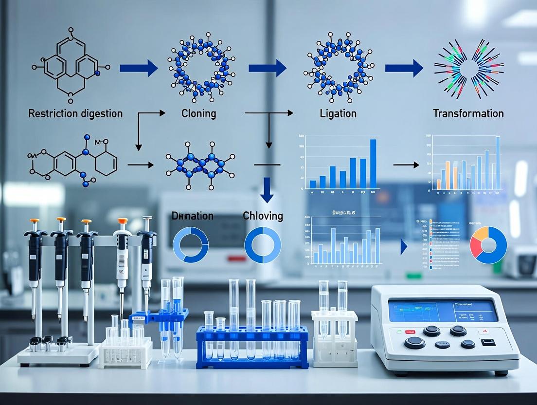
Visual Workflow and Logical Relationships
Title: Golden Gate Assembly Workflow
Title: Type IIS Enzyme Mechanism & Ligation
Application Notes
Golden Gate cloning is a powerful, one-pot, restriction-ligation method that enables the seamless and scarless assembly of multiple DNA fragments with high efficiency and fidelity. Its precision stems from the use of Type IIS restriction enzymes, which cleave DNA outside their recognition sequences, generating user-defined overhangs. This allows for the ordered assembly of fragments in a single reaction, with the final product lacking the original enzyme recognition sites—hence "scarless." Within the broader thesis on advanced DNA assembly techniques, Golden Gate represents a cornerstone methodology for synthetic biology, metabolic engineering, and the construction of complex genetic circuits, particularly valuable for drug development professionals engineering pathways for therapeutic compound production.
Protocols
Protocol 1: Standard Golden Gate Assembly for Multiple Fragments
Objective: Assemble 4-8 DNA fragments into a linearized destination vector in a single reaction.
Materials:
- DNA fragments and vector with appropriate Type IIS overhangs (e.g., BsaI-HFv2 or BbsI sites).
- T4 DNA Ligase Buffer (or isothermal buffer).
- Type IIS Restriction Enzyme (e.g., BsaI-HFv2, 10 U/μL).
- High-concentration T4 DNA Ligase (400 U/μL).
- Nuclease-free water.
- Thermocycler.
Method:
- Reaction Setup: In a single tube, combine:
- 50-100 ng of linearized destination vector.
- Equimolar amounts of each insert fragment (typical fragment:vector molar ratio 2:1).
- 1.5 μL 10X T4 DNA Ligase Buffer.
- 1 μL BsaI-HFv2 (10 U/μL).
- 1 μL T4 DNA Ligase (400 U/μL).
- Nuclease-free water to 15 μL total.
- Thermocycling: Run the following program:
- 25-37 cycles of: 30-60 seconds at 37°C (digestion) + 2-3 minutes at 16°C (ligation).
- Final digestion: 5 minutes at 50°C.
- Enzyme inactivation: 5-10 minutes at 80°C.
- Hold at 4°C.
- Transformation: Transform 2-5 μL of the reaction directly into competent E. coli. Plate on selective media.
- Screening: Screen colonies by colony PCR or restriction digest for correct assembly.
Protocol 2: Golden Gate for Modular Library Construction
Objective: Create a variant library by assembling a fixed backbone with variable, modular cassettes.
Materials: As per Protocol 1, with pre-validated modular fragment libraries.
Method:
- Design modular fragments with standardized, compatible overhangs (e.g., using the MoClo or GoldenBraid standards).
- Set up multiple Golden Gate reactions, each combining the backbone with a different set of modular cassettes.
- Use a shortened thermocycle (e.g., 25 cycles) to minimize bias.
- Pool all assembly reactions before transformation to generate a comprehensive library.
- Isolate plasmid DNA from the pooled colonies for downstream screening or selection.
Data Presentation
Table 1: Comparison of Type IIS Enzymes for Golden Gate Assembly
| Enzyme (Vendor Example) | Recognition Site (5'→3') | Cleavage Offset | Optimal Temp. | Common Use |
|---|---|---|---|---|
| BsaI-HFv2 (NEB) | GGTCTC | 1/5 | 37°C | Standard modular assembly |
| BbsI (NEB) | GAAGAC | 2/6 | 37°C | Alternative to BsaI |
| SapI (NEB) | GCTCTTC | 1/4 | 37°C | Assembly of repetitive sequences |
| PaqCI | CACCTGC | 4/8 | 37°C | High-fidelity, thermostable assembly |
Table 2: Efficiency of Golden Gate Assembly by Fragment Number
| Number of Fragments* | Average Transformation (CFU/μg) | Assembly Success Rate (%) | Recommended Cycles |
|---|---|---|---|
| 2-4 | 1.0 x 10⁴ - 1.0 x 10⁵ | >95 | 25-30 |
| 5-8 | 1.0 x 10³ - 1.0 x 10⁴ | 80-95 | 30-37 |
| 9-12 | 1.0 x 10² - 1.0 x 10³ | 60-80 | 37-50 |
*Including destination vector. CFU and success rates are typical examples; actual results depend on fragment length and quality.
Visualizations
Title: Golden Gate One-Pot Reaction Workflow
Title: Golden Gate Role in DNA Assembly Thesis
The Scientist's Toolkit: Research Reagent Solutions
| Item (Example Vendor) | Function in Golden Gate Assembly |
|---|---|
| BsaI-HFv2 Restriction Enzyme (NEB) | High-fidelity Type IIS enzyme for precise digestion; minimizes star activity. |
| T4 DNA Ligase (400 U/μL) (Thermo Fisher) | High-concentration ligase for efficient joining of digested fragments in the same buffer. |
| 10X T4 DNA Ligase Buffer | Provides ATP and optimal ionic conditions for both restriction and ligation activities. |
| Nuclease-Free Water (Invitrogen) | Solvent free of contaminants that could degrade DNA or inhibit enzymes. |
| Chemically Competent E. coli (NEB 5-alpha) | For efficient transformation of the assembled plasmid DNA after the reaction. |
| DNA Oligonucleotides (IDT) | For PCR amplification of fragments with added Type IIS recognition sites. |
| High-Fidelity DNA Polymerase (Q5, NEB) | For error-free PCR amplification of assembly fragments. |
| DNA Clean & Concentrator Kits (Zymo) | For purifying PCR fragments and final assembled plasmids. |
Application Notes & Protocols: A Thesis Framework for Golden Gate Assembly
This document details the critical components and standardized protocols for Golden Gate assembly, a scarless, restriction-ligation-based method for seamless assembly of multiple DNA fragments. Its efficiency and fidelity make it indispensable for synthetic biology, pathway engineering, and modular vector construction in drug development.
Core Enzymes: Type IIs Restriction Endonucleases
The foundation of Golden Gate cloning is the use of Type IIs restriction endonucleases. These enzymes cut DNA outside of their recognition sequences, generating user-defined 4-base pair (bp) overhangs.
Table 1: Common Type IIs Enzymes for Golden Gate Assembly
| Enzyme | Recognition Sequence (5'→3')^ | Cut Site (↓) | Optimal Temp. | Typical Incubation Time | Key Feature |
|---|---|---|---|---|---|
| BsaI | GGTCTC | 1/5 | 37°C | 1-2 hours | Gold standard; most common system (e.g., MoClo). |
| BsmBI | CGTCTC | 1/5 | 55°C | 5-15 mins | Thermostable; enables rapid cycling. |
| BbsI | GAAGAC | 2/6 | 37°C | 1-2 hours | Common in early systems (e.g., GoldenBraid). |
| SapI | GCTCTTC | 1/4 | 37°C | 1-2 hours | Creates asymmetric overhangs for directional assembly. |
| Aaiv | GAGGAG | 10/14 | 37°C | 1-2 hours | Recognizes longer sequence for higher specificity. |
^ Nicking variants (e.g., BsaI-HFv2, BsmBI-v2) are available to reduce star activity.
Protocol 1.1: Standard Golden Gate Reaction Setup
- Reagents:
- DNA fragments (10-100 fmol each)
- Destination vector (50-100 fmol)
- T4 DNA Ligase Buffer (1X final)
- Type IIs Restriction Enzyme (e.g., BsaI-HFv2, 5-10 units)
- High-concentration T7 DNA Ligase (400 units/µL, e.g., NEB)
- Nuclease-free water
- Method:
- Assemble reaction on ice: 50-100 ng vector, equimolar inserts, 1µL BsaI-HFv2, 1µL T7 DNA Ligase, 1X ligase buffer. Total volume: 10-20 µL.
- Cycle in a thermocycler: (37°C for 2-5 mins + 16°C for 5 mins) x 25-50 cycles.
- Final digestion: 50°C for 5 mins, 80°C for 10 mins (enzyme inactivation).
- Transform 2-5 µL into competent E. coli.
Vector Backbones
Vectors are engineered to contain the enzyme recognition sites flanking the cloning cassette. They often include negative selection markers (e.g., ccdB) for counter-selection against empty vectors.
Table 2: Common Golden Gate Vector Features
| Feature | Function | Example (Addgene #) |
|---|---|---|
| Destination Cassette | Contains two inward-facing Type IIs sites to accept inserts. | pYTK001 ( #'s vary) |
| ccdB Suicide Gene | Positive selection; only successful assembly removes ccdB, allowing cell survival. | pDest (e.g., #'s vary) |
| Mobility (oriT) | Enables conjugation into other bacterial hosts or fungi. | pUC-based vectors |
| Promoter/Reporter | Drives expression or provides visual screening (e.g., GFP). | pGGAselect (GFP) |
Protocol 1.2: Preparation of Modular Golden Gate Vector (Level 0)
- Objective: Clone a basic part (e.g., promoter, CDS, terminator) into a Level 0 acceptor vector.
- Method:
- Design primers with appropriate 4-bp overhangs (from standard toolkits like MoClo) for your part and vector.
- Amplify part via PCR using a high-fidelity polymerase.
- Purify PCR product and vector backbone (digested with appropriate enzyme).
- Perform Golden Gate assembly as in Protocol 1.1 using BsaI.
- Screen colonies by colony PCR or restriction digest.
Oligonucleotide Design
Oligonucleotides (primers) define the assembly junctions. The 5' extensions must correspond to the desired 4-bp overhangs and must be free of the enzyme's recognition sequence.
Key Design Rules:
- Overhang Uniqueness: Each 4-bp overhang in an assembly must be unique and complementary only to its intended partner.
- Avoidance: The recognition sequence (e.g., GGTCTC for BsaI) must be absent from the final assembled sequence and all intermediate overhangs.
- Standardization: Use community-accepted overhang sets (e.g., MoClo TK: GGAG, AATG, GCTT, etc.) for interoperability.
The Scientist's Toolkit: Essential Research Reagents
Table 3: Essential Materials for Golden Gate Experiments
| Item | Function | Example Product/Brand |
|---|---|---|
| High-Fidelity PCR Mix | Amplifies DNA parts with minimal errors. | Q5 High-Fidelity (NEB), Phusion (Thermo) |
| T7 DNA Ligase | High-efficiency ligase critical for cycled ligation. | T7 DNA Ligase (400 U/µL, NEB) |
| Competent E. coli | High-efficiency cells for transformation of assembly reactions. | NEB 5-alpha, DH5α, Mach1 |
| PCR Purification Kit | Cleans up PCR products and assembly reactions. | Qiagen QIAquick, Monarch PCR & DNA Cleanup Kit |
| Gel Extraction Kit | Isolates correctly sized DNA fragments from agarose gels. | Zymoclean Gel DNA Recovery Kit |
| Plasmid Miniprep Kit | Rapid isolation of plasmid DNA for screening. | GeneJET Plasmid Miniprep Kit |
| DNA Size Ladder | Accurate sizing of DNA fragments for validation. | 1 kb Plus DNA Ladder (Invitrogen) |
Visualizing Workflows
Title: Golden Gate Assembly Experimental Workflow
Title: BsaI Recognition, Cleavage, and Scarless Ligation Mechanism
Why Choose Golden Gate? Key Advantages Over Traditional Cloning for Multi-Gene Constructs
Golden Gate cloning has emerged as a cornerstone technique for the seamless assembly of multiple DNA fragments, driven by the use of Type IIS restriction enzymes. This Application Note details its core advantages over traditional restriction enzyme/ligase cloning within the context of advanced research in synthetic biology and multi-gene pathway engineering.
Quantitative Advantages: Golden Gate vs. Traditional Cloning
Table 1: Comparative Analysis of Cloning Methodologies
| Parameter | Traditional Cloning (Single RE) | Golden Gate Cloning (Type IIS) |
|---|---|---|
| Assembly Efficiency | Low for >2 fragments; rapid exponential drop. | High; routinely 90-95% efficiency for 4-6 fragment assemblies. |
| Typical Assembly Time | Multi-step, often >3 days for complex constructs. | Single-tube, one-day reaction (digestion & ligation). |
| Seamlessness | Leaves behind scars (restriction site sequences). | Truly scarless; removes recognition site from final construct. |
| Directional Control | Limited; often requires multiple, incompatible enzymes. | Inherently directional due to designed, asymmetric overhangs. |
| Multiplexing Capacity | Very limited, typically 1-2 fragments. | High; standard assemblies of 5-10 fragments, with advanced systems (MoClo) enabling 20+. |
| Cost per Correct Clone | High due to extensive screening needed. | Low; high efficiency minimizes screening (often 1 colony PCR). |
Table 2: Example Assembly Success Rates (Recent Data)
| Number of DNA Fragments | Golden Gate Efficiency (% Correct Clones) | Traditional Cloning Efficiency* |
|---|---|---|
| 2 | >98% | ~70% |
| 4 | 90-95% | <10% |
| 6 | 80-90% | ~1% |
| 10 (Modular System) | 60-80% | Negligible |
*Estimates based on sequential cloning steps.
Detailed Protocol: One-Pot Golden Gate Assembly
Objective: Assemble 4 transcriptional units into a single plasmid backbone.
Research Reagent Solutions & Essential Materials:
| Item | Function |
|---|---|
| Type IIS Enzyme (e.g., BsaI-HFv2, Esp3I) | Cuts outside its recognition site, generating unique 4bp overhangs. |
| T4 DNA Ligase (High-Concentration) | Joins annealed DNA fragments with compatible overhangs. |
| 10x T4 DNA Ligase Buffer | Provides optimal ionic conditions for simultaneous restriction and ligation. |
| Designed Entry Vectors / PCR Fragments | DNA parts with appropriate prefix/suffix sequences containing enzyme sites. |
| Competent E. coli (High-Efficiency) | For transformation of the assembled plasmid. |
| Agar Plates with Selective Antibiotic | For selection of successful transformants containing the assembled plasmid. |
Workflow:
- Design: Define 4bp overhangs for each fragment junction. Ensure inward-facing BsaI sites (e.g., GGAGACC for fragment left, AAAC for fragment right).
- Setup Reaction: In a single tube on ice, combine:
- 50 ng of linearized backbone.
- Each insert fragment at a 2:1 molar ratio to backbone.
- 1µL BsaI-HFv2 (10U).
- 1µL T4 DNA Ligase (400U).
- 2µL 10x T4 DNA Ligase Buffer.
- Nuclease-free water to 20µL.
- Thermocycling: Place tube in a thermocycler: 30 cycles of (37°C for 2-5 minutes + 16°C for 5 minutes), then 50°C for 5 minutes, 80°C for 10 minutes.
- Transformation: Transform 2µL of the reaction into 50µL competent E. coli, plate on selective agar, and incubate overnight.
- Screening: Pick 1-3 colonies for colony PCR or analytical digestion. Due to high efficiency, most will be correct.
Visualization: Workflow and Logic
Golden Gate Assembly Workflow
Golden Gate vs. Traditional Cloning Logic
Application Notes
Golden Gate cloning, a highly efficient, seamless DNA assembly method, has evolved from a novel concept in 2008 to a cornerstone technique for synthetic biology and metabolic engineering. Its core principle utilizes Type IIS restriction endonucleases, which cut outside their recognition sites, generating user-defined cohesive overhangs. This enables the precise, ordered, and scarless assembly of multiple DNA fragments in a single-tube reaction.
Table 1: Evolution of Key Golden Gate Assembly Systems
| System (Year Introduced) | Key Enzyme(s) | Typical Fragment Capacity | Primary Advantage | Reference/Kit |
|---|---|---|---|---|
| Original Method (2008) | BsaI-HF | 2-10 fragments | Proof of concept for scarless, one-pot assembly | Engler et al., 2008 |
| MoClo (2012) | BsaI | ~10 fragments | Standardized, hierarchical modular cloning system for plants | Weber et al., 2012 |
| GoldenBraid (2013) | BsaI, BsmBI | High (iterative) | Standardized, iterative assembly for plant biotechnology | Sarrion-Perdigones et al., 2013 |
| NEBridge Golden Gate Assembly (2018) | BsaI-HFv2, BsmBI-v2, etc. | 2-20+ fragments | Commercial kit with high-fidelity, pre-optimized buffers | New England Biolabs |
| Modular Cloning (MoClo) Toolkit for Mammalian Cells (2020+) | BsaI, BbsI | Varies by kit | Extended standardization to mammalian systems | Various consortia |
Table 2: Quantitative Performance Metrics of Modern Golden Gate Kits
| Kit/System | Typical Assembly Efficiency (Correct Colonies) | Optimal Fragment Number | Incubation Time | Compatibility |
|---|---|---|---|---|
| NEBridge Golden Gate (BsaI) | >90% (4-6 fragments) | 2-10 | 1 hour (cycling) | High-throughput, automated workflows |
| MoClo Plant Toolkit | >80% (5-10 fragments) | 5-10+ | 2-6 hours | Hierarchical, multi-gene construction |
| Commercial "Mix-and-Go" Kits | 70-95% (2-4 fragments) | 2-6 | 10-30 minutes | Fast, simple routine cloning |
Detailed Protocols
Protocol 1: Standard One-Pot Golden Gate Assembly for 4 Fragments
Objective: Assemble 4 DNA fragments into a linearized plasmid backbone in a single reaction.
Materials: See "The Scientist's Toolkit" below.
Procedure:
- Fragment Preparation: Dilute each purified DNA fragment (inserts and backbone) to 10-20 fmol/µL. Ensure each fragment has unique, complementary 4-bp overhangs designed in silico.
- Reaction Setup: Assemble the following on ice:
- 2.5 µL 2X Golden Gate Reaction Mix (containing buffer, ATP, BsaI-HFv2, and T7 DNA Ligase).
- 1 µL Vector Backbone (10 fmol).
- 1 µL of each Insert Fragment (10 fmol each).
- Nuclease-free water to 5 µL total.
- Thermocycling: Place tube in a thermocycler with the following program:
- 25-37 cycles of: 37°C for 2-5 minutes (digestion), 16°C for 3-5 minutes (ligation).
- Final digestion: 37°C for 5 minutes.
- Enzyme inactivation: 60°C for 5-10 minutes.
- Hold at 4°C.
- Transformation: Transform 2 µL of the reaction into 50 µL of competent E. coli cells via heat shock or electroporation. Plate on selective media.
- Screening: Screen colonies by colony PCR or diagnostic restriction digest.
Protocol 2: Hierarchical MoClo Assembly
Objective: Assemble multiple transcription units into a final destination vector.
Procedure:
- Level 0 (Basic Parts): Assemble individual promoters, coding sequences, and terminators into Level 0 acceptor vectors using BsaI. Screen for correct clones.
- Level 1 (Transcription Unit): Assemble Level 0 parts (e.g., Promoter + CDS + Terminator) into a Level 1 vector using BsaI. This creates a functional gene unit.
- Level 2+ (Multi-Gene Construct): Assemble multiple Level 1 transcription units into a Level 2 or higher destination vector using a second Type IIS enzyme (e.g., BbsI or BsmBI), which recognizes different sites flanking the Level 1 modules.
- Verification: Confirm each hierarchical step by sequencing across all new junctions.
Visualizations
Title: Golden Gate Assembly Core Workflow
Title: Hierarchical MoClo Assembly Strategy
The Scientist's Toolkit
Table 3: Essential Research Reagent Solutions for Golden Gate Cloning
| Item | Function & Key Features |
|---|---|
| Type IIS Restriction Enzyme (e.g., BsaI-HFv2, BsmBI-v2) | High-fidelity enzyme that cleaves outside its recognition site, generating designed 4-base overhangs. HF variants reduce star activity. |
| T7 DNA Ligase | DNA ligase with high activity at cycling temperatures (16-25°C), compatible with Type IIS enzyme buffers, enabling one-pot digestion/ligation. |
| 2X Golden Gate Master Mix | Pre-optimized commercial mix containing buffer, ATP, enzyme, and ligase. Simplifies reaction setup and improves reproducibility. |
| Nuclease-Free Water | Essential for diluting DNA and setting up reactions without degrading components. |
| Chemically Competent E. coli (High Efficiency) | For transformation of the assembled plasmid. >1×10⁸ cfu/µg efficiency is recommended for complex, multi-fragment assemblies. |
| Selection Antibiotics & Agar Plates | For selective growth of colonies containing the correctly assembled plasmid with the desired resistance marker. |
| PCR Reagents for Screening | Polymerase, dNTPs, and junction-spanning primers for rapid colony PCR verification of correct assembly. |
| DNA Purification Kits (Gel & PCR) | For purification of individual fragments (e.g., from PCR or restriction digest) prior to assembly. |
Step-by-Step Golden Gate Protocol: Designing, Assembling, and Transforming Multi-Fragment Constructs
Application Notes
Golden Gate cloning, utilizing Type IIS restriction enzymes, is the foundation of modern modular DNA assembly systems. Its robustness for assembling multiple fragments in a single reaction hinges on precise primer and fragment design, particularly regarding overhangs. This protocol, framed within a thesis on high-throughput multi-fragment assembly for synthetic biology and therapeutic construct development, details the critical rules and standardized practices for the MoClo (Modular Cloning) and GoldenBraid ecosystems.
Core Design Principles:
- Overhang Design: The 4-base pair (bp) overhangs generated by enzymes like BsaI, BpiI, or Esp3I must be unique and non-palindromic within an assembly to prevent misassembly and ensure directional ligation. Standardized toolkits define fixed overhang sets.
- Insulator Sequences: To prevent unwanted cleavage, the recognition site of the Type IIS enzyme must be absent from the final assembled sequence. "Insulator" or "spacer" nucleotides are added between the overhang and the internal sequence.
- Standardized Positions: In systems like MoClo, parts (promoters, CDS, terminators) are stored in Level 0 plasmids with standardized flanking positions. Assembly follows a strict positional grammar (e.g., A-Linker-B).
- PCR Primer Design: Primers must append the correct enzyme recognition site, overhang sequence, and any required insulator bases to the amplicon. Melting temperature (Tm) calculations should focus on the annealing region.
Quantitative Data Summary:
Table 1: Common Type IIS Enzymes and Their Properties
| Enzyme | Recognition Site (5'→3') | Cleavage Offset | Overhang Length | Common System |
|---|---|---|---|---|
| BsaI | GGTCTC(N)₁↓ | 1 nt downstream | 4 bp | MoClo, Golden Gate |
| BpiI (BbsI) | GAAGAC(N)₂↓ | 2 nt downstream | 4 bp | GoldenBraid, MoClo |
| Esp3I | CGTCTC(N)₁↓ | 1 nt downstream | 4 bp | MoClo-derivatives |
| SapI | GCTCTTC(N)₁↓ | 1 nt downstream | 3 bp | Advanced assemblies |
Table 2: Standardized MoClo Overhang Sets for Basic Assembly
| Position | Standard Overhang (5'→3') | Complementary To | Purpose |
|---|---|---|---|
| Prefix | GGAG | CCTC | Links to previous part |
| Suffix | AATG | TTAC | Links to next part |
| Start (CDS) | AAGC | TTCG | Links promoter to CDS |
| End (CDS) | GCTT | CGAA | Links CDS to terminator |
Experimental Protocols
Protocol 1: Designing a PCR Fragment for MoClo Level 0 Assembly
Objective: Amplify a coding sequence (CDS) for entry into a MoClo Level 0 acceptor vector.
Materials: See "The Scientist's Toolkit" below.
Method:
- Identify CDS Sequence: Obtain the pure CDS (start to stop codon) from a trusted database (e.g., NCBI, Ensembl).
- Design Forward Primer (Fw):
- 5' Add-on:
5'-[BsaI Site]-[Overhang]-[Insulator]- 3' - Example for CDS Start (AAGC):
5'- ATATGGTCTC AAGC ATG... -3'ATATG: Additional bases for efficient enzyme digestion.GGTCTC: BsaI recognition site.AAGC: Standardized start overhang.ATG: Insulator + start codon (ensures no internal BsaI site).
- The 3' end (18-22 bp) anneals to the CDS start.
- 5' Add-on:
- Design Reverse Primer (Rv):
- 5' Add-on:
5'-[BsaI Site]-[Overhang]-[Insulator]- 3' - Example for CDS End (GCTT), reverse complement:
5'- ATATGGTCTC GCTT CTA... -3'GCTT: Standardized end overhang.CTA: Insulator (stop codon is part of the annealed CDS).
- The 3' end anneals to the CDS end, omitting the stop codon if a C-terminal tag is planned.
- 5' Add-on:
- PCR Amplification: Use a high-fidelity polymerase. Typical thermocycler program: 98°C 30s; 35 cycles of [98°C 10s, 60-72°C 20s, 72°C 15-30s/kb]; 72°C 5 min.
- Purification: Gel-purify the PCR product to remove primers and non-specific bands.
Protocol 2: Golden Gate Assembly for Multi-Fragment Level 1 Construction
Objective: Assemble 4 transcriptional units (Level 0 parts) into a Level 1 destination vector.
Materials: See "The Scientist's Toolkit" below.
Method:
- Calculate Stoichiometry: Use a molar ratio of ~1:1 for all inserts and a 1:2 ratio for vector:total insert. For 4 inserts, a common ratio is vector:insert1:insert2:insert3:insert4 = 1:1:1:1:1.
- Set Up Reaction:
- In a 0.2 mL tube, combine:
- 50-100 ng Level 1 destination vector (e.g., pL1R).
- Equimolar amounts of each Level 0 plasmid (Promoter, CDS, Terminator) per assembly.
- 1.5 μL 10x T4 DNA Ligase Buffer (contains ATP).
- 1 μL (10 U) BsaI-HFv2 or equivalent.
- 1 μL (400 U) T4 DNA Ligase.
- Nuclease-free water to 15 μL.
- In a 0.2 mL tube, combine:
- Run Thermocycling Program:
- Standard Program: 25-50 cycles of [37°C (2-5 min) → 16°C (2-5 min)], followed by 50°C for 5 min, 80°C for 10 min.
- Fast Program: 5-10 cycles of [37°C 3 min → 16°C 4 min] is often sufficient for 4-5 fragments.
- Transformation: Transform 2-5 μL of the reaction into competent E. coli (e.g., DH5α), plate on selective media, and incubate overnight.
Diagrams
Title: Golden Gate Assembly Workflow for MoClo
Title: Primer Add-on and Fragment Structure
The Scientist's Toolkit
Table 3: Essential Research Reagent Solutions
| Item | Function & Critical Features |
|---|---|
| Type IIS Restriction Enzymes (BsaI-HFv2, BpiI) | High-fidelity (HF) versions minimize star activity. They simultaneously digest PCR fragments/vectors and create compatible overhangs for assembly. |
| T4 DNA Ligase | Ligates the compatible 4-bp overhangs created by Type IIS digestion. Requires ATP (usually supplied in buffer). |
| High-Fidelity DNA Polymerase (e.g., Q5, Phusion) | For error-free amplification of parts for Level 0 cloning. Essential for maintaining sequence integrity. |
| MoClo/GoldenBraid Toolkit Vectors | Standardized acceptor (Level 0) and destination (Level 1+) vectors with predefined overhangs and resistance markers. |
| DNA Clean-Up & Gel Extraction Kits | For purifying PCR products and isolating correctly sized fragments from agarose gels to prevent carryover of primers or template. |
| Chemically Competent E. coli (DH5α, NEB Stable) | For transformation of assembly reactions. High efficiency (>1x10⁸ cfu/μg) is recommended for complex multi-fragment assemblies. |
Within the broader thesis on Golden Gate cloning for assembling multiple DNA fragments, this protocol details a streamlined workflow from PCR amplification to a one-pot assembly reaction. This method enables high-efficiency, seamless assembly of multiple inserts into a destination vector in a single step, crucial for constructing complex genetic circuits, metabolic pathways, and synthetic biology applications in drug development.
Research Reagent Solutions
Table 1: Essential Reagents and Materials for PCR-to-Assembly Workflow
| Reagent/Material | Function | Key Consideration |
|---|---|---|
| High-Fidelity DNA Polymerase | Amplifies DNA fragments from templates with minimal errors. Essential for generating mutation-free inserts. | Use polymerases with proofreading activity (e.g., Q5, Phusion). |
| Type IIS Restriction Enzyme (e.g., BsaI-HFv2, Esp3I) | Cleaves DNA at specific sequences outside its recognition site, generating unique, user-defined 4bp overhangs for seamless assembly. | The workhorse of Golden Gate assembly. Ensure it is compatible with the reaction buffer. |
| T4 DNA Ligase | Joins DNA fragments with complementary overhangs in the same reaction as digestion. | High concentration (e.g., 400U/µL) is critical for one-pot success. |
| ATP | Essential cofactor for T4 DNA Ligase activity. | Often supplied with the ligase buffer. Verify concentration. |
| Thermocycler | Precisely cycles temperature for PCR and the Golden Gate assembly reaction. | Must support rapid temperature cycling between 37°C and 16°C. |
| Destination Vector | Carries antibiotic resistance and replication origin; contains the Type IIS enzyme sites flanking the cloning site. | Typically prepared with two inverted Type IIS sites to remove the "scar" sequence. |
Detailed Experimental Protocols
PCR Amplification of DNA Fragments with Added Overhangs
Objective: Amplify target DNA fragments while appending the necessary Type IIS restriction enzyme sites and desired 4bp overhangs.
Materials:
- Template DNA (genomic, plasmid, synthetic)
- Forward and Reverse Primers (with 5' extensions)
- High-fidelity PCR Master Mix
- Nuclease-free water
- Thermocycler
Protocol:
- Primer Design: Design primers with the following structure (5'->3'):
- Forward Primer:
5'-[4bp overhang][BsaI site (GGTCTC)][Gene-specific sequence]-3' - Reverse Primer:
5'-[4bp overhang][BsaI site (GAGACC)][Gene-specific sequence]-3' - Ensure the 4bp overhangs are unique and complementary between adjacent fragments.
- Forward Primer:
PCR Setup (50 µL reaction):
- Nuclease-free water: to 50 µL
- 2X High-Fidelity Master Mix: 25 µL
- Forward Primer (10 µM): 2.5 µL
- Reverse Primer (10 µM): 2.5 µL
- Template DNA: 1-100 ng Mix gently and centrifuge briefly.
PCR Cycling Conditions:
- Initial Denaturation: 98°C for 30 sec
- 30 Cycles:
- Denaturation: 98°C for 10 sec
- Annealing: (Tm of gene-specific sequence +5°C) for 20 sec
- Extension: 72°C for 20-30 sec/kb
- Final Extension: 72°C for 2 min
- Hold: 4°C
Purification: Clean up PCR products using a spin-column PCR purification kit. Elute in 20 µL nuclease-free water. Quantify via spectrophotometry.
One-Pot Golden Gate Assembly Reaction
Objective: Digest PCR fragments and the destination vector with BsaI and ligate them together in a single, cyclical reaction.
Materials:
- Purified PCR fragments (inserts)
- Linearized destination vector
- BsaI-HFv2 (or equivalent Type IIS enzyme)
- T4 DNA Ligase (400 U/µL)
- 10X T4 DNA Ligase Reaction Buffer (contains ATP)
- Thermocycler
Protocol:
- Molar Ratio Calculation: Calculate the volume of each insert to use based on equimolar amounts relative to the vector. A typical starting molar ratio is 2:1 (insert:vector).
Reaction Assembly: Assemble components on ice in the order listed (water, buffer, DNA, enzymes). Mix gently by pipetting. Centrifuge briefly.
Thermocycling for Assembly:
- Place tube in a thermocycler.
- Run the following program:
- Cycles: 25-30 cycles of:
- Digestion: 37°C for 2-3 minutes
- Ligation: 16°C for 3-5 minutes
- Final Digestion: 50°C for 5 minutes (optional, to inactivate BsaI)
- Enzyme Inactivation: 80°C for 10 minutes
- Hold: 4°C
- Cycles: 25-30 cycles of:
Transformation: Transform 2-5 µL of the assembly reaction into competent E. coli cells via heat shock or electroporation. Plate on selective media.
Data Presentation
Table 3: Expected Outcomes and Troubleshooting Guide
| Parameter | Expected Result | Deviation & Possible Cause |
|---|---|---|
| PCR Yield | >50 ng/µL, single band on agarose gel. | Low yield: poor primer design, suboptimal annealing temp. Multiple bands: primer dimers, nonspecific binding. |
| Assembly Efficiency (Colonies) | 10-1000+ CFU per reaction, depending on complexity. | Very few colonies: incorrect molar ratios, inactive enzymes, non-complementary overhangs. |
| Correct Assembly Rate | >80% positive clones for 2-4 fragment assemblies. | High background (empty vector): incomplete digestion of destination vector. Incorrect assemblies: mis-ordered overhangs. |
Visualized Workflows
Diagram 1: PCR to One-Pot Assembly Workflow (76 chars)
Diagram 2: One-Pot Reaction Mechanism (42 chars)
Choosing the Right Backbone Vector and Preparing Entry/Level 0 Modules
Application Notes
The Role of the Backbone Vector in Golden Gate Assembly Systems
The backbone vector (often referred to as the destination or Level 1+ vector) is the final acceptor plasmid that will contain the fully assembled multi-gene construct. Current research in synthetic biology and metabolic engineering for drug development emphasizes modular cloning systems like MoClo or GoldenBraid. Key selection criteria include:
- Compatibility: Must contain the correct Type IIS restriction enzyme sites (e.g., BsaI, BbsI) flanking the cloning cassette, complementary to those in the Entry modules.
- Antibiotic Resistance: A selection marker distinct from those used in Entry modules to enable selective pressure for the final assembly.
- Destination Marker: A visually selectable marker (e.g., ccdB death gene, fluorescent protein) that is excised upon successful insertion of the assembly, allowing for "clone-of-interest" selection.
- Host Range & Copy Number: Suitability for intermediate (E. coli) and final host organisms (e.g., yeast, mammalian cells for protein production).
Design and Preparation of Level 0 (Entry) Modules
Entry modules are standardized, sequence-validated basic biological parts (promoters, ORFs, terminators, tags) cloned into a standardized plasmid backbone. Their precise preparation is critical for efficient hierarchical assembly.
- Standardization: All parts are flanked by defined, non-palindromic four-base pair overhangs (fusion sites) generated by Type IIS enzyme digestion.
- Validation: Sanger sequencing of each Entry clone is mandatory to prevent propagation of errors through the assembly hierarchy.
- Storage: Organized in a physical and digital library (e.g., using Plasmid ID numbers) to facilitate rapid construct design.
Quantitative Comparison of Common Backbone Systems
The table below summarizes key characteristics of prevalent backbone systems used in modern Golden Gate-based research.
Table 1: Comparison of Common Golden Gate Assembly Backbone Vectors
| Backbone System | Type IIS Enzyme | Destination Marker | Common Antibiotic Resistance | Typical Application Context | Assembly Efficiency (Reported Range)* |
|---|---|---|---|---|---|
| MoClo (Level 1) | BsaI-HFv2 | ccdB (death gene) | Spectinomycin | Plant biotechnology, Metabolic pathways | 80-95% |
| GoldenBraid 2.0 | BsaI, BsmBI | LacZα (blue-white screening) | Kanamycin | Plant synthetic biology | 70-90% |
| EcoFlex (Modular) | BsaI | GFP (excised) | Chloramphenicol | Bacterial circuit engineering | >90% |
| Mammalian MoClo | BbsI | DestRFP (excised) | Ampicillin, Puromycin | Mammalian gene expression, Drug target validation | 75-85% |
*Efficiency data aggregated from recent literature (2022-2024) and represents the percentage of correct clones obtained from a standard 6-part assembly.
Experimental Protocols
Protocol 1: Preparation of Level 0 Entry Modules from PCR Products
Objective: To clone a basic genetic part (e.g., a promoter) into a standardized Entry vector.
Materials:
- Purified PCR product with appropriate overhangs (added via primers).
- pL0 Entry Vector (e.g., pICH41308 from MoClo toolkit).
- BsaI-HFv2 restriction enzyme & compatible ligase (e.g., T4 DNA Ligase).
- Chemically competent E. coli (DH5α).
Methodology:
- Digestion-Ligation Reaction (Golden Gate Assembly):
- Set up a 20 µL reaction on ice:
- 50 ng pL0 Entry Vector.
- 20-40 ng purified PCR product (∼2:1 insert:vector molar ratio).
- 1 µL BsaI-HFv2 (10 U/µL).
- 1 µL T4 DNA Ligase (400 U/µL).
- 2 µL 10X T4 DNA Ligase Buffer.
- Nuclease-free water to 20 µL.
- Set up a 20 µL reaction on ice:
- Thermocycling:
- Run the following program: (37°C for 5 min; 16°C for 5 min) x 30 cycles → 60°C for 10 min → 80°C for 10 min. Hold at 12°C.
- Transformation:
- Transform 5 µL of the reaction into 50 µL of competent E. coli.
- Plate on LB agar with the appropriate antibiotic (e.g., Kanamycin for pL0 vectors).
- Incubate overnight at 37°C.
- Validation:
- Pick 3-5 colonies for colony PCR and analytical digestion.
- Perform Sanger sequencing of the cloned insert using plasmid-specific primers.
Protocol 2: Multi-Fragment Assembly into a Level 1 Backbone Vector
Objective: To assemble 4-6 Level 0 modules into a chosen backbone vector.
Materials:
- Purified Level 0 plasmids (∼50 ng/µL each).
- Chosen Backbone Vector (e.g., a MoClo Level 1 vector with ccdB).
- BsaI-HFv2 and T4 DNA Ligase.
Methodology:
- Assembly Reaction:
- Set up a 20 µL reaction on ice:
- 50 ng Backbone Vector.
- Equimolar amounts of each Level 0 plasmid (typically 20-40 ng each).
- 1 µL BsaI-HFv2 (10 U/µL).
- 1 µL T4 DNA Ligase (400 U/µL).
- 2 µL 10X T4 DNA Ligase Buffer.
- Water to 20 µL.
- Set up a 20 µL reaction on ice:
- Thermocycling:
- Use the same thermocycling program as in Protocol 1.
- Transformation and Selection:
- Transform 5-10 µL into competent E. coli.
- Plate on LB agar containing the antibiotic for the backbone (e.g., Spectinomycin for MoClo Level 1). Note: The ccdB death gene is excised upon successful assembly, allowing only correct clones to grow.
- Screening:
- Screen 4-8 colonies by analytical restriction digest or colony PCR using primers spanning assembly junctions.
- Confirm the final construct by diagnostic digest or sequencing of key junctions.
Diagrams
Golden Gate Hierarchical Assembly Workflow
Backbone Vector Selection Logic Tree
The Scientist's Toolkit
Table 2: Essential Research Reagent Solutions for Golden Gate Cloning
| Reagent/Material | Function & Rationale |
|---|---|
| Type IIS Restriction Enzymes (BsaI-HFv2, BbsI-HF) | High-fidelity versions minimize star activity, essential for creating precise, defined overhangs for seamless ligation. The workhorses of Golden Gate assembly. |
| T4 DNA Ligase | Catalyzes the ligation of the complementary overhangs generated by Type IIS digestion. Its activity in the same buffer as restriction enzymes enables one-pot reactions. |
| ccDB-Toxin Expressing Competent Cells (e.g., DB3.1) | Required for the propagation of destination vectors containing the ccdB death gene. Standard cloning strains (DH5α) cannot survive. |
| Chemically Competent E. coli (DH5α, NEB Stable) | For transformation of assembly reactions. High-efficiency (>1x10⁸ CFU/µg) cells are recommended for complex multi-fragment assemblies. |
| Validation Primers (Insert-Flanking, Junction-Spanning) | Custom oligonucleotides for colony PCR and sequencing to verify the identity and correct assembly of each Level 0 module and final construct. |
| Standardized Level 0 Entry Vector (e.g., pICH41308) | A uniform acceptor plasmid for basic parts. Contains fixed flanking BsaI sites that generate the required four-base overhangs upon digestion. |
| Agarose Gel DNA Recovery Kit | For reliable purification of PCR products and digested plasmid fragments to remove enzymes, salts, and primers that can interfere with assembly efficiency. |
| Plasmid Miniprep Kit (High-Yield) | For rapid purification of sufficient quantities of high-quality Level 0 plasmids and final constructs for sequencing, archiving, and downstream applications. |
Application Notes
Golden Gate cloning, particularly using Type IIS restriction enzymes like BsaI-HFv2 or Esp3I, has become the cornerstone of modern synthetic biology for assembling multiple DNA fragments in a single, one-pot reaction. Its precision, efficiency, and modularity enable the construction of complex biological systems. This document details applications and protocols framed within a thesis on high-throughput, multi-fragment Golden Gate assembly.
Building Programmable Gene Circuits
Gene circuits are engineered networks of regulators that process cellular signals. Golden Gate assembly excels here by enabling the rapid combinatorial assembly of promoters, coding sequences (CDS), and terminators into standardized vectors.
Key Quantitative Data: Table 1: Efficiency Metrics for Gene Circuit Assembly (6-fragment assembly using *BsaI-HFv2).*
| Parameter | Typical Performance | Notes |
|---|---|---|
| Assembly Efficiency | 85-95% correct colonies | With optimized modular parts (4bp overhangs). |
| One-Pot Capacity | Up to 15 fragments | Efficiency decreases beyond ~10 fragments. |
| Reaction Time | 1-2 hours (digestion-ligation) | Followed by standard transformation. |
| Background | <5% | Using destination vector with lethal gene (e.g., ccdB). |
Constructing Metabolic Pathways
Heterologous metabolic pathway engineering requires the stable co-expression of multiple enzymes. Golden Gate allows the sequential or hierarchical assembly of large operons or multigene constructs into genomic integration vectors.
Key Quantitative Data: Table 2: Pathway Assembly Outcomes for a 5-gene Operon.
| Assembly Strategy | Success Rate | Throughput Advantage |
|---|---|---|
| Single-step (all fragments) | ~70% | Fastest; requires highly efficient parts. |
| Hierarchical (sub-assemblies first) | >95% | Most reliable for >7 genes. |
| MoClo-Compatible | >90% | Enables library generation from interchangeable parts. |
Generating Large Plasmid Libraries
For directed evolution or combinatorial screening, Golden Gate is used to shuffle modular parts (e.g., promoter variants, enzyme mutants) to create vast plasmid libraries.
Key Quantitative Data: Table 3: Library Construction Scale.
| Library Component | Number of Variants | Theoretical Library Size | Practical Colony Yield |
|---|---|---|---|
| Promoter (P) | 10 | 10^3 | ~5 x 10^5 CFU/µg vector |
| RBS (R) | 10 | ||
| CDS (G) | 10 |
Experimental Protocols
Protocol 1: One-Pot Assembly of a 6-part Gene Circuit
Objective: Assemble a circuit with architecture: Promoter - Transcription Factor A - Linker - Promoter - Reporter Gene - Terminator.
Materials: Purified DNA fragments (with Type IIS overhangs), BsaI-HFv2 (NEB), T4 DNA Ligase (HC, NEB), 10x T4 Ligase Buffer, thermocycler.
Procedure:
- Fragment Preparation: Dilute all plasmid part donors and destination vector to 50 fmol/µL.
- Reaction Setup:
- In a PCR tube, mix:
- 50 fmol of each DNA fragment (equimolar).
- 1 µL BsaI-HFv2 (NEB).
- 1 µL T4 DNA Ligase (HC, NEB).
- 2 µL 10x T4 Ligase Buffer.
- Nuclease-free water to 20 µL.
- In a PCR tube, mix:
- Cycling Conditions:
- 37°C for 5 minutes (digestion).
- 16°C for 5 minutes (ligation).
- Repeat for 25 cycles.
- Final: 50°C for 5 minutes; 80°C for 10 minutes.
- Transformation: Transform 2 µL into competent E. coli (DH5α). Plate on selective media.
- Screening: Colony PCR or diagnostic restriction digest.
Protocol 2: Hierarchical Assembly of a Metabolic Pathway (8 genes)
Objective: Construct a plasmid for a 8-enzyme pathway.
Materials: As Protocol 1. Additional Level 1 acceptor vectors.
Procedure:
- Level 0: All basic parts in standard entry vectors.
- Level 1 (Sub-assembly):
- Perform 4 separate Golden Gate reactions to assemble two 2-gene operons (e.g., Operon A: Gene1-Gene2; Operon B: Gene3-Gene4).
- Transform, isolate plasmids, and sequence-verify.
- Level 2 (Final Assembly):
- Use the verified Level 1 operon plasmids and the remaining gene fragments as donors.
- Perform a final Golden Gate reaction to assemble all operons and genes into the final destination vector (e.g., a yeast integration vector).
- Verification: Use analytical gel electrophoresis and long-read sequencing (e.g., Nanopore) for final validation.
Visualizations
Golden Gate Hierarchical Assembly Workflow
Combinatorial Library Generation Logic
The Scientist's Toolkit
Table 4: Essential Research Reagent Solutions for Golden Gate Applications
| Reagent/Kit/Material | Function & Rationale | Example (Supplier) |
|---|---|---|
| Type IIS Restriction Enzyme | Creates unique, user-defined 4bp overhangs for seamless assembly. | BsaI-HFv2, Esp3I (NEB, Thermo). |
| High-Concentration T4 DNA Ligase | Efficiently ligates annealed overhangs in the same pot as digestion. | T4 DNA Ligase (HC) (NEB). |
| Golden Gate Assembly Kit | Pre-optimized buffers and vectors for specific standards (MoClo, Phytobricks). | MoClo Toolkit (Addgene). |
| CcdB Survival-Competent Cells | Allows direct selection of correct assemblies using destination vectors with ccdB toxin. | DB3.1, Stbl3 E. coli strains. |
| High-Fidelity DNA Polymerase | For amplification of parts without mutations, crucial for functional circuits. | Q5, Phusion (NEB). |
| Modular Cloning Parts Library | Collection of standardized, sequence-verified Level 0 parts for rapid design. | Plant, Yeast, Mammalian MoClo Parts (Addgene). |
| Long-Read Sequencing Service | Essential for verifying large, repetitive, or complex multigene assemblies. | Nanopore (Oxford), PacBio. |
This application note details a case study for the rapid, one-pot assembly of a multi-gene biosynthetic pathway using Golden Gate cloning. This methodology is central to accelerating synthetic biology approaches in drug discovery, enabling the heterologous expression of complex natural product pathways from uncultivable microbes in tractable host organisms like Streptomyces coelicolor or Saccharomyces cerevisiae. The specific case involves reconstructing the 6-gene thaxtomin A biosynthetic pathway, a phytotoxin with potential applications as a herbicide lead. The protocol is framed within a broader thesis on Golden Gate assembly for high-throughput, combinatorial pathway construction, which is foundational for modern drug discovery pipelines.
Application Notes
Key Advantages of Golden Gate for Pathway Assembly
- High Efficiency & Fidelity: Type IIS restriction enzymes (e.g., BsaI, Esp3I) cleave outside their recognition sites, enabling seamless, scarless assembly of multiple fragments in a defined order.
- Modularity and Standardization: Utilizes standardized, interchangeable genetic parts (promoters, genes, terminators) formatted in compatible vectors (e.g., MoClo, Phytobrick standards).
- Scalability: Hierarchical assembly allows for the construction of operons or whole pathways from basic parts, then into larger constructs like multi-gene clusters.
- One-Pot Reaction: Multiple DNA fragments can be assembled in a single-tube reaction, saving time and reagents.
Quantitative Assembly Success Metrics
The following table summarizes typical success rates and key metrics for Golden Gate assembly of multi-gene constructs, based on recent literature and internal data.
Table 1: Performance Metrics for Multi-Fragment Golden Gate Assembly
| Parameter | 4-Fragment Assembly (1 Operon) | 6-Fragment Assembly (Full Pathway) | Notes / Conditions |
|---|---|---|---|
| Cloning Efficiency (CFU/µg) | 1.2 x 10⁴ | 3.5 x 10³ | Using NEB Golden Gate Assembly Mix, 37°C for 1 hr, then 50 cycles of 37°C/16°C. |
| Correct Assembly Rate (%) | 92% | 78% | Verified by analytical digestion and Sanger sequencing of junctions. |
| Optimal Fragment Size | 0.5 - 3.0 kb | 0.5 - 4.0 kb | Larger fragments (>5 kb) can reduce efficiency. |
| Typical Plasmid Yield (ng/µL) | 120 - 250 | 80 - 150 | Miniprep yield from E. coli after transformation. |
| Total Hands-On Time | ~3 hours | ~4 hours | Excludes incubation, transformation, and colony screening time. |
Detailed Protocols
Protocol: One-Pot Golden Gate Assembly of a 6-Gene Pathway
Objective: Assemble six individual gene expression units (each with promoter, CDS, terminator) into a single destination vector (e.g., pETDuet-1 modified with Golden Gate sites) for heterologous expression.
Materials (Research Reagent Solutions):
- BsaI-HF v2 (NEB): High-fidelity Type IIS restriction enzyme for digestion.
- T4 DNA Ligase (NEB): For seamless ligation of compatible overhangs.
- 10x T4 DNA Ligase Buffer: Provides ATP and optimal ionic conditions.
- NEB Golden Gate Assembly Kit (BsaI-HF v2): A pre-optimized mix of enzyme and buffer.
- Chemically Competent E. coli (NEB 10-beta): High-efficiency cells for transformation.
- Agar plates with appropriate antibiotic (e.g., Carbenicillin): For selection.
- Destination Vector (e.g., pGG-AC): Level 1 acceptor vector with chloramphenicol resistance and a counter-selectable marker (e.g., ccdB).
- Entry Vectors (Level 0): Individual plasmids containing each standardized gene part with appropriate flanking BsaI sites (e.g., prefix-B1-B2-gene-B3-B4-suffix).
Procedure:
- Reaction Setup: In a sterile 0.2 mL PCR tube, combine the following on ice:
- 50 ng linearized destination vector.
- Equimolar amounts (typically 20-40 fmol each) of the six entry vectors (Level 0 parts).
- 1 µL of BsaI-HF v2 (10 U/µL).
- 1 µL of T4 DNA Ligase (400 U/µL).
- 2 µL of 10x T4 DNA Ligase Buffer.
- Nuclease-free water to a final volume of 20 µL.
- Thermocycling: Place the tube in a thermocycler and run the following program:
- 37°C for 5 minutes (digestion).
- 16°C for 5 minutes (ligation).
- Repeat steps 1 & 2 for 30 cycles.
- 50°C for 5 minutes (final digestion).
- 80°C for 10 minutes (enzyme inactivation).
- Hold at 4°C.
- Transformation:
- Add 2 µL of the assembly reaction to 50 µL of chemically competent E. coli cells. Incubate on ice for 30 minutes.
- Heat-shock at 42°C for 30 seconds, then place on ice for 2 minutes.
- Add 950 µL of SOC medium and incubate at 37°C with shaking for 1 hour.
- Plate 100 µL onto LB agar plates containing the appropriate antibiotic (e.g., carbenicillin for the assembled plasmid, chloramphenicol resistance is lost upon successful assembly).
- Screening: Pick 8-12 colonies for colony PCR using primers that span the inserted fragment junctions. Analyze by agarose gel electrophoresis. Confirm correct clones by Sanger sequencing.
Protocol: Analytical Restriction Digest for Assembly Verification
Objective: Quickly verify the correct assembly and size of the constructed pathway plasmid.
Procedure:
- Isolate plasmid DNA from 5 mL overnight cultures of candidate clones using a standard miniprep kit.
- Set up a 20 µL restriction digest using 2-3 different enzymes that cut in the vector backbone and at strategic points within the inserted pathway (e.g., one enzyme that cuts once in the vector and once in the middle of the insert).
- Incubate at the optimal temperature for 1 hour.
- Run the digested products on a 0.8-1.0% agarose gel alongside an appropriate DNA ladder.
- Compare the observed fragment sizes to the expected pattern from in silico digestion (using software like SnapGene).
The Scientist's Toolkit
Table 2: Essential Research Reagent Solutions for Golden Gate Pathway Assembly
| Item | Function / Explanation | Example Product |
|---|---|---|
| Type IIS Restriction Enzyme | Cleaves DNA outside recognition site, generating unique, user-defined 4 bp overhangs for scarless assembly. | BsaI-HF v2, Esp3I, AarI |
| High-Activity DNA Ligase | Joins DNA fragments with complementary overhangs generated by Type IIS enzymes. | T4 DNA Ligase |
| Optimized Assembly Mix | Pre-mixed enzyme/buffer solution simplifying reaction setup and improving reproducibility. | NEB Golden Gate Assembly Kit |
| Standardized Genetic Parts (Level 0) | Basic functional units (promoters, CDS, terminators) in standardized vectors with uniform flanking sites, enabling modularity. | MoClo Toolkit, Phytobricks |
| Acceptor/Backbone Vectors | Destination plasmids containing selectable markers and lethal gene (ccdB) for negative selection of empty vectors. | pICH477xx series, pGGA |
| High-Efficiency Competent Cells | Essential for transforming large, complex plasmid assemblies with high yield. | NEB Stable, NEB 10-beta, E. cloni |
| Colony PCR Master Mix | Allows for rapid, direct screening of bacterial colonies without prior plasmid extraction. | OneTaq Quick-Load Master Mix |
| Gateway BP Clonase (Optional) | Enables recombination of the assembled Golden Gate construct into expression vectors for different host organisms (e.g., yeast, Streptomyces). | Thermo Fisher Scientific |
Visualizations
Title: Golden Gate Assembly of Multiple DNA Fragments
Title: Workflow for Assembling a Biosynthetic Pathway
Solving Golden Gate Challenges: Expert Troubleshooting and Optimization Strategies for High Efficiency
Golden Gate assembly is a cornerstone technique for the modular, seamless assembly of multiple DNA fragments. However, its efficiency in high-fragment-number assemblies is often compromised by several recurring experimental pitfalls, leading to low yield, incorrect assemblies, and a lack of clear interpretation from negative controls. This application note, framed within broader research on robust multiplex DNA assembly, details these challenges and provides optimized protocols to overcome them.
Pitfall 1: Low Assembly Yield
Low transformation efficiency and few correct colonies are frequent issues. The root causes are often related to suboptimal template quality, ineffective digestion-ligation cycling, or poor transformation practices.
Quantitative Analysis of Yield Factors
Table 1: Impact of Common Factors on Golden Gate Assembly Yield
| Factor | Low/Incorrect Condition | Optimized Condition | Typical Yield Change (Colony Count) | Key Reference/Note |
|---|---|---|---|---|
| Template Purity | PCR product with carryover primers/dNTPs | Purified (column/SPRI) PCR product | Increase of 10-50x | Engler et al., 2008; Potapov et al., 2018 |
| Enzyme-to-Substrate Ratio | 1:1 (unit:pmol fragment ends) | 5:1 to 10:1 (unit:pmol fragment ends) | Increase of 5-20x | NEBioCalculator recommendations |
| Cycle Number | 10 cycles of (37°C/5 min + 16°C/5 min) | 30-50 cycles of (37°C/3 min + 16°C/4 min) | Increase of 3-10x | Standard for >5 fragment assemblies |
| Ligation Time | Short ligation phase (<1 min per cycle) | Extended ligation phase (4-10 min per cycle) | Increase of 2-5x | Prioritizes ligation kinetics |
| Post-Assembly Treatment | Direct transformation | Proteinase K treatment (15 min, 37°C) to inactivate enzymes | Increase of 2-3x | Reduces vector re-circularization |
Protocol 1: High-Yield Golden Gate Assembly
Objective: Assemble 4-8 DNA fragments into a destination vector in a single reaction.
Materials:
- Purified DNA fragments (25-50 fmol each, with appropriate Type IIS overhangs).
- Destination vector (50-100 fmol, linearized with complementary overhangs).
- T4 DNA Ligase Buffer (10X).
- Research Reagent Solutions: See Toolkit Table.
- Thermocycler.
- Chemically competent E. coli (high efficiency, >1e8 CFU/µg).
Procedure:
- Reaction Setup: In a thin-walled PCR tube, combine on ice:
- 50-100 fmol of each DNA fragment.
- 50-100 fmol of destination vector.
- 1.0 µL of 10X T4 DNA Ligase Buffer.
- 10 U of Type IIS Restriction Enzyme (e.g., BsaI-HFv2, Esp3I).
- 400 U of T4 DNA Ligase.
- Nuclease-free water to a final volume of 10 µL.
- Digestion-Ligation Cycling: Place the tube in a thermocycler and run the following program:
- Step 1: 30-50 cycles of:
- 37°C for 3 minutes (digestion).
- 16°C for 4 minutes (ligation).
- Step 2: 60°C for 5 minutes (final ligation).
- Step 3: 80°C for 10 minutes (enzyme inactivation).
- Hold: 4°C.
- Step 1: 30-50 cycles of:
- Post-Assembly Cleanup: Add 1 µL of Proteinase K (20 mg/mL) to the reaction. Incubate at 37°C for 15 minutes to digest the restriction enzyme and ligase.
- Transformation: Transform 2 µL of the Proteinase K-treated reaction into 50 µL of chemically competent E. coli using standard heat-shock methods. Plate the entire transformation volume on selective agar plates. Incubate overnight at 37°C.
Pitfall 2: Incorrect Assemblies
Incorrect assemblies (deletions, scrambles, empty vectors) arise from poor fragment design, star activity of enzymes, or misannealing of homologous overhangs.
Quantitative Analysis of Accuracy Factors
Table 2: Strategies to Minimize Incorrect Assemblies
| Strategy | Problem Addressed | Implementation | Expected Outcome (Correct Colony %) |
|---|---|---|---|
| Overhang Design | Misannealing and scrambles | Use non-palindromic, unique 4-nt overhangs for each junction; tools like MoClo Designer | >90% accuracy for 4-6 fragment assemblies |
| Fragment PCR Cleanup | Primer dimer carryover | Strict size-selective cleanup (e.g., SPRI beads) post-PCR | Eliminates >95% of competing short fragments |
| Backbone Pre-treatment | Empty vector background | 5'-Phosphorylation of insert fragments only; vector dephosphorylation (if using single enzyme) | Reduces background by 1-2 orders of magnitude |
| Thermostable Ligase | Misligation at RT | Use Taq DNA Ligase for isothermal assembly steps | Improved specificity for perfect base pairing |
| Additives | Star activity | Use DTT-free buffers or add spermidine to stabilize enzyme specificity | Minimizes off-site cutting |
Protocol 2: Verifying Assembly Accuracy
Objective: Screen colonies by rapid PCR to confirm correct insert size and composition.
Materials:
- Colony PCR Master Mix.
- Insert-flanking and internal verification primers.
- Gel electrophoresis equipment.
Procedure:
- Pick 8-12 colonies into separate 10 µL sterile water droplets on a PCR plate.
- Prepare a master mix containing:
- 1X PCR Buffer.
- 200 µM dNTPs.
- 0.5 µM forward primer (binding in backbone upstream of insert).
- 0.5 µM reverse primer (binding in backbone downstream of insert or at an internal junction).
- 0.025 U/µL DNA Polymerase.
- Aliquot master mix into PCR tubes. Use a pipette tip to transfer a small inoculum from each colony resuspension into a tube.
- Run a standard colony PCR program (e.g., 95°C 2 min; 30 cycles of [95°C 20s, 55°C 20s, 72°C 1 min/kb]; 72°C 5 min).
- Analyze 5 µL of each product by agarose gel electrophoresis. Correct assemblies will show a single band of the expected size. Sequence positive candidates.
Pitfall 3: Negative Controls
Poorly designed negative controls lead to uninterpretable results. Effective controls are essential for diagnosing where an assembly failed.
Experimental Design with Diagnostic Controls
Table 3: Essential Negative Controls for Golden Gate Assembly
| Control Name | Reaction Composition | Expected Result (No Colonies) | If Colonies Grow, It Indicates: |
|---|---|---|---|
| Vector-Only Control | Vector + Enzymes + Buffer | Strong growth | Incomplete digestion of vector or insufficient phosphatase treatment. |
| Single-Insert Control | Vector + One Insert + Enzymes + Buffer | No growth (if overhangs are incompatible) | Star activity creating compatible ends, or insert self-circularization. |
| No-Ligase Control | All fragments/vector + RE only | No growth | Ligation is essential; background from undigested vector. |
| No-RE Control | All fragments/vector + Ligase only | No growth | Digestion is essential; background from pre-cut/linearized vector contamination. |
Protocol 3: Implementing and Interpreting Negative Controls
Objective: Run a complete set of controls alongside the main assembly reaction.
Procedure:
- When setting up the main Golden Gate reaction (Protocol 1, Step 1), prepare four additional 5 µL control reactions as outlined in Table 3. Scale all components proportionally.
- Run all control reactions in the same thermocycler block as the main assembly.
- Transform 1-2 µL of each control reaction identically to the main assembly, plating on the same antibiotic selection.
- Compare colony counts after overnight incubation.
- If Vector-Only control has many colonies: The backbone is not being efficiently cut or is re-circularizing. Solution: Check restriction enzyme activity, use fresh ATP, or phosphatase-treat the vector.
- If No-Ligase control has colonies: The DNA is ligating inefficiently, or the vector is pre-linearized. Solution: Check ligase activity and ensure vector is purified from a dam/ dcm host if using methylation-sensitive enzymes.
- If No-RE control has colonies: Contamination with pre-cut vector. Solution: Use freshly prepared vector.
Visualization of Workflows and Pitfalls
Title: Golden Gate Cloning Pitfalls and Optimization Pathways
Title: Optimized Golden Gate Assembly and Screening Workflow
The Scientist's Toolkit: Research Reagent Solutions
Table 4: Essential Reagents for Robust Golden Gate Assembly
| Item | Function & Rationale | Example Product/Buffer |
|---|---|---|
| High-Fidelity Type IIS RE | Clean, complete digestion with minimal star activity. Essential for defining overhangs. | BsaI-HFv2, Esp3I, AarI (Thermo Scientific FastDigest) |
| High-Concentration T4 DNA Ligase | Efficient ligation of annealed overhangs during thermocycling. | 400,000 U/mL T4 DNA Ligase (NEB M0202) |
| 10X T4 DNA Ligase Buffer | Provides ATP and optimal ionic conditions for both restriction and ligation. | Supplied with ligase (contains DTT, which can sometimes be problematic) |
| DTT-Free RE Buffer | Alternative for enzymes prone to star activity in DTT-containing buffers. | NEB CutSmart Buffer |
| ATP Supplement (25mM) | Replenishes ATP degraded during thermocycling, crucial for high-cycle-number assemblies. | NEB B0200 |
| Proteinase K (20 mg/mL) | Post-assembly enzyme inactivation to prevent re-cutting/ligation in E. coli. | Invitrogen 25530049 |
| Size-Selective Cleanup Beads | For stringent purification of PCR fragments to remove primers, dimers, and non-full-length products. | SPRIselect Beads (Beckman Coulter) |
| Chemically Competent E. coli | High-efficiency cells are critical for obtaining sufficient colonies from low-volume assemblies. | NEB 5-alpha (>1e8 CFU/µg), Stbl3 (for repetitive sequences) |
| Colony PCR Master Mix | For rapid, direct screening of transformants without plasmid purification. | 2X PCR Master Mix with standard buffer |
Thesis Context: This document provides a detailed technical appendix for a thesis on high-efficiency Golden Gate assembly of multiple DNA fragments (>10 fragments), focusing on the precise optimization of reaction parameters to achieve >95% assembly efficiency. This work underpins scalable vector construction for synthetic biology and drug development pipelines.
Table 1: Optimization Matrix for Golden Gate Assembly
| Parameter | Standard/Baseline Condition | Optimized Condition (Multi-Fragment) | Key Finding & Rationale |
|---|---|---|---|
| T4 DNA Ligase Concentration | 400 cohesive end units/µL | 600-800 cohesive end units/µL | Higher ligase concentration counteracts potential phosphatase activity from BsaI-HFv2 and maintains ligation kinetics in complex, multi-part assemblies. |
| Type IIS Enzyme (BsaI-HFv2) | 2.5 units/µL | 1.0-1.5 units/µL | Lower, but sufficient, enzyme concentration reduces star activity and substrate depletion, improving fidelity for large assemblies. |
| Cycling Parameters | (37°C 5 min + 16°C 5 min) x 25, then 60°C 10 min, 80°C 10 min | (37°C 3 min + 16°C 4 min) x 50-60 cycles, then 60°C 10 min, 80°C 10 min | Increased cycle number with shorter steps ensures complete digestion and ligation for high fragment numbers, driving the reaction to near-completion. |
| Molar Insert:Vector Ratio | 2:1 per fragment | 1.5:1 for fragments >3 kb; 2.5:1 for fragments <500 bp | Adjusted ratios compensate for differential annealing kinetics and purification recovery, ensuring equimolar participation of all parts. |
| Total DNA Amount | 100-200 ng | 150-300 ng | Higher total DNA mass increases molecular collisions in the final assembly mix without inhibiting enzyme activity. |
| Additives (e.g., PEG-8000) | Not present | 5-10% (w/v) | Macromolecular crowding agent significantly enhances ligation efficiency by increasing effective concentration of DNA ends. |
Protocol 1: Optimized Multi-Fragment Golden Gate Assembly Reaction Setup
Objective: To assemble 12 DNA fragments into a single plasmid vector in a one-pot reaction.
Materials (The Scientist's Toolkit):
| Reagent/Material | Function/Benefit |
|---|---|
| BsaI-HFv2 (10 U/µL) | High-fidelity Type IIS restriction enzyme. Reduced star activity is critical for complex assemblies. |
| T4 DNA Ligase (400,000 U/mL) | Provides robust ligation of cohesive ends generated by BsaI. High concentration is vital. |
| 10mM ATP | Essential cofactor for T4 DNA ligase activity. Must be fresh. |
| PEG-8000 (50% w/v stock) | Crowding agent. Drives the ligation equilibrium toward product formation. |
| NEBuffer 3.1 | Optimal buffer for combined BsaI and T4 DNA Ligase activity. |
| DpnI (20 U/µL) | Used post-assembly to digest methylated template DNA from PCR amplifications. |
| Chemically Competent E. coli (High Efficiency) | ≥ 1 x 10^9 cfu/µg for transformation of scarce, large composite plasmids. |
Procedure:
- Calculate DNA Amounts: For a 12-fragment + vector assembly, prepare each fragment at 1.5-2.5 fmol (see Table 1). Use an online molar ratio calculator.
- Prepare Master Mix on ice:
- 2 µL 10x NEBuffer 3.1
- 1 µL BsaI-HFv2 (10 U/µL)
- 1.5 µL T4 DNA Ligase (400,000 U/mL)
- 1 µL 10mM ATP
- 2 µL 50% PEG-8000 (Final 10%)
- X µL Nuclease-free water to bring final volume to 20 µL
- Add DNA: Combine the calculated volumes of each purified DNA fragment (PCR-amplified with appropriate overhangs) and linearized vector backbone into a PCR tube.
- Assemble Reaction: Add the master mix to the DNA. Mix gently by pipetting. Centrifuge briefly.
- Thermocycling: Place tube in a thermocycler with the following optimized program:
- 50 cycles of:
- 37°C for 3 minutes (Digestion)
- 16°C for 4 minutes (Ligation)
- 60°C for 10 minutes (enzyme inactivation)
- 80°C for 10 minutes (final denaturation)
- Hold at 4°C.
- 50 cycles of:
- Post-Assembly Processing: Add 1 µL of DpnI directly to the 20 µL reaction. Incubate at 37°C for 1 hour to digest parental PCR templates.
- Transformation: Transform 2-5 µL of the DpnI-treated reaction into 50 µL of high-efficiency competent cells. Plate on appropriate antibiotic selection.
- Analysis: Screen at least 8-12 colonies by colony PCR and/or restriction digest. Sanger sequence the final junction regions and full assembly for validation.
Protocol 2: Empirical Determination of Optimal Insert:Vector Ratios via Test Assembly
Objective: To empirically determine the ideal molar ratio for fragments of varying sizes prior to a large assembly.
Procedure:
- Select Test Fragments: Choose one short (<500 bp) and one long (>3 kb) fragment representative of your assembly set.
- Setup Test Matrix: Set up a series of 10 µL Golden Gate reactions (standard enzyme concentrations) containing the vector and the two test fragments. Vary the molar ratio of the short fragment from 1:1 to 4:1 and the long fragment from 0.5:1 to 2:1 relative to the vector.
- Control: Include a reaction with the standard 2:1 ratio for both.
- Run & Analyze: Perform a shortened thermocycle (30 cycles). Transform 2 µL of each reaction into standard competent cells. Count colonies. The ratio yielding the highest colony count with correct clones (verified by PCR) is optimal for that fragment type and can be extrapolated to similar fragments in the full assembly.
Diagram 1: GGD Workflow and Optimization Points
Diagram 2: Enzyme Kinetics in Multi-Cycle GGD
Within the broader thesis on high-efficiency Golden Gate cloning for assembling multiple DNA fragments, a significant technical hurdle is the reliable incorporation of "difficult" DNA sequences. These fragments—characterized by high GC-content, stable secondary structures, or large size—routinely impede assembly efficiency by disrupting the activity of the Type IIS restriction enzymes (e.g., BsaI, BsmBI) and the DNA ligase central to the Golden Gate reaction. This application note provides targeted strategies, optimized protocols, and reagent solutions to overcome these challenges, enabling robust and reproducible one-pot assembly of complex constructs from diverse genetic parts.
Quantitative Challenges of Difficult Fragments
The following table summarizes the documented impact of difficult sequences on Golden Gate assembly efficiency, based on recent literature and internal validation studies.
Table 1: Impact of Fragment Characteristics on Golden Gate Assembly Efficiency
| Fragment Characteristic | Typical Metric | Observed Efficiency Reduction (vs. Control) | Primary Mechanism of Interference |
|---|---|---|---|
| High GC Content | >70% GC | 40-80% | Increased melting temperature (Tm) impedes enzyme binding; promotes non-specific annealing. |
| Secondary Structure | ΔG < -8 kcal/mol in overhang region | 50-90% | Hairpins/structures block enzyme access to cleavage sites; prevent proper ligation junction alignment. |
| Large Insert Size | >3 kbp per fragment | 30-60% (increases with size) | Reduced diffusion/locus concentration; increased likelihood of internal secondary structure. |
| Repeat Sequences | Direct repeats >20 bp | Up to 95% | Promotes misalignment and recircularization of partial assemblies. |
Protocol: Enhanced Golden Gate Assembly for Difficult Fragments
This protocol modifies the standard Golden Gate reaction to mitigate issues with GC-rich regions, secondary structure, and large inserts. It uses a thermocycled, two-step approach.
Materials & Reagents
- DNA Fragments: PCR-amplified or synthesized with appropriate 4-nt Type IIS overhangs.
- Type IIS Restriction Enzyme: High-fidelity BsaI-HFv2 or BsmBI-v2 (NEB).
- DNA Ligase: T7 DNA Ligase (high-concentration) or HiFi T4 DNA Ligase (NEB).
- Additives: Betaine (5M stock), DMSO (100% stock), PEG-8000 (50% w/v stock).
- Reaction Buffer: Proprietary buffer or T4 DNA Ligase Buffer.
- Thermocycler with heated lid.
Detailed Procedure
Step 1: Primer & Fragment Preparation
- For GC-rich/Structured Regions: Design primers to create overhangs with lower secondary structure propensity (avoid GC at terminal position). Consider ordering fragments as single-stranded oligonucleotides for assembly or from a provider using enzymatic synthesis.
- PCR Amplification: Use a high-fidelity polymerase mix (e.g., Q5 Hot Start, NEB) supplemented with 1M betaine or 3-5% DMSO. Optimize annealing temperature using a gradient.
- Purification: Gel-purify all fragments, especially large inserts (>3 kbp), to remove misamplified products and primer dimers. Quantify via fluorometry.
Step 2: Optimized Golden Gate Reaction Setup Prepare the following reaction mix on ice:
| Component | Volume (µL) | Final Amount/Concentration |
|---|---|---|
| DNA Fragments (each) | Variable | 20-50 fmol per fragment |
| 10X T4 DNA Ligase Buffer | 2.0 | 1X |
| Betaine (5M) | 3.2 | 0.8 M |
| DMSO (100%) | 0.6 | 3% (v/v) |
| PEG-8000 (50%) | 0.8 | 2% (w/v) |
| BsaI-HFv2 (or BsmBI-v2) | 1.0 | 20 units |
| T7 DNA Ligase (or HiFi T4 Ligase) | 1.0 | 800-2000 units |
| Nuclease-free Water | To 20 µL |
Notes: Betaine acts as a thermo-protectant and destabilizer of GC-rich duplexes. DMSO reduces secondary structure. PEG increases macromolecular crowding, favoring ligation of large fragments.
Step 3: Thermocycling Protocol Run the following program in a thermocycler:
- Digestion/Ligation Cycling: (40 cycles)
- 37°C for 2 minutes (Digestion & Ligation)
- 16°C for 3 minutes (Ligation)
- For fragments >5 kbp or extreme GC, extend ligation to 5 minutes.
- Final Digestion: 60°C for 10 minutes (enzyme inactivation).
- Hold: 4°C.
Step 4: Transformation and Analysis
- Transform 2 µL of the reaction into 50 µL of high-efficiency chemically competent E. coli (>1×10⁹ cfu/µg).
- Plate on selective media and incubate overnight.
- Screen at least 8 colonies by colony PCR or restriction digest. For complex assemblies, sequence the entire insert.
The Scientist's Toolkit: Research Reagent Solutions
Table 2: Essential Reagents for Difficult Fragment Cloning
| Reagent/Kit | Supplier Examples | Function in Protocol |
|---|---|---|
| Q5 Hot Start High-Fidelity 2X Master Mix | New England Biolabs (NEB) | PCR amplification with high fidelity and yield, especially for GC-rich templates. |
| GC Enhancer/Betaine Solution | QIAGEN, Sigma-Aldrich | Added to PCR or assembly to equalize Tm of GC-rich and AT-rich regions. |
| BsaI-HFv2 & BsmBI-v2 | NEB, Thermo Fisher | High-fidelity Type IIS enzymes with reduced star activity, crucial for complex mixes. |
| T7 DNA Ligase (high-conc.) | NEB | Highly efficient ligase, active in cycling conditions, superior for structured junctions. |
| In-Fusion HD Cloning Kit | Takara Bio | Alternative, ligase-free method for large or challenging single-fragment inserts. |
| Phusion Blood Direct PCR Master Mix | Thermo Fisher | For direct colony PCR screening of large, potentially toxic constructs. |
| Chemically Competent E. coli (High Efficiency) | NEB, Zymo Research | Essential for recovering low-yield assemblies; use strains like NEB Stable or Stbl4 for repeats. |
Visualization of Strategies and Workflows
Title: Strategy & Protocol for Difficult Fragment Assembly
Title: Molecular Action of Additives in Golden Gate Assembly
Application Notes and Protocols
Within the broader thesis framework of Golden Gate cloning for multi-fragment DNA assemblies, rigorous quality control (QC) is paramount. This document details analytical digest protocols and next-generation sequencing (NGS) strategies to validate complex, high-order constructs essential for synthetic biology and therapeutic protein development.
1. Analytical Restriction Digest for Rapid Assembly Screening
Protocol 1.1: Diagnostic Digest of Plasmid Assemblies Objective: To rapidly confirm the success of a Golden Gate assembly by verifying insert presence and size.
Materials:
- Purified plasmid DNA (miniprep, 50-100 ng/µL).
- Restriction Enzymes: Select 1-2 enzymes that yield a distinct fingerprint pattern. Common choices include enzymes that cut once within the vector backbone and once within the assembled insert, or enzymes that release the entire insert.
- Appropriate 10X restriction enzyme buffer.
- Nuclease-free water.
- Gel electrophoresis system (1% agarose gel, SYBR Safe DNA stain).
Procedure:
- Prepare reaction on ice:
- 10 µL: Purified plasmid DNA (~200 ng total)
- 2 µL: 10X restriction buffer
- 1 µL: Each restriction enzyme (5-10 units)
- Nuclease-free water to 20 µL total volume.
- Mix gently and centrifuge briefly.
- Incubate at the optimal temperature for the enzymes (typically 37°C) for 1 hour.
- Heat-inactivate enzymes if required (e.g., 65°C for 20 minutes).
- Load the entire reaction alongside an appropriate DNA ladder on a 1% agarose gel.
- Run electrophoresis at 100V for 45 minutes and visualize.
Expected Results & Troubleshooting: Compare fragment sizes to expected in silico digest using software like SnapGene. A correct assembly will match the predicted pattern. A pattern matching the empty vector indicates assembly failure.
Table 1: Expected Fragment Sizes from Diagnostic Digest of a 6-Fragment Assembly (Vector: 3.0 kb, Inserts: 0.5 kb each)
| Enzyme Pair | Correct Assembly Pattern | Empty Vector Pattern |
|---|---|---|
| EcoRI + BamHI | 3.5 kb, 2.5 kb | 3.0 kb |
| HindIII (single cutter in insert array) | 4.0 kb, 2.0 kb | 3.0 kb |
2. Sequencing Strategies for Complex Constructs
Protocol 2.1: Illumina MiSeq Amplicon Sequencing for Assembly Validation Objective: To achieve deep, quantitative sequence verification of the entire assembled construct and identify minor populations of errors.
Materials:
- Purified plasmid DNA (miniprep or maxiprep).
- PCR Primers: Designed to anneal to vector regions flanking the assembly site, including Illumina adapter overhangs.
- High-fidelity PCR master mix (e.g., Q5).
- Illumina Nextera XT or equivalent library preparation kit.
- AMPure XP beads.
- MiSeq Reagent Kit v3 (600-cycle).
Procedure:
- Amplification: PCR-amplify the entire assembled region using high-fidelity polymerase. Cycle number should be minimized (≤25) to reduce PCR errors.
- Library Preparation: Purify the amplicon with AMPure XP beads. Use the Illumina kit to fragment, index, and add sequencing adapters according to manufacturer instructions.
- Sequencing: Pool libraries and sequence on an Illumina MiSeq platform using a 2x300 bp paired-end run to ensure overlap and high-quality coverage across junctions.
- Analysis: Demultiplex reads. Map to the reference assembly sequence using a stringent aligner (e.g., BWA-MEM). Use a variant caller (e.g., GATK) to identify single-nucleotide polymorphisms (SNPs) or indels. Calculate percentage of reads matching the perfect reference sequence.
Table 2: NGS QC Metrics for a Validated 8-Fragment Golden Gate Assembly
| Metric | Acceptance Threshold | Typical Result |
|---|---|---|
| Mean Coverage Depth | >500X | 1,200X |
| % Reads Mapping to Reference | >95% | 99.5% |
| % Perfect Assembly Reads | >85% | 94.7% |
| Major Error Types | N/A | Point mutations at junction sites |
Protocol 2.2: Oxford Nanopore Sequencing for Large Assembly Verification Objective: To confirm the correct order and orientation of many fragments, especially in assemblies >10 kb, and detect structural variants.
Procedure:
- High-Molecular-Weight DNA Preparation: Isolate plasmid using a maxiprep kit with minimal vortexing or pipetting shear.
- Library Prep: Use the Native Barcoding Kit (SQK-NBD114.96) following the standard protocol, without fragmentation.
- Sequencing: Load the library onto a MinION R10.4.1 or PromethION flow cell.
- Analysis: Basecall with Guppy, map reads to reference with minimap2, and visualize assembly graph and alignments in a tool like Bandage or IGV to confirm structure.
3. The Scientist's Toolkit: Research Reagent Solutions
Table 3: Essential Materials for QC of Complex Golden Gate Assemblies
| Item | Function |
|---|---|
| High-Fidelity Restriction Enzymes (e.g., BsaI-HFv2) | Ensure complete digestion in Golden Gate reactions with minimal star activity. |
| Q5 High-Fidelity DNA Polymerase | Accurate PCR amplification of assembly regions for NGS library prep. |
| AMPure XP Beads | Solid-phase reversible immobilization (SPRI) for precise DNA size selection and purification. |
| Illumina Nextera XT DNA Library Preparation Kit | Facilitates streamlined, multiplexed NGS library construction from amplicons. |
| Oxford Nanopore Ligation Sequencing Kit (SQK-LSK114) | Enables long-read sequencing for structural verification of large constructs. |
| SnapGene or Geneious Prime Software | In silico simulation of restriction digests and NGS read mapping for QC analysis. |
4. Visualized Workflows
Title: QC Workflow for Complex DNA Assemblies
Title: NGS Data Analysis Pipeline for Assembly QC
Within the broader thesis on Golden Gate cloning for complex DNA assembly, scaling from 4-fragment to 20+-fragment assemblies represents a critical inflection point. Success at this scale enables the rapid construction of entire metabolic pathways, large genetic circuits, or multi-gene expression vectors, which is essential for advanced research in synthetic biology and drug development. This application note details the systematic protocols and strategic adjustments required to achieve robust, high-efficiency assemblies at high fragment numbers.
The primary challenges when scaling fragment numbers involve a precipitous drop in efficiency due to an exponential increase in byproducts. The data below, synthesized from current literature and empirical studies, quantifies these challenges and the efficacy of standard solutions.
Table 1: Scalability Challenges and Mitigation Strategies
| Challenge | Impact on 4-fragment Assembly | Impact on 20+ fragment Assembly | Proven Mitigation Strategy | Expected Efficiency Gain |
|---|---|---|---|---|
| Ligation Cycling | Moderate; 2-3 cycles sufficient. | Critical; insufficient cycling leads to partial assemblies. | Increased cycles & optimized thermocycling. | From <1% to 20-60% (correct colonies). |
| Vector:Insert Ratio | Forgiving; standard 1:3 ratio works. | Highly sensitive; incorrect ratios amplify incorrect assemblies. | Optimized Molar Ratios (see Protocol 2.1). | Up to 10-fold increase in correct clones. |
| PCR Fragment Purity | Low purity often tolerated. | Cumulative impurities inhibit ligation. | Post-PCR Purification (see Protocol 2.2). | Essential; without it, efficiency nears 0%. |
| BsaI Enzyme Stability | Standard 1-hour digestion sufficient. | Activity loss over long reactions reduces yield. | Enzyme & ATP Stabilization (see Protocol 2.3). | Prevents drop from >50% to <10% efficiency. |
| E. coli Transformation | Standard competent cells (10⁷ cfu/µg) adequate. | High molecular weight assemblies transform poorly. | Use of high-efficiency electrocompetent cells (≥10⁹ cfu/µg). | 10-100x more transformants for large constructs. |
Detailed Experimental Protocols
Protocol 2.1: Optimized Molar Ratio Calculation and Setup
Principle: For n fragments, the optimal molar ratio is not uniform. The vector and each insert should be calculated to favor the complete, correct assembly over partial ligation products.
- Quantification: Accurately measure the concentration (ng/µL) of each purified DNA fragment (vector and inserts) via fluorometry (e.g., Qubit).
- Length Determination: Determine the length (bp) of each fragment.
- Molarity Calculation: Calculate the molar concentration (fmol/µL) for each fragment using the formula:
Molarity (fmol/µL) = [Concentration (ng/µL) * 10⁶] / [Length (bp) * 650 Da] - Ratio Setup: Use a stoichiometric ratio of 1:2 for vector:each internal fragment. For a 20-fragment assembly (1 vector + 19 inserts), the recommended reaction setup is:
- Vector: 1 fmol
- Each internal insert: 2 fmol
- Note: Some protocols for >15 fragments further optimize by increasing the ratio of the two terminal inserts to 4 fmol to drive assembly completion.
- Master Mix Assembly: Combine calculated volumes of each fragment in a single tube. Dry down in a speed-vac and resuspend in the Golden Gate reaction mix to ensure precise, equal participation of all fragments.
Protocol 2.2: High-Throughput PCR Fragment Purification
Principle: Scalable, consistent purification of many PCR fragments is necessary to remove primers, nucleotides, and polymerase inhibitors.
- PCR Amplification: Amplify all fragments using a high-fidelity polymerase with 3´→5´ exonuclease activity.
- Post-PCR Treatment: Add 1 µL of DpnI restriction enzyme directly to each 50 µL PCR reaction. Incubate at 37°C for 1 hour to digest the methylated template plasmid.
- Magnetic Bead Cleanup:
- Add 1.8x volumes of room-temperature magnetic bead suspension (e.g., SPRIselect beads) to each reaction. Mix thoroughly.
- Incubate at room temperature for 5 minutes.
- Place on a magnetic stand until the supernatant is clear.
- Discard the supernatant.
- Wash beads twice with 80% ethanol without disturbing the pellet.
- Air-dry for 5 minutes.
- Elute DNA in nuclease-free water or low-EDTA TE buffer.
- Quality Control: Verify concentration and purity (A260/A280 ~1.8) for each fragment before assembly.
Protocol 2.3: Stabilized Golden Gate Reaction Assembly
Principle: Extended reaction times required for high-fragment-number assemblies necessitate stabilization of the Type IIS enzyme and ATP.
- Prepare Reaction Mix (10 µL total volume):
- 1 µL 10x T4 DNA Ligase Buffer (contains ATP)
- Critical Additive: 0.5 µL 100mM Fresh ATP (final 5mM). This replenishes degraded ATP.
- Critical Additive: 0.5 µL 1M DTT (final 50mM). Stabilizes BsaI enzyme activity.
- 20-50 fmol total DNA (from Protocol 2.1, dried and resuspended).
- 40 units BsaI-HFv2 or similar high-fidelity enzyme.
- 400 units T4 DNA Ligase.
- Nuclease-free water to 10 µL.
- Thermocycling Program:
- Cycles: 50 cycles for >15 fragments.
- Steps per cycle: 37°C for 2 minutes (digestion) → 16°C for 3 minutes (ligation).
- Final Steps: 60°C for 5 minutes (enzyme inactivation) → 80°C for 5 minutes (ligase inactivation) → Hold at 4°C.
- Transformation: Use 2 µL of the reaction to transform 25 µL of high-efficiency electrocompetent cells (≥10⁹ cfu/µg). Recover in SOC medium for 1-2 hours before plating on selective agar.
The Scientist's Toolkit: Research Reagent Solutions
Table 2: Essential Materials for Scalable Golden Gate Assembly
| Item | Function & Criticality for Scaling | Example Product/Note |
|---|---|---|
| High-Fidelity Type IIS Enzyme | Catalyzes precise fragment digestion. Must retain activity over 50+ cycles. | BsaI-HFv2, Esp3I. Avoid standard, less stable versions. |
| T4 DNA Ligase (High-Concentration) | Ligates compatible overhangs. High concentration drives rapid ligation in each cycle. | 400,000 coh./µL ligase. |
| Fresh ATP Solution (100mM) | Replenishes degraded ATP in ligase buffer during long reactions. Critical for >10 fragments. | Prepare aliquots, freeze at -20°C, avoid freeze-thaw. |
| Magnetic Bead Purification Kit | Enables high-throughput, consistent purification of many PCR fragments. | SPRIselect beads, AMPure XP. |
| Electrocompetent E. coli Cells | Essential for transforming large, high molecular weight assemblies. | NEB 10-beta Electrocompetent, ≥1 x 10⁹ cfu/µg. |
| Fragment Design Software | Automates overhang design and enforces unique fusion sites for large assemblies. | SnapGene, Benchling, Genetic Constructor. |
| Fluorometric Quantifier | Provides accurate DNA concentration measurements essential for molar ratio calculations. | Qubit Flex with dsDNA HS Assay. |
Visualization of Workflows
Diagram 1: High-Fragment Assembly Workflow
Diagram 2: Challenge vs. Solution Mapping
Golden Gate vs. Other Methods: Validating Efficiency and Choosing the Right Cloning Strategy
This application note is framed within a thesis investigating Golden Gate Assembly (GGA) as a core methodology for the high-throughput, one-pot construction of complex genetic circuits and multi-gene pathways. The ability to seamlessly and efficiently assemble multiple DNA fragments is pivotal for synthetic biology, metabolic engineering, and therapeutic protein development. This document provides a direct, quantitative comparison of three dominant cloning strategies to guide experimental design.
Quantitative Comparison of Cloning Methods
Table 1: Core Characteristics and Performance Metrics
| Parameter | Traditional Restriction Cloning | Gibson Assembly | Golden Gate Assembly |
|---|---|---|---|
| Principle | Restriction enzyme digestion & ligation | 5' exonuclease, polymerase, ligase | Type IIs restriction enzyme & ligase |
| Fragments per Reaction | 1-2 (typically) | 2-10+ | 4-10+ (in a single pot) |
| Assembly Time | 2-3 days | 1-2 days | 1 day (digestion/ligation in one step) |
| Cloning Efficiency (CFU/µg) | ~10^3 - 10^4 | ~10^3 - 10^6 | ~10^4 - 10^6 |
| Sequence Dependency | Requires specific, non-internal sites | No sequence constraints (overlap designed) | Requires 4-bp fusion site; scarless |
| Scar Sequence | Yes (restriction site remnant) | Typically scarless | Scarless (by design) |
| Cost per Reaction | Low (enzymes) | High (proprietary master mix) | Medium (commercial mixes available) |
| Multipart Assembly | Cumbersome, sequential | Excellent (one-pot) | Excellent (one-pot, modular) |
| Key Advantage | Universal, low cost | Flexible, seamless, fast | High fidelity, modular, standardized |
Table 2: Optimal Use Case Scenarios
| Method | Ideal Application | Primary Limitation |
|---|---|---|
| Traditional | Simple insert-vector cloning; using existing characterized vectors. | Scar sequence, limited multi-fragment capability. |
| Gibson | Assembling PCR fragments with no fixed sites; pathway construction from PCR products. | Cost, potential for misassembly with repeats. |
| Golden Gate | High-throughput, modular assembly (e.g., MoClo, GoldenBraid); library construction; scarless cloning. | Requires forward planning of 4-bp fusion sites. |
Detailed Experimental Protocols
Protocol 1: Golden Gate Assembly for a 4-Fragment Construct
Objective: One-pot assembly of four standardized modules into a destination vector. Key Reagent Solutions: See "The Scientist's Toolkit" below.
- Design: Ensure each fragment is flanked by compatible BsaI recognition sites (GGTCTC) with unique 4-bp overhangs for directional assembly.
- Reaction Setup: In a 20 µL total volume:
- 50 ng destination vector (linearized with BsaI).
- Equimolar ratio of each insert fragment (typical final concentration 10-50 fmol each).
- 1x T4 DNA Ligase Buffer.
- 10 U BsaI-HFv2.
- 400 U T4 DNA Ligase.
- Nuclease-free water to volume.
- Thermocycling:
- 37°C for 1 hour (digestion).
- 16°C for 4 hours (ligation).
- 50°C for 10 minutes (enzyme inactivation).
- 80°C for 10 minutes.
- Transformation: Transform 2-5 µL of the reaction into 50 µL of competent E. coli (DH5α or similar). Plate on selective media.
- Screening: Colony PCR using primers flanking the insertion site.
Protocol 2: Gibson Assembly for a 3-Fragment Construct
Objective: Assembly of three PCR-amplified fragments with 20-40 bp homologous overlaps.
- Design: Design PCR primers to generate fragments with 20-40 bp overlaps between adjacent pieces.
- Fragment Preparation: Purify PCR products via gel extraction or PCR cleanup kit.
- Reaction Setup: In a 20 µL total volume:
- Combine fragments in an equimolar ratio (total DNA 0.02-0.5 pmol).
- Add 10 µL of 2x Gibson Assembly Master Mix (commercial).
- Incubate at 50°C for 15-60 minutes.
- Transformation & Screening: Transform 2 µL directly into competent cells. Screen via colony PCR or diagnostic digest.
Protocol 3: Traditional Restriction/ Ligation Cloning
Objective: Insert a single fragment into a plasmid vector.
- Digestion: Digest 200-500 ng of vector and insert with the appropriate restriction enzymes (e.g., EcoRI and HindIII) for 1 hour at 37°C. Purify digested DNA.
- Ligation: In a 10 µL volume:
- 50 ng digested vector.
- 3:1 molar ratio of insert:vector.
- 1x T4 DNA Ligase Buffer.
- 400 U T4 DNA Ligase.
- Incubate at 16°C for 1 hour or room temperature for 10 minutes.
- Transformation & Screening: Transform all ligation, plate on selective media. Screen colonies by restriction digest of miniprep DNA.
Visualization of Workflows
Golden Gate One-Pot Assembly
Gibson Seamless Assembly
Traditional Restriction Cloning
The Scientist's Toolkit: Key Research Reagent Solutions
| Reagent / Solution | Function in Experiment | Key Consideration |
|---|---|---|
| BsaI-HFv2 | Type IIs restriction enzyme for Golden Gate; cuts outside recognition site to generate unique overhangs. | High-fidelity (HF) reduces star activity. Must be isoschizomer-compatible with T4 Ligase buffer. |
| T4 DNA Ligase | Catalyzes phosphodiester bond formation between adjacent fragments. | Critical for one-pot GGA; requires ATP. |
| Gibson Assembly Master Mix | Proprietary blend of 5' exonuclease, DNA polymerase, and DNA ligase. | Enables seamless assembly in a single isothermal step. Cost-effective for high-throughput. |
| Phusion HF DNA Polymerase | High-fidelity PCR amplification of fragments for Gibson or Golden Gate. | Essential for generating error-free inserts with designed overlaps. |
| Electrocompetent E. coli (e.g., NEB 10-beta) | High-efficiency transformation for complex assemblies or large plasmids. | Crucial for recovering low-yield multipart assemblies. Efficiency >10^9 CFU/µg recommended. |
| Modular Cloning (MoClo) Toolkit Vectors | Standardized set of Level 0-2 plasmids for hierarchical Golden Gate assembly. | Enables rapid, reusable, and highly parallelized construction of genetic circuits. |
| DNA Clean & Concentrator Kits | Rapid purification of PCR products or digested DNA. | Essential for removing enzymes, salts, and primers prior to assembly reactions. |
Within the broader thesis on advancing Golden Gate cloning methodologies for complex multi-fragment DNA assembly, the rigorous quantification of success metrics is paramount. This application note details the protocols and analytical frameworks for measuring the critical triad of Success Rates, Throughput, and Fidelity in multi-fragment Golden Gate assemblies. These quantitative metrics are essential for researchers, scientists, and drug development professionals to standardize practices, optimize protocols, and reliably construct complex genetic circuits, pathways, and synthetic biology components.
Quantitative Metrics: Definitions and Data
The performance of a multi-fragment Golden Gate assembly is evaluated using three interdependent metrics.
Table 1: Core Quantitative Metrics for Multi-Fragment Assembly
| Metric | Definition | Typical Measurement Method | Ideal Range for High-Efficiency Cloning |
|---|---|---|---|
| Success Rate | The percentage of correct, full-length constructs obtained from the total number of cloning attempts or analyzed colonies. | Colony PCR followed by diagnostic restriction digest or Sanger sequencing. | >80% for 4-6 fragment assemblies. |
| Throughput | The number of correct constructs assembled per unit time and/or cost, reflecting experimental efficiency. | Count of verified constructs / (hands-on time + incubation time). | Maximized by automation and optimized master mixes. |
| Fidelity | The accuracy of the assembly, measured by the absence of mutations (e.g., indels, point mutations) at the fusion junctions and within the fragments. | Next-Generation Sequencing (NGS) of the assembled plasmid or Sanger sequencing of all junctions. | 100% sequence identity at junctions; error rate < 1 bp/kb assembled. |
Recent data (2023-2024) from optimized protocols indicates that for a one-pot Golden Gate assembly of 6 fragments using Type IIS enzymes like Esp3I or BsaI-HFv2, researchers can consistently achieve:
- Success Rate: 75-95% for 4-6 fragments with carefully designed fragments and optimized molar ratios.
- Throughput: Dozens of simultaneous assemblies can be processed in a single day using modular master mixes and automated liquid handlers.
- Fidelity: Junction error rates below 0.5% when using high-fidelity polymerases for fragment generation and premium, fidelity-verified restriction enzymes.
Experimental Protocols
Protocol 1: Standardized Multi-Fragment Golden Gate Assembly
Objective: To assemble 4-6 DNA fragments into a linearized acceptor vector in a single reaction. Materials: See "The Scientist's Toolkit" below. Procedure:
- Fragment Preparation: Generate DNA fragments via PCR with primers containing the appropriate 4-bp overhangs specific to your Golden Gate design. Purify fragments using a spin-column or magnetic bead-based kit. Quantify using a fluorometer.
- Molar Ratio Calculation: Calculate the concentration of each fragment in fmol/µL. Aim for a 1:1 molar ratio of all inserts to each other, and a 1:2 vector:total insert ratio. A typical starting point is 10-20 fmol of each fragment per 10 µL reaction.
- Reaction Setup: Assemble on ice:
- 10 fmol each purified DNA fragment
- 20 fmol linearized, dephosphorylated acceptor vector
- 1 µL T4 DNA Ligase buffer (10X, containing ATP)
- 10 U Type IIS Restriction Enzyme (e.g., BsaI-HFv2)
- 400 U T7 DNA Ligase
- Nuclease-free water to 10 µL.
- Positive Control: A validated 2-fragment assembly kit control.
- Negative Control: Reaction without the restriction enzyme.
- Thermocycling: Place in a thermocycler: (25-37 cycles of: [37°C for 2-5 min + 16°C for 2-5 min], then 50°C for 5 min, 80°C for 10 min). Hold at 4°C.
- Transformation: Dilute reaction 2-5x with water or purify it. Transform 2 µL into 25 µL of chemically competent E. coli (e.g., NEB 5-alpha). Recover, plate on selective agar, and incubate overnight at 37°C.
Protocol 2: Quantitative Analysis of Success Rate & Fidelity
Objective: To quantify the percentage of correct clones and verify sequence integrity. Procedure:
- Colony PCR (Success Rate Initial Screen): Pick 8-12 colonies. Resuspend in 10 µL water. Use 1 µL as template in a 20 µL PCR with primers flanking the insert region. Analyze products by agarose gel electrophoresis for correct size.
- Diagnostic Digest (Success Rate Confirmation): Inoculate positive colonies from step 1 in liquid culture. Perform plasmid mini-preparation. Digest 200 ng of plasmid with 1-2 restriction enzymes that release a characteristic fragment pattern. Analyze by gel electrophoresis.
- Sequencing Analysis (Fidelity Validation): For plasmids with correct digest patterns, perform Sanger sequencing with primers reading across each assembly junction. For ultimate fidelity assessment (e.g., for therapeutic DNA assembly), prepare a single, correct clone and submit the plasmid for Next-Generation Sequencing (NGS) to survey the entire population for low-frequency errors.
Experimental Workflow Visualization
Title: Multi-Fragment Golden Gate Assembly & Analysis Workflow
The Scientist's Toolkit
Table 2: Essential Research Reagent Solutions for High-Efficiency Multi-Fragment Assembly
| Item | Function & Rationale |
|---|---|
| High-Fidelity DNA Polymerase (e.g., Q5, Phusion) | Generates PCR fragments with ultra-low error rates, ensuring high starting material fidelity. |
| Type IIS Restriction Enzyme (e.g., BsaI-HFv2, Esp3I) | Cleaves outside recognition site to generate unique, user-defined 4-bp overhangs for seamless assembly. "HF" denotes reduced star activity. |
| High-Concentration T7 or T4 DNA Ligase | Efficiently ligates the complementary overhangs created by the Type IIS enzyme during the thermocycling protocol. |
| Chemically Competent E. coli (High Efficiency) | For transformation of the assembled plasmid; efficiencies >1x10⁸ CFU/µg ensure capture of complex assemblies. |
| Modular Assembly Master Mix | Pre-mixed, optimized solution of ligase buffer, ATP, and enzyme(s) to improve reproducibility and throughput. |
| Magnetic Bead-based Purification Kit | For rapid and efficient cleanup of PCR fragments and reaction mixtures, crucial for removing contaminants that inhibit assembly. |
| Automated Liquid Handling System | Enables high-throughput, precise setup of dozens of assembly reactions, maximizing throughput and minimizing human error. |
| Next-Generation Sequencing (NGS) Service/Platform | Provides the gold standard for assessing assembly fidelity by sequencing entire construct populations. |
In the context of a broader thesis on Golden Gate cloning for complex multi-fragment DNA assemblies, rigorous validation is paramount. A single, efficient assembly reaction can yield hundreds of colonies, but a significant proportion may contain errors such as incorrect inserts, internal deletions, or sequence rearrangements. This application note details a tiered validation strategy—from rapid, high-throughput screening to definitive sequence confirmation—ensuring reliable isolation of perfect clones for downstream research and drug development applications.
A Tiered Validation Strategy
The validation workflow progresses from fast, low-cost methods suitable for screening many colonies to comprehensive, definitive sequencing of final candidates.
Validation Workflow for Golden Gate Clones
Protocols & Application Notes
Tier 1: Colony PCR for Insert Presence & Size
Purpose: Rapidly screen 10s-100s of colonies for the presence and approximate size of the assembled insert.
Protocol:
- Primer Design: Design primers that anneal to the backbone vector just outside the cloning site (e.g., forward primer upstream of the promoter, reverse primer downstream of the terminator for expression constructs).
- PCR Master Mix (25 µL reaction):
- 12.5 µL: 2X High-Fidelity PCR Master Mix
- 1.0 µL: Forward Primer (10 µM)
- 1.0 µL: Reverse Primer (10 µM)
- 10.5 µL: Nuclease-Free Water
- Template Addition: Using a sterile pipette tip, touch a colony, then streak a fresh master plate for archive. Swirl the same tip in the PCR tube to inoculate the reaction mix.
- Thermocycling Conditions:
- 98°C for 2 min (initial denaturation & cell lysis)
- 35 cycles of:
- 98°C for 15 sec (denaturation)
- 60°C for 15 sec (annealing; Tm-dependent)
- 72°C for 60 sec/kb of expected full insert (extension)
- 72°C for 5 min (final extension)
- Analysis: Run 5-10 µL on a 0.8-1.2% agarose gel. Colonies with the correct insert size proceed to Tier 2.
Tier 2: Diagnostic Restriction Digest (Analytical Digest)
Purpose: Verify the internal assembly structure by checking for the presence of expected restriction sites within the assembled fragments.
Protocol:
- Miniprep: Perform plasmid miniprep on 3-5 mL overnight cultures from PCR-positive colonies.
- In Silico Design: Using sequence analysis software, choose 1-2 restriction enzymes that cut at specific sites within the assembled construct to generate a unique fingerprint pattern. Avoid enzymes used in the Golden Gate assembly.
- Digest Setup (20 µL reaction):
- 200-500 ng: Purified Plasmid DNA
- 2.0 µL: 10X Appropriate Restriction Enzyme Buffer
- 5-10 U: Each Restriction Enzyme
- Nuclease-Free Water to 20 µL
- Incubation: Incubate at the enzyme's optimal temperature for 1-2 hours.
- Analysis: Run the entire digest on a 0.8-1.2% agarose gel alongside an uncut plasmid control and a DNA ladder. Compare the fragment pattern to the in silico simulated digest of the correct construct.
Table 1: Comparison of Validation Methods
| Method | Throughput | Speed | Cost | Information Gained | Key Limitation |
|---|---|---|---|---|---|
| Colony PCR | High (96-well) | ~2 hours | Very Low | Insert presence/size | No internal sequence info |
| Diagnostic Digest | Medium (12-24) | 4-6 hours | Low | Internal restriction map | Blind to point mutations |
| Long-Read Sequencing | Low (1-6) | 1-3 days | High | Complete sequence, structural variants | Higher cost per sample |
Tier 3: Long-Read Sequencing for Definitive Confirmation
Purpose: Obtain complete, single-molecule sequence data to confirm perfect assembly, especially critical for large, repetitive, or complex multi-fragment constructs.
Protocol (Oxford Nanopore Technology - ONT):
- High-Quality Plasmid Prep: Isolate plasmid DNA from 30-50 mL culture using a maxiprep or high-purity miniprep kit. Quantify via fluorometry.
- Library Preparation: Follow manufacturer protocol (e.g., ONT Ligation Sequencing Kit).
- DNA Repair & End-Prep: Repair nicks and prepare blunt ends.
- Adapter Ligation: Ligate sequencing adapters to the DNA.
- Sequencing: Load the library onto a primed R9.4.1 or R10.4.1 flow cell. Run on a MinION or GridION for 1-24 hours, targeting 50-100x coverage.
- Data Analysis:
- Basecalling: Use Guppy or Dorado to convert raw signals to FASTQ.
- Alignment: Map reads to the reference assembly sequence using minimap2.
- Variant Calling: Use tools like Medaka or Clair3 to call consensus sequence and identify variants (SNPs, indels).
- Visualization: View aligned reads in a genome browser (e.g., IGV) to assess coverage and structural integrity.
Table 2: Typical Long-Read Sequencing Outcomes for Golden Gate Validation
| Observation | Implication | Action |
|---|---|---|
| Uniform coverage, 0 variants | Perfect assembly. | Proceed with clone. |
| Localized coverage drop | Possible deletion or rearrangement. | Reject clone. |
| Consistent single-nucleotide variant | Point mutation from synthesis/PCR. | Reject or repair. |
| Mixed signal at assembly junctions | Heterogeneous population (incomplete assembly). | Re-pick colony or re-assemble. |
The Scientist's Toolkit: Research Reagent Solutions
| Item | Function & Application Note |
|---|---|
| High-Fidelity PCR Mix (2X) | For colony PCR; provides robust amplification from crude template and minimizes PCR-induced mutations. |
| Broad-Host-Range E. coli Cloning Strain | (e.g., DH5α, NEB Stable). Essential for high-efficiency transformation and stable maintenance of complex plasmids. |
| Restriction Enzymes with Unique Buffers | For diagnostic digests. Choose enzymes with high fidelity and activity in a universal buffer for double digests. |
| Plasmid Miniprep Kit | For rapid, silica-membrane-based purification of plasmid DNA for Tier 2 and 3 analysis. |
| Fluorometric DNA Quantifier | Essential for accurately quantifying DNA for long-read library prep (e.g., Qubit, Picogreen). |
| ONT Ligation Sequencing Kit | Comprehensive kit containing all enzymes and buffers for preparing sequencing libraries from plasmid DNA. |
| Analysis Software Suite | (e.g., Geneious, SnapGene). For in silico digest design, sequence alignment, and visualization of long-read data. |
This application note is framed within a broader thesis investigating high-efficiency Golden Gate cloning for the assembly of multiple DNA fragments (e.g., 5-10+ parts). While Golden Gate cloning, utilizing Type IIS restriction enzymes like BsaI-HFv2 or BsmBI-v2, is renowned for its high fidelity and one-pot assembly capability, a practical cost-benefit analysis is essential for project planning. This document quantifies the trade-offs between time investment, direct reagent costs, and the required technical expertise when comparing traditional cloning methods, standard Golden Gate, and advanced modular assembly systems (e.g., MoClo, GoldenBraid). The goal is to provide researchers and development professionals with a clear framework to select the optimal strategy for their specific construct assembly needs.
Quantitative Cost-Benefit Comparison
The following tables summarize key metrics for different cloning approaches relevant to multi-fragment assembly.
Table 1: Comparative Analysis of Cloning Strategies for Multi-Fragment Assembly
| Parameter | Traditional (Serial) Cloning (e.g., Restriction/ligation) | Standard Golden Gate Assembly | Modular Golden Gate Systems (e.g., MoClo) |
|---|---|---|---|
| Typical Hands-On Time (for 5-part assembly) | 15-20 hours (over 1-2 weeks) | 3-5 hours | 2-4 hours (after library creation) |
| Time to Final Construct (from parts) | 2-4 weeks | 3-7 days | 4-10 days (including library cloning) |
| Success Rate per Assembly | Low to Moderate (10-50%) | High (70-95%) | Very High (>90% with validated parts) |
| Required Expertise Level | Moderate to High | Moderate | High (initial setup), Low (routine use) |
| Upfront Planning/Design | Low | High (critical) | Very High (standardization required) |
| Scalability for Many Constructs | Poor | Good | Excellent |
Table 2: Estimated Reagent Cost Breakdown per 10 µL Golden Gate Reaction (Current Prices)
| Reagent | Vendor Example | Catalog # | Cost per Reaction | Notes |
|---|---|---|---|---|
| Type IIS Enzyme (BsaI-HFv2) | NEB | R3733S | ~$2.50 - $3.50 | Most significant single cost. High-fidelity (HF) variants recommended. |
| T4 DNA Ligase | NEB | M0202S | ~$1.00 - $1.50 | Often used at high concentration. |
| 10X T4 Ligase Buffer | NEB | B0202S | Included | Contains ATP. |
| PCR Fragments/Vector (100-200 ng total) | Prepared in-lab | - | Variable | Cost of polymerase, nucleotides, purification kits. |
| Agarose Gel Electrophoresis | Standard | - | ~$1.00 | For analysis. |
| Competent Cells (Chemically) | Various | - | ~$2.00 - $3.00 | Per transformation. |
| Total Estimated Direct Cost | ~$7.00 - $10.00 | Excludes labor and overhead. |
Detailed Experimental Protocols
Protocol: Standard Single-Pot Golden Gate Assembly (BsaI-based)
Objective: To assemble 4-6 DNA fragments into a destination vector in a single reaction.
Materials:
- Purified DNA fragments (PCR-amplified or from plasmids) with 4-base overhangs designed per the BsaI cleavage site.
- BsaI-HFv2 restriction enzyme (NEB R3733S).
- T4 DNA Ligase (NEB M0202S).
- 10x T4 DNA Ligase Reaction Buffer.
- Nuclease-free water.
- Thermocycler.
- Chemically competent E. coli (e.g., DH5α).
Methodology:
- Reaction Setup: In a 0.2 mL PCR tube on ice, combine:
- 50-100 ng of linearized destination vector.
- Molar equivalent of each insert fragment (typical 1:2 vector:insert molar ratio per fragment).
- 1 µL BsaI-HFv2 (10 units).
- 1 µL T4 DNA Ligase (400 units).
- 2 µL 10x T4 DNA Ligase Buffer.
- Nuclease-free water to 20 µL final volume.
- Thermocycling: Place the tube in a thermocycler and run the following program:
- Cycle (25-50 cycles):
- 37°C for 2-5 minutes (digestion).
- 16°C for 2-5 minutes (ligation).
- Final Steps:
- 60°C for 10 minutes (enzyme inactivation).
- 4°C hold.
- Cycle (25-50 cycles):
- Transformation: Transform 2-5 µL of the reaction into 50 µL of chemically competent E. coli cells via heat shock, following standard protocols. Plate on selective agar.
- Screening: Pick 3-6 colonies for colony PCR or diagnostic restriction digest to identify correct clones. Sequence-verify at least one correct clone.
Protocol: Modular Cloning (MoClo) Level 1 Assembly
Objective: To assemble multiple Level 0 basic parts into a Level 1 transcription unit using a standardized toolkit.
Materials:
- Level 0 plasmids containing basic parts (promoter, CDS, terminator) with standardized BsaI overhangs.
- Level 1 destination vector (empty).
- BsaI-HFv2 and T4 DNA Ligase (as above).
- Appropriate antibiotics for selection.
Methodology:
- Reaction Setup: Assemble as in Protocol 3.1, but using the standardized MoClo part molar ratios (e.g., 1:1 for all parts). The overhang design is pre-defined by the toolkit.
- Thermocycling: Use a shortened cycling protocol (e.g., 30 cycles of 37°C 2 min / 16°C 3 min), as efficiencies are typically higher with pre-validated parts.
- Transformation & Screening: Transform and plate on the appropriate antibiotic for the Level 1 vector. Screening is often simplified due to the standardized system and can be done via PCR with universal vector primers flanking the insert site.
Visualizations
Golden Gate Method Decision Workflow
Cost Factors Driving Golden Gate Benefits
The Scientist's Toolkit: Research Reagent Solutions
Table 3: Essential Reagents for Golden Gate Cloning
| Item | Example Product (Vendor) | Function in Experiment |
|---|---|---|
| Type IIS Restriction Enzyme | BsaI-HFv2 (NEB), BsmBI-v2 (NEB) | Cleaves DNA outside its recognition site, generating designed, sticky-ended overhangs for precise fragment assembly. HF variants reduce star activity. |
| High-Concentration Ligase | T4 DNA Ligase (400,000 U/mL) (NEB) | Catalyzes the covalent joining of DNA fragments with complementary overhangs. High concentration is crucial for efficient one-pot reactions. |
| Optimized Buffer | 10X T4 DNA Ligase Buffer (NEB) | Provides optimal ionic strength and pH for both restriction enzyme and ligase activity, and includes essential ATP for ligation. |
| Thermostable Polymerase | Q5 High-Fidelity DNA Polymerase (NEB) | For high-fidelity amplification of DNA fragments (parts) from templates, ensuring error-free sequences for assembly. |
| DNA Purification Kits | PCR Clean-up Kit, Gel Extraction Kit (Qiagen) | For purifying PCR-amplified parts and isolating correctly digested vector/inserts, removing enzymes, salts, and primers. |
| Modular Cloning Toolkit | Golden Gate MoClo Toolkit (Addgene) | A curated, standardized collection of Level 0 parts and destination vectors with compatible overhangs, enabling scalable, hierarchical assembly. |
| Competent E. coli Cells | NEB 5-alpha, DH5α Competent Cells | For transformation of the assembled plasmid post-reaction. High transformation efficiency (>1e8 CFU/µg) is recommended for complex assemblies. |
Application Note
Golden Gate assembly, characterized by its use of Type IIS restriction enzymes and seamless ligation, has evolved from a specialized technique to a central workflow component. Its true power is unlocked through seamless integration with modern synthetic biology platforms. This note details its compatibility with CRISPR-based genome editing, yeast homologous recombination assembly, and combinatorial protein engineering, emphasizing quantitative efficiency gains.
Integration with CRISPR/Cas9 Workflows: Golden Gate is the predominant method for constructing plasmid-based CRISPR expression vectors and donor DNA templates. The modular assembly of U6-gRNA expression cassettes and Cas9 variants enables rapid library generation for multiplexed editing.
Synergy with Yeast Assembly (e.g., MoClo/Yeast Toolkit): Golden Gate-prepared transcriptional units are directly compatible with yeast assembly methods like the Yeast Toolkit (YTK), which uses in vivo homologous recombination to assemble multiple fragments into chromosomes or episomal vectors.
Enabling Protein Engineering Pipelines: The hierarchical nature of Golden Gate cloning is ideal for constructing complex gene libraries for protein engineering. It facilitates the assembly of variant domains, promoter-gene-terminator circuits, and multi-gene pathways for directed evolution or metabolic engineering.
Table 1: Comparative Assembly Efficiencies for Integrated Workflows
| Platform Integration | Typical Fragment Number | Reported Assembly Efficiency (%) | Primary Application |
|---|---|---|---|
| CRISPR gRNA Library Build | 4-8 | >95 | Multiplexed gRNA expression vector construction |
| Yeast Toolkit (Hierarchical) | 5-10 per level | >80 (in vivo) | Metabolic pathway assembly in S. cerevisiae |
| Golden Gate + Gibson | 10+ | 70-85 | Large construct assembly by combining modular GGD and Gibson/Seamless subassemblies |
| Protein Domain Swapping | 3-6 per gene | 85-98 | Chimeric protein generation for enzyme engineering |
Table 2: Key Type IIS Enzymes and Their Use Cases
| Enzyme | Recognition Site | Overhang Length | Ideal For |
|---|---|---|---|
| BsaI | GGAGAC(N)₂ | 4 bp | Standard MoClo, CRISPR vector assembly |
| BsmBI | CGTCTC(N)₅ | 4 bp | Golden Gate Assembly v2.0; useful for avoiding internal BsaI sites |
| AarI | CACCTG(N)₇ | 5-7 bp | Increased specificity for complex, high-fragment number assemblies |
| SapI | GCTCTTC(N)₄ | 3 bp | Reduced likelihood of internal site occurrence, used in YTK |
Protocols
Protocol 1: Hierarchical Assembly of a Multiplexed CRISPR/gRNA Expression Vector
Objective: Assemble a plant transformation vector containing a Cas9 expression cassette and 4 distinct gRNA expression units using a two-level Golden Gate assembly.
Research Reagent Solutions:
| Item | Function |
|---|---|
| pMOD_BsaI Level 0 Vectors | Donor vectors containing standardized promoters, gRNA scaffolds, terminators. |
| BsaI-HFv2 (NEB) | High-fidelity Type IIS enzyme for digestion and ligation in a one-pot reaction. |
| T4 DNA Ligase (HC) | High-concentration ligase for efficient sticky-end ligation in Golden Gate mix. |
| NEB Golden Gate Assembly Kit (BsaI) | Optimized buffer and enzyme mix for robust assembly. |
| Stbl3 E. coli Cells | Chemically competent cells for stable propagation of repeat-containing plasmids. |
Methodology:
- Level 0 (Module Generation): Clone individual gRNA spacer sequences (target-specific 20bp) into a pMOD_BsaI gRNA scaffold vector via BsaI Golden Gate. Cycle: 25 cycles of (37°C for 2 min, 16°C for 5 min), then 50°C for 5 min, 80°C for 5 min.
- Level 1 (Multiplexing): Perform a second Golden Gate reaction using BsaI to combine 4 validated Level 0 gRNA transcription units, a Level 0 Cas9 expression unit, and a Level 1 acceptor plasmid. Use a 1:2 molar ratio of acceptor:donor vectors.
- Transformation: Transform 2 µL of the final reaction into Stbl3 competent cells, plate on appropriate antibiotic, and incubate overnight at 37°C.
- Screening: Screen colonies by colony PCR using primers flanking the insertion sites. Sequence-confirm correct assemblies.
Protocol 2: Integration with Yeast Toolkit for Pathway Assembly
Objective: Assemble a 6-gene biosynthetic pathway into a yeast episomal vector using Golden Gate to create transcriptional units, followed by yeast homologous recombination.
Methodology:
- Golden Gate for Transcriptional Unit (TU) Assembly: For each gene, assemble a Level 1 TU from Level 0 parts (promoter, gene CDS, terminator) using SapI or BsaI in a 20 µL reaction. Purify each correct TU plasmid.
- PCR Amplification with Homology Arms: Amplify each Level 1 TU using primers that add 40 bp homology arms to the adjacent TUs and the linearized yeast destination vector backbone.
- Yeast Transformation-Assisted Assembly: Co-transform 100 ng of linearized backbone and equimolar amounts (∼200 ng each) of the 6 PCR-amplified TU fragments into competent S. cerevisiae (e.g., BY4741) using the LiAc/SS carrier DNA/PEG method.
- Selection and Validation: Plate on appropriate synthetic dropout media. Screen yeast colonies for pathway presence by direct colony PCR across junctions. Recover plasmid for sequence confirmation.
Protocol 3: Combinatorial Assembly for Protein Domain Library Construction
Objective: Generate a library of chimeric enzymes by shuffbling 3 promoter variants, 5 signal peptide domains, and 4 catalytic domain variants.
Research Reagent Solutions:
| Item | Function |
|---|---|
| Domesticated Gene Fragments | Protein domain sequences codon-optimized and devoid of internal Type IIS sites. |
| AarI Restriction Enzyme | Provides longer overhangs (5-7bp) for higher specificity in multi-fragment assembly. |
| PCR Purification Kit | For clean-up of digested fragments prior to library assembly. |
| Electrocompetent Cells | For high-efficiency transformation of large, complex library DNA. |
Methodology:
- Fragment Preparation: Digest donor vectors containing each part with AarI. Gel-purify or PCR-purify the fragments containing the promoter, signal peptide, and catalytic domain with their complementary overhangs.
- One-Pot Combinatorial Assembly: Set up a 20 µL Golden Gate reaction with AarI-HF, T4 DNA Ligase, and a 1:1:1 molar ratio of the pooled parts for each category (promoter, signal, domain). Use a destination vector with appropriate antibiotic resistance. Cycling: 30 cycles of (37°C for 3 min, 16°C for 4 min), then 50°C for 10 min, 80°C for 10 min.
- Library Transformation and Analysis: Desalt the reaction and electroporate into high-efficiency E. coli cells. Plate serial dilutions to estimate library size. Sequence 20-30 random colonies to assess diversity and accuracy.
Visualizations
Title: CRISPR Multiplexed Vector Assembly Workflow
Title: Golden Gate-Yeast Hybrid Pathway Assembly
Title: Combinatorial Protein Domain Library Construction
Conclusion
Golden Gate cloning has emerged as an indispensable, robust, and highly efficient method for the seamless assembly of multiple DNA fragments. Its foundational strength lies in the precision of Type IIS enzymes, enabling scarless, one-pot reactions that far surpass traditional methods in scalability and reliability. Through optimized methodological protocols and expert troubleshooting, researchers can reliably construct complex genetic circuits and pathways critical for synthetic biology and drug development. When validated against alternatives, Golden Gate often provides superior fidelity and speed for high-throughput projects. Looking forward, continued optimization and integration with automation and next-generation sequencing will further solidify its role in accelerating biomedical research, from foundational genetic studies to the development of novel multi-gene therapies and engineered biologics.