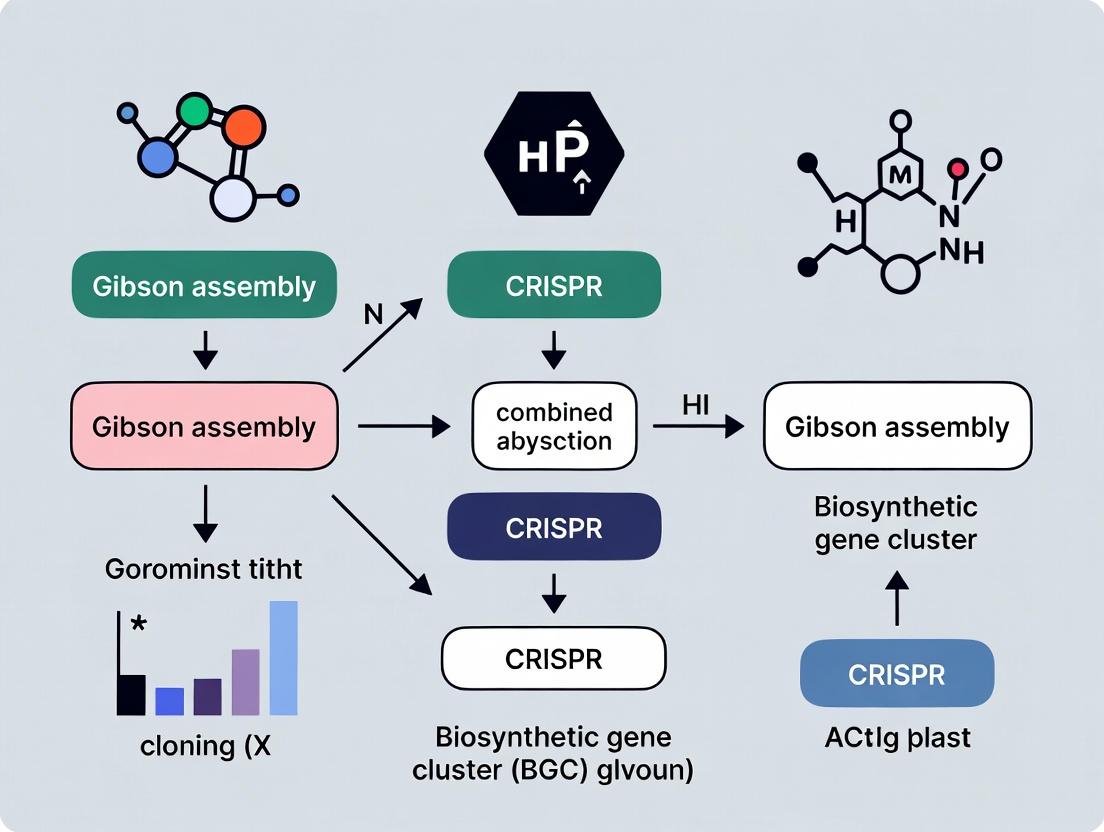
CRISPR-Gibson Assembly: A Powerful Synergy for Biosynthetic Gene Cluster Cloning and Drug Discovery
This article provides a comprehensive guide for researchers and drug development professionals on the synergistic combination of Gibson assembly and CRISPR-Cas technologies for the targeted cloning of biosynthetic gene clusters...
CRISPR-Gibson Assembly: A Powerful Synergy for Biosynthetic Gene Cluster Cloning and Drug Discovery
Abstract
This article provides a comprehensive guide for researchers and drug development professionals on the synergistic combination of Gibson assembly and CRISPR-Cas technologies for the targeted cloning of biosynthetic gene clusters (BGCs). We explore the foundational principles of each technology, detail step-by-step methodological workflows for precise BGC excision and assembly, address common troubleshooting and optimization strategies, and validate the approach through comparisons with traditional methods. This integrated technique accelerates the discovery and engineering of novel natural products for biomedical applications.
Understanding the Synergy: The Foundational Principles of CRISPR and Gibson Assembly for BGCs
Introduction to Biosynthetic Gene Clusters (BGCs) and Their Role in Natural Product Discovery
Application Notes: BGCs in Modern Drug Discovery
Biosynthetic Gene Clusters (BGCs) are sets of co-localized genes in microbial genomes that orchestrate the production of a secondary metabolite or natural product. These compounds are a primary source of bioactive molecules, forming the basis for many antibiotics, antifungals, anticancer agents, and immunosuppressants. The conventional approach of activating silent BGCs in native hosts is often inefficient. The integration of Gibson assembly for precise, multi-fragment DNA cloning with CRISPR-Cas systems for targeted genome editing represents a transformative strategy for BGC refactoring and heterologous expression, accelerating the discovery pipeline.
Table 1: Impact of Major Natural Product Classes Derived from BGCs
| Natural Product Class | Example Drug | BGC Type (e.g., PKS, NRPS) | Primary Therapeutic Use |
|---|---|---|---|
| Polyketides | Erythromycin | Type I PKS | Antibiotic |
| Nonribosomal Peptides | Penicillin | NRPS | Antibiotic |
| Hybrid (PKS-NRPS) | Rapamycin | Type I PKS/NRPS | Immunosuppressant |
| Terpenes | Artemisinin | Terpene Synthase | Antimalarial |
| Ribosomally synthesized and post-translationally modified peptides (RiPPs) | Nisin | LanB/LanC | Antimicrobial (Food Preservative) |
Protocols for BGC Cloning and Engineering
The following protocol details a core methodology for capturing and refactoring BGCs using CRISPR-Cas9 coupled with Gibson assembly, suitable for expression in a heterologous host like Streptomyces coelicolor or Aspergillus nidulans.
Protocol: CRISPR-Cas9 Mediated BGC Capture and Gibson Assembly Refactoring Objective: To excise a target BGC from a genomic DNA (gDNA) source and clone it into a refactored expression vector.
Materials (Research Reagent Solutions):
- Reagent/Tool: CRISPR-Cas9 System (SpCas9). Function: Creates double-strand breaks at specific sites flanking the BGC for precise excision.
- Reagent/Tool: Gibson Assembly Master Mix. Function: One-step, isothermal assembly of multiple linear DNA fragments with homologous overlaps.
- Reagent/Tool: BGC-Specific sgRNAs. Function: Guides Cas9 to the precise genomic loci defining the BGC boundaries.
- Reagent/Tool: Refactored Expression Vector (e.g., pCAP01). Function: Heterologous expression platform containing strong promoters, terminators, and selectable markers optimized for the host.
- Reagent/Tool: Yeast Artificial Chromosome (YAC) or Bacterial Artificial Chromosome (BAC). Function: Maintains large (>50 kb) BGC inserts in a surrogate host pre-assembly.
- Reagent/Tool: Phusion High-Fidelity DNA Polymerase. Function: Amplifies BGC fragments and vector backbones with high accuracy for assembly.
Procedure: Part A: BGC Excision and Capture
- Design & Synthesis: Design two sgRNAs targeting sequences immediately upstream and downstream of the BGC. Synthesize these in vitro.
- In Vitro Digestion: Set up a reaction containing purified source gDNA (1 µg), SpCas9 enzyme (10 units), and the two sgRNAs (each 50 nM) in the provided buffer. Incubate at 37°C for 2 hours.
- Size Selection: Run the digest on a low-melting-point agarose gel. Excise the gel slice corresponding to the expected size of the excised BGC fragment. Purify the DNA.
- Capture: Using Gibson Assembly, clone the purified fragment into a linearized YAC/BAC vector. Transform into yeast or E. coli and screen for correct clones.
Part B: BGC Refactoring and Assembly
- Refactoring PCR: Amplify the captured BGC from the YAC/BAC as 3-5 overlapping sub-fragments (each 10-15 kb). Simultaneously, amplify the desired strong promoters and terminators from template plasmids.
- Vector Preparation: Linearize the destination expression vector (e.g., pCAP01) by PCR or restriction digest.
- Gibson Assembly: Combine ~100 ng of linearized vector, equimolar amounts of each BGC sub-fragment and refactoring part (promoters/terminators), and Gibson Assembly Master Mix in a 20 µL total volume. Incubate at 50°C for 60 minutes.
- Transformation & Screening: Transform the assembly reaction into capable E. coli cells. Screen colonies by PCR and analyze positive clones by restriction digest and sequencing (e.g., PacBio long-read) to confirm correct assembly.
Visualizations
Diagram 1: BGC Discovery & Engineering Workflow
Diagram 2: CRISPR-Gibson BGC Refactoring
Within the advanced framework of biosynthetic gene cluster (BGC) cloning and engineering, the fusion of Gibson Assembly with CRISPR-based technologies has emerged as a transformative strategy. This synergy accelerates the assembly and refactoring of large, complex pathways by enabling precise, scarless, and multi-fragment cloning coupled with targeted genomic modifications. This application note details the mechanism, protocol, and resources for employing Gibson Assembly in this critical research context.
Mechanism and Key Enzymes
Gibson Assembly is a single-tube, isothermal method that utilizes three synergistic enzymatic activities to assemble multiple overlapping DNA fragments in one step.
- 5' Exonuclease: Selectively chews back 5' ends to generate single-stranded 3' overhangs. This allows complementary overlaps between fragments to anneal.
- DNA Polymerase: Fills in gaps within the annealed region using the dNTPs provided.
- DNA Ligase: Seals the nicks in the assembled DNA backbone, creating a covalently closed, double-stranded molecule.
The core innovation lies in the coordinated activity of these enzymes at a single optimal temperature (typically 50°C), enabling simultaneous overlap generation, annealing, gap filling, and ligation.
Advantages for Multi-Fragment Cloning
| Advantage | Quantitative/Qualitative Benefit in BGC Cloning |
|---|---|
| Speed & Efficiency | Assembly of 5-10 fragments in 1 hour with >90% efficiency common. |
| Seamlessness | Creates scarless junctions, critical for maintaining open reading frames in operons. |
| High-Fidelity | Uses high-fidelity polymerase, preserving complex BGC sequences. |
| Modularity | Ideal for combinatorial library generation by swapping modular parts. |
| Scalability | Can assemble very large constructs (e.g., 50+ kb BGCs) from multiple sub-fragments. |
| CRISPR Compatibility | Assembled donors pair with CRISPR/Cas9 for precise chromosomal integration. |
Research Reagent Solutions Toolkit
| Reagent/Solution | Function in Gibson Assembly/CRISPR for BGCs |
|---|---|
| Gibson Assembly Master Mix | Pre-mixed, optimized blend of the three key enzymes (exonuclease, polymerase, ligase) and reaction buffer. |
| High-Fidelity DNA Polymerase | For PCR amplification of fragments with minimal error; essential for large BGC fragment prep. |
| T5 Exonuclease | The specific 5' exonuclease used in the standard Gibson Assembly method. |
| Phusion or Q5 Polymerase | Industry-standard high-fidelity polymerases for insert and vector preparation. |
| RecA-deficient E. coli Strains | (e.g., DH5α, Stbl3) Prevent unwanted recombination of repetitive sequences common in BGCs. |
| Cas9 Nuclease & sgRNA | For generating targeted double-strand breaks in the host genome for BGC integration. |
| Homology-Directed Repair (HDR) Donor | The Gibson-assembled linear DNA fragment containing the BGC flanked by homology arms. |
Detailed Protocol: Gibson Assembly for CRISPR-mediated BGC Integration
Step 1: Fragment Design and Preparation
- Design: Design all fragments (BGC modules, vector backbone) with 20-40 bp homologous overlaps to adjacent fragments. For CRISPR integration, include 500-1000 bp homology arms targeting the genomic locus.
- PCR Amplify: Amplify each fragment using a high-fidelity polymerase. Purify via spin column or gel extraction.
Step 2: Assembly Reaction
- Setup: Combine 50-100 ng of linearized vector with equimolar amounts of each insert fragment. A typical starting point is a 2:1 insert:vector molar ratio for multi-fragment assemblies.
- Master Mix: Add equal volume of 2x Gibson Assembly Master Mix. Total reaction volume: 10-20 µL.
- Incubate: Incubate in a thermal cycler at 50°C for 15-60 minutes (15 min for 2-3 fragments, 60 min for >5 fragments).
Typical Reaction Setup Table:
| Component | Volume (for 10 µL reaction) | Final Amount |
|---|---|---|
| Linearized Vector | x µL | 50-100 ng |
| Insert Fragment 1 | y µL | Equimolar to vector* |
| Insert Fragment N | z µL | Equimolar to vector* |
| 2x Gibson Master Mix | 5 µL | 1x |
| Nuclease-free Water | To 10 µL | - |
Note: Use an online molar ratio calculator to determine volumes.
Step 3: Transformation and Screening
- Transform: Dilute reaction 2-5x with water or buffer. Transform 2-5 µL into competent E. coli. Recover cells and plate on appropriate antibiotic selection.
- Screen: Screen colonies by colony PCR or restriction digest. Sequence confirmed constructs for downstream CRISPR steps.
Step 4: CRISPR/Cas9 Integration (Example Workflow)
- Co-transform or electroporate the Gibson-assembled BGC plasmid (HDR donor) along with a Cas9-sgRNA expression plasmid targeting the desired genomic locus into the host strain.
- Select for clones where successful homologous recombination has integrated the BGC.
- Validate integration via junction PCR and phenotypic screening (e.g., metabolite production).
Visual Workflows
Diagram Title: Gibson Assembly and CRISPR Workflow for BGC Cloning
Diagram Title: Gibson Assembly Enzyme Mechanism
CRISPR-Cas systems, particularly Cas9 and Cas12, have revolutionized functional genomics and metabolic engineering. Within the context of a broader thesis on Gibson assembly combined with CRISPR for Biosynthetic Gene Cluster (BGC) cloning, these nucleases serve as precision tools for the targeted excision of large genomic regions. This facilitates the capture and heterologous expression of BGCs in tractable host organisms for natural product discovery and drug development.
Key Application Notes:
- Cas9: A dual-RNA guided nuclease producing blunt-ended double-strand breaks (DSBs). Ideal for precise, targeted excision when paired with two sgRNAs flanking a BGC. Its high fidelity and efficiency make it suitable for complex genomic operations in Actinomycetes and fungi.
- Cas12a (Cpf1): A single-RNA guided nuclease producing staggered, 5'-overhang DSBs. Its simpler ribonucleoprotein complex and ability to process its own crRNA arrays are advantageous for multiplexed excision strategies. The staggered ends can be designed to be compatible with Gibson assembly overhangs.
- Integration with Gibson Assembly: The DSBs generated by Cas9 or Cas12a can be repaired via homology-directed repair (HDR) using a linear cloning vector assembled via Gibson assembly. This vector contains homology arms (HA) matching the sequences flanking the excised BGC, enabling seamless capture and circularization.
Quantitative Comparison of Cas9 and Cas12a for Genomic Excision
Table 1: Functional Comparison of Cas9 and Cas12a Nucleases
| Feature | Cas9 (SpCas9) | Cas12a (LbCas12a) |
|---|---|---|
| Guide RNA | Dual-tracrRNA:crRNA or chimeric sgRNA | Single crRNA |
| PAM Sequence | 5'-NGG-3' (SpCas9) | 5'-TTTV-3' (LbCas12a) |
| Cleavage Type | Blunt-ended DSB | Staggered DSB (5' overhangs) |
| Cleavage Site | 3 bp upstream of PAM | Distal to PAM, 18-23 bp apart |
| RNA Processing | No inherent activity; requires pre-processed RNA | Self-processes pre-crRNA arrays |
| Size (aa) | ~1368 | ~1228 |
| Typical Excision Efficiency* | 65-85% (in model Actinomycetes) | 45-75% (in model Actinomycetes) |
| Key Advantage for BGC Cloning | High efficiency, well-characterized | Simplified multiplexing, staggered ends for direct cloning |
*Efficiency depends on host organism, delivery method, and target locus.
Table 2: Key Parameters for CRISPR-Cas Mediated BGC Excision & Cloning
| Parameter | Typical Range or Value | Protocol Section |
|---|---|---|
| Homology Arm Length (for HDR) | 500 - 2000 bp | 3.1 |
| Gibson Assembly Overlap Length | 20 - 40 bp | 3.2 |
| BGC Size Limit for Efficient Excision | Up to 150 kbp (varies by system) | 3.3 |
| Typical Transformation Efficiency Required | >10⁵ CFU/µg DNA (for screening) | 3.4 |
| Recommended Screening Method | PCR & Antibiotic Selection | 3.5 |
Detailed Experimental Protocols
Protocol 3.1: Design and Synthesis of CRISPR RNA Guides and HDR Template
Objective: To create components for the targeted excision of a BGC and its capture via a cloning vector.
- Identify Flanking Regions: Using genomic sequence data, identify unique 20-23 bp target sequences immediately outside the BGC boundaries. Ensure the presence of a compatible PAM (NGG for SpCas9, TTTV for LbCas12a).
- Design sg/crRNAs: Design two guides (Guide A, Guide B) targeting opposite strands upstream and downstream of the BGC. Use tools like CHOPCHOP or Benchling. Order as synthetic DNA oligos with appropriate promoter overhangs (e.g., for T7 polymerase).
- Generate HDR Template (Gibson Assembly Vector):
- Design homology arms (HA-L and HA-R) as 500-2000 bp sequences identical to the regions just outside the Guide A and Guide B cut sites.
- Design a linear cloning vector backbone (containing an origin of replication and selection marker) with 20-40 bp overlaps complementary to the ends of the homology arms.
- Assemble the vector via Gibson Assembly: Mix 100 ng of each PCR-amplified fragment (HA-L, Backbone, HA-R) with 2x Gibson Assembly Master Mix. Incubate at 50°C for 15-60 minutes. Transform into competent E. coli, isolate plasmid, and sequence-verify.
Protocol 3.2: Delivery of CRISPR-Cas Components and Excision in Streptomyces
Objective: To introduce CRISPR-Cas components into the BGC host and isolate clones with the excised BGC captured on an episomal vector.
- Prepare RNP Complexes (for Cas9): For each guide, combine 10 pmol of purified Cas9 protein with 30 pmol of synthetic sgRNA in NEBuffer 3.1. Incubate at 25°C for 10 minutes.
- Protoplast Preparation & Transformation:
- Grow the host Streptomyces to mid-exponential phase in liquid culture with 0.5% glycine.
- Harvest mycelia, wash, and digest with lysozyme (1 mg/mL) in osmotically stabilized P buffer for 60 minutes at 30°C.
- Filter through sterile cotton, pellet protoplasts gently, and wash twice with P buffer.
- Co-transformation:
- Resuspend ~10⁹ protoplasts in 500 µL P buffer.
- Add 10 µL of pre-formed RNP complex (for each guide) OR 2 µg of plasmid DNA expressing Cas12a and crRNAs.
- Add 2 µg of the linear HDR template (Gibson-assembled vector from 3.1).
- Add 500 µL of 50% PEG 6000, mix gently, and incubate for 2 minutes.
- Plate on osmotically stabilized R2YE plates. Overlay with soft agar containing appropriate antibiotics (e.g., apramycin for selection of the captured BGC vector) after 12-16 hours of recovery.
Protocol 3.3: Screening and Validation of Excision Clones
Objective: To confirm successful BGC excision and circularization into the cloning vector.
- Primary Colony PCR: Pick 20-50 antibiotic-resistant colonies. Using primers binding within the vector backbone and within the BGC, perform PCR to verify the presence of junction fragments.
- Plasmid Rescue: Isolate plasmid from PCR-positive clones via alkaline lysis miniprep.
- Restriction Analysis & Sequencing: Digest the rescued plasmid with 1-2 restriction enzymes (e.g., HindIII, EcoRI) and analyze by gel electrophoresis against the native genomic DNA. Confirm the structure by long-read sequencing (e.g., Nanopore, PacBio).
Visualizations
Diagram 1: CRISPR-Gibson BGC Cloning Workflow (86 chars)
Diagram 2: Cas9 vs Cas12a Cleavage Mechanism (47 chars)
The Scientist's Toolkit: Key Research Reagent Solutions
Table 3: Essential Materials for CRISPR-Cas BGC Excision
| Reagent/Material | Function & Description | Example Vendor/Cat. No. (if common) |
|---|---|---|
| High-Fidelity DNA Polymerase | PCR amplification of homology arms and vector fragments with low error rates. | NEB Q5, Thermo Fisher Phusion |
| Gibson Assembly Master Mix | Enzymatic mix for seamless, one-pot assembly of linear DNA fragments. | NEB Gibson Assembly, In-Fusion Snap Assembly |
| Purified Cas9 Nuclease | Recombinant protein for forming Ribonucleoprotein (RNP) complexes for delivery. | IDT Alt-R S.p. Cas9 Nuclease |
| Synthetic sgRNA/crRNA | Chemically synthesized guide RNAs for high-efficiency targeting. | Synthego, IDT Alt-R CRISPR RNA |
| Cas12a Expression Plasmid | Plasmid for in vivo expression of Cas12a and crRNA arrays in the host. | Addgene (#69982) |
| Osmotically Stabilized Media (P Buffer, R2YE) | Essential for protoplast formation, transformation, and regeneration in Streptomyces. | Prepare in-house per standard protocols. |
| Polyethylene Glycol (PEG) 6000 | Facilitates DNA uptake during protoplast transformation. | Sigma-Aldrich 81240 |
| Antibiotics for Selection | Select for the cloning vector and counter-select against the parent genome. | Apramycin, Thiostrepton, Hygromycin |
| Long-Read Sequencing Service | Validate the structure of large, excised BGC plasmids. | Oxford Nanopore, PacBio |
The targeted capture of Biosynthetic Gene Clusters (BGCs) from complex genomic DNA remains a bottleneck in natural product discovery. This application note details a transformative methodology combining CRISPR-Cas9-mediated precise excision with Gibson Assembly for seamless, scarless, and high-throughput cloning of large BGCs (>10 kb). Framed within a thesis on advanced DNA assembly techniques, this protocol enables researchers to directly clone BGCs into expression-ready vectors in a single, isothermal reaction, dramatically accelerating the pipeline from genome mining to compound production.
Traditional methods for BGC capture, such as cosmids, BAC libraries, or PCR-based approaches, are often labor-intensive, size-limited, or prone to errors. The integration of CRISPR-guided excision provides single-nucleotide precision in defining BGC boundaries, while Gibson Assembly (isothermal, 50°C) offers a highly efficient, multi-fragment assembly system. This combination bypasses the need for restriction sites, allows for in vitro assembly free from host recombination machinery, and facilitates the direct construction of expression vectors in a single step.
Key Advantages & Quantitative Data
Table 1: Performance Comparison of BGC Capture Methods
| Method | Typical Max Insert Size (kb) | Throughput | Precision (Boundary Control) | Hands-on Time | Success Rate (%) |
|---|---|---|---|---|---|
| Cosmid/BAC Library & Screening | 30-40 | Low | Low | Weeks | 60-80 |
| PCR + Yeast Recombination | < 15 | Medium | High | Days | 30-60 |
| TAR Capture in Yeast | > 100 | Low | Medium | Weeks | 20-50 |
| CRISPR/Gibson (This Protocol) | 10-50 | High | Single-Base | 1-2 Days | > 85 |
Table 2: Representative Gibson Assembly Reaction Efficiency for BGC Constructs
| BGC Size (kb) | Vector Backbone (kb) | Total Assembly Length (kb) | Transformation Efficiency (CFU/µg) | Correct Assembly Verification Rate (%) |
|---|---|---|---|---|
| 12 | 8 | 20 | 3.5 x 10⁴ | 92 |
| 25 | 8 | 33 | 8.2 x 10³ | 87 |
| 40 | 8 | 48 | 1.1 x 10³ | 78 |
Detailed Protocol: CRISPR Excision & Gibson Assembly for BGC Capture
Part I: CRISPR-Cas9 Design and Excision of BGC from Genomic DNA
Objective: Generate linear DNA fragments containing the target BGC with precise, overlapping ends compatible with Gibson Assembly.
- Design sgRNAs: Using bioinformatics (e.g., antiSMASH), design two sgRNAs that flank the BGC. Target sites should be as close as possible to the cluster boundaries to minimize extraneous DNA.
- In vitro Cas9 Cleavage Reaction:
- Reagents:
- Genomic DNA (100-500 ng/µL)
- Cas9 Nuclease (10 µM)
- Synthesized sgRNAs (10 µM each)
- 10X Cas9 Reaction Buffer
- Protocol:
- Set up a 50 µL reaction: 5 µL 10X Buffer, 1 µg gDNA, 1.5 µL Cas9, 1.5 µL of each sgRNA (final 300 nM each), nuclease-free water.
- Incubate at 37°C for 2 hours.
- Run the reaction on a low-melting point agarose gel (0.8%). Excise the gel slice containing the linear BGC fragment.
- Purify DNA using a gel extraction kit. Elute in 20 µL nuclease-free water. This is Fragment A.
- Reagents:
Part II: Vector Preparation via CRISPR-Cas9 or PCR
Objective: Generate a linearized vector with ends homologous to the termini of Fragment A.
- Linearize Expression Vector:
- Option A (CRISPR): Perform a Cas9 cleavage on the circular vector plasmid using a single sgRNA targeting within the multiple cloning site. Purify the linear backbone.
- Option B (PCR): Amplify the entire vector backbone using primers whose 5' ends contain 40-bp homology arms matching the ends of the BGC fragment (Fragment A).
- Purify the linear vector backbone (Fragment B) using a PCR purification kit.
Part III: Gibson Assembly for Seamless BGC Integration
Objective: Assemble the BGC fragment into the linearized vector in a single, isothermal reaction.
- Gibson Assembly Master Mix (2X) Preparation (can be commercially sourced):
- T5 Exonuclease (0.04 U/µL)
- Phusion DNA Polymerase (0.05 U/µL)
- Taq DNA Ligase (0.125 U/µL)
- dNTPs (0.5 mM each)
- PEG-8000 (5% w/v)
- Tris-HCl, pH 7.5 (50 mM)
- MgCl₂ (10 mM)
- DTT (1 mM)
- NAD (0.2 mM)
- Assembly Reaction:
- Mix on ice: 10 µL 2X Gibson Master Mix, 20-100 ng Fragment A (BGC), 50-100 ng Fragment B (vector). Adjust total volume to 20 µL with nuclease-free water.
- Incubate at 50°C for 60 minutes.
- Transformation and Screening:
- Transform 2-5 µL of the assembly reaction into competent E. coli (e.g., DH10B).
- Plate on appropriate antibiotic plates.
- Screen colonies by colony PCR or diagnostic digest. For large constructs, verify by long-read sequencing (e.g., Nanopore, PacBio).
The Scientist's Toolkit: Research Reagent Solutions
Table 3: Essential Materials and Reagents
| Item | Function & Critical Feature | Example Product/Type |
|---|---|---|
| High-Fidelity Polymerase | Amplify vector backbone with long homology arms; minimal error rate. | Phusion U Green, Q5 High-Fidelity |
| Cas9 Nuclease (Wild-type) | Generates precise double-strand breaks at BGC boundaries guided by sgRNAs. | Alt-R S.p. Cas9 Nuclease |
| In vitro-transcribed or synthetic sgRNA | Guides Cas9 to specific genomic loci for excision. | Alt-R CRISPR-Cas9 sgRNA |
| Gibson Assembly Master Mix | All-in-one enzymatic mix for seamless, isothermal assembly. | NEBuilder HiFi DNA Assembly Master Mix |
| Low-Melt Agarose | For gentle recovery of large, fragile DNA fragments post-CRISPR excision. | SeaPlaque GTG Agarose |
| Electrocompetent E. coli | High-efficiency transformation of large, complex plasmid constructs (>40 kb). | ElectroTen-Blue Electrocompetent Cells |
| Positive Selection Vector | Backbone with antibiotic resistance and inducible promoter for heterologous expression. | pET-based, pCAP-based vectors |
Workflow and Pathway Visualizations
Title: CRISPR-Gibson BGC Capture Workflow
Title: Gibson Assembly Enzymatic Mechanism
Key Applications in Pharmaceutical Research and Synthetic Biology
This application note details the synergistic use of Gibson Assembly and CRISPR-Cas9 for the cloning and engineering of Bacterial Genomic Clusters (BGCs), a cornerstone of modern pharmaceutical discovery. BGCs encode pathways for a vast array of bioactive natural products, including antibiotics, antifungals, and anticancer agents. The combination of seamless DNA assembly and precise genome editing accelerates the refactoring, heterologous expression, and optimization of these valuable genetic loci for drug development and synthetic biology.
Application Notes
Targeted Capture and Assembly of Large BGCs
The primary challenge in BGC research is the capture of large (often >50 kb), high-GC content sequences from complex genomic DNA. Traditional methods are inefficient. Our integrated protocol uses CRISPR-Cas9 to generate specific double-strand breaks flanking the target BGC in situ, followed by Gibson Assembly to seamlessly clone the excised fragment into a replicative vector in a single, isothermal reaction.
Table 1: Comparison of BGC Cloning Methods
| Method | Typical Max Insert Size (kb) | Efficiency (%) | Hands-on Time (hrs) | Primary Use Case |
|---|---|---|---|---|
| Traditional PCR & Ligation | < 10 | 5-20 | 8-12 | Small gene clusters, subcloning |
| Fosmid/Cosmid Libraries | 30-40 | Varies (library-dependent) | 24+ (screening) | Untargeted library construction |
| CRISPR-Cas9 Excision + Gibson Assembly | > 100 | 60-85 | 6-8 | Targeted capture of large BGCs |
| Transformation-Associated Recombination (TAR) | > 100 | 30-70 | 10-14 | Yeast-based assembly of very large clusters |
Refactoring BGCs for Heterologous Expression
Silent or poorly expressed BGCs in native hosts can be activated by refactoring—replacing native regulatory elements with standardized synthetic parts. CRISPR-Cas9 facilitates the precise deletion of native promoters and terminators, while Gibson Assembly enables the high-throughput insertion of synthetic biological parts (e.g., constitutive promoters, RBSs) to optimize expression in industrial chassis like Streptomyces coelicolor or Pseudomonas putida.
Table 2: Key Performance Metrics in BGC Refactoring
| Parameter | Pre-Refactoring Titer (mg/L) | Post-Refactoring Titer (mg/L) | Fold Increase | Chassis Organism |
|---|---|---|---|---|
| Antibiotic A (Nonribosomal Peptide) | 0.5 | 15.2 | 30.4 | S. coelicolor M1152 |
| Anticancer Compound B (Polyketide) | Undetectable | 8.7 | N/A | P. putida KT2440 |
| Antifungal C (Terpene) | 1.2 | 22.1 | 18.4 | S. albus J1074 |
Protocols
Protocol 1: CRISPR-Cas9-Mediated Excision of a BGC from Genomic DNA
Objective: Generate linear vector and target BGC fragment with homologous ends for subsequent Gibson Assembly. Materials:
- Bacterial genomic DNA (gDNA) containing target BGC.
- pCRISPR-Cas9-sgRNA plasmid (Addgene #62655) or equivalent.
- High-fidelity PCR enzymes (e.g., Q5 Hot Start).
- T7 Endonuclease I for validation.
Procedure:
- Design sgRNAs: Using bioinformatics tools (e.g., Benchling), design two sgRNAs targeting sequences immediately upstream and downstream of the BGC. Ensure minimal off-targets.
- Clone sgRNAs: Clone each sgRNA sequence into the pCRISPR plasmid. Transform into an appropriate E. coli strain.
- Delivery and Excision: Introduce the two pCRISPR plasmids (or a single plasmid expressing both sgRNAs) into the native BGC host strain via conjugation or electroporation.
- Validate Excision: Isolve genomic DNA from exconjugants. Perform PCR across the new junction created by the double-strand break repair. Confirm by pulsed-field gel electrophoresis for large fragments.
- Fragment Recovery: Amplify the excised linear BGC fragment using primers that add 40-bp overlaps homologous to the destination vector.
Protocol 2: Gibson Assembly for BGC Cloning and Refactoring
Objective: Assemble the excised/amplified BGC fragment into a pre-digested shuttle vector (e.g., pRSF1010-based) in a single reaction. Materials:
- Gibson Assembly Master Mix (commercial, e.g., NEB HiFi DNA Assembly Mix, or prepared in-house).
- Linearized vector backbone (200 ng).
- Purified BGC insert fragment(s) (at a 2:1 or 3:1 molar ratio of insert:vector).
- Chemically competent E. coli (e.g., NEB Stable).
Procedure:
- Prepare Vector: Linearize the destination vector by restriction enzyme digestion or inverse PCR. Gel-purify.
- Set Up Assembly Reaction: Combine in a thin-walled PCR tube:
- 100-200 ng linearized vector
- BGC insert(s) (calculated molar excess)
- 1X Gibson Assembly Master Mix
- Total volume: 20 µL
- Incubate: Place reaction in a thermal cycler at 50°C for 15-60 minutes (15 min for assemblies <20 kb; 60 min for >50 kb).
- Transform: Add 2-5 µL of the assembly reaction to 50 µL of competent E. coli. Recover, plate on selective media, and incubate.
- Screen Colonies: Screen via colony PCR using check primers spanning assembly junctions. Confirm positive clones by restriction digest and Sanger sequencing of junctions.
Diagrams
Title: CRISPR-Gibson BGC Cloning Workflow
Title: Modular Polyketide Synthase Pathway
The Scientist's Toolkit
Table 3: Essential Research Reagent Solutions for CRISPR-Gibson BGC Cloning
| Item | Function | Example Product/Supplier |
|---|---|---|
| High-Fidelity DNA Polymerase | Error-free PCR amplification of BGC fragments and vector backbones. | Q5 Hot Start High-Fidelity DNA Polymerase (NEB) |
| Gibson Assembly Master Mix | One-step, isothermal assembly of multiple DNA fragments with homologous ends. | NEBuilder HiFi DNA Assembly Master Mix (NEB) |
| CRISPR-Cas9 Plasmid System | For delivery of Cas9 nuclease and customizable sgRNA to target cells. | pCRISPR-Cas9-sgRNA (Addgene) |
| Shuttle Vector with Selectable Markers | Replicates in both E. coli and the heterologous expression host. | pRSF1010-derivative (AmpR/KanR, oriT for conjugation) |
| Chemically Competent Cells (E. coli) | Efficient transformation of large, complex plasmid assemblies. | NEB Stable Competent E. coli |
| Conjugation Donor Strain | Enables transfer of assembled constructs from E. coli to Actinomycetes. | E. coli ET12567/pUZ8002 |
| Antibiotics for Selection | Selective pressure for maintaining plasmids and engineered constructs. | Apramycin, Kanamycin, Chloramphenicol |
| DNA Purification Kits (Gel & PCR) | Critical for obtaining high-purity fragments for assembly. | Zymoclean Gel DNA Recovery Kit (Zymo Research) |
Step-by-Step Protocol: A Detailed Workflow for CRISPR-Gibson BGC Cloning
This Application Note details the first critical step in a methodology for cloning Bacterial Biosynthetic Gene Clusters (BGCs) via a combined CRISPR-Gibson Assembly pipeline. Precise excision of large genomic regions requires the design of highly specific single guide RNAs (sgRNAs) targeting the flanks of the BGC. In silico design minimizes off-target effects and ensures compatibility with downstream enzymatic processing and assembly.
Key Design Parameters and Quantitative Data
The success of CRISPR-mediated excision hinges on optimizing the following parameters during sgRNA design.
Table 1: Key Parameters for In Silico sgRNA Design Targeting BGC Flanks
| Parameter | Optimal Value / Feature | Rationale & Impact on Efficiency |
|---|---|---|
| Target Location | Within 500 bp upstream of start codon (5' flank) and downstream of stop codon (3' flank). | Ensures sufficient homologous overlap for Gibson Assembly while maintaining functional integrity of cluster genes. |
| sgRNA Length | 20-nt spacer sequence (NGG PAM not part of spacer). | Standard length for SpCas9 binding and cleavage specificity. |
| Protospacer Adjacent Motif (PAM) | 5'-NGG-3' (for SpCas9). | Mandatory sequence for Cas9 recognition. Must be present on the genomic target strand. |
| GC Content | 40-60%. | Influences sgRNA stability and binding efficiency. <20% or >80% can reduce activity. |
| On-Target Efficiency Score | >50 (per Doench et al., 2016 algorithm). | Predictive score for guide knockout efficiency. Higher score correlates with higher success probability. |
| Off-Target Potential | Zero off-target sites with ≤3 mismatches. | Critical for precise cutting. Mismatches in the "seed" region (PAM-proximal 8-12 bp) are most disruptive. |
| Self-Complementarity | Minimal hairpin formation (low risk of secondary structure). | Prevents sgRNA folding that would impede Cas9 binding. |
| Genomic Context | Avoids repetitive elements, high polymorphism regions. | Ensures specificity and reproducibility across strains. |
Table 2: Comparison of Common sgRNA Design Tools (2024 Update)
| Tool | Access | Key Algorithm Features | Best For | Outputs |
|---|---|---|---|---|
| CHOPCHOP | Web, standalone | Efficiency & specificity scoring, visualizes in JBrowse. | Broad organisms, gene editing & BGC targeting. | Ranked sgRNAs, primer suggestions, off-target lists. |
| Benchling | Web/Cloud | Integrated with molecular biology suite, custom genomes. | Collaborative, end-to-end workflow design. | Efficiency scores, detailed off-target analysis. |
| CRISPick (Broad) | Web | Rule Set 2 scoring (Doench et al.), excellent off-target search. | Rigorous, publication-grade design for human/mouse, adaptable for microbes. | Ranked list, off-target summary with mismatch details. |
| CRISPRscan | Web | Model trained on zebrafish, good for non-model organisms. | Designing sgRNAs for less-characterized microbial genomes. | Efficiency score, predicted activity. |
| Cas-Designer | Standalone | Detailed off-target analysis with bulges. | Deep dive into potential off-target effects. | Comprehensive off-target report. |
Detailed Protocol:In SilicosgRNA Design for BGC Flanks
Protocol 3.1: Target Identification and sgRNA Selection
Objective: To identify and rank 2-4 candidate sgRNAs for each flank (5' and 3') of the target BGC.
Materials & Reagents:
- Genomic Sequence: FASTA file of the host bacterial genome.
- BGC Coordinates: Defined start and end points of the target gene cluster.
- sgRNA Design Software: Access to one or more tools from Table 2 (e.g., CHOPCHOP).
Procedure:
- Define Flank Regions: Extract 1-2 kb sequences immediately upstream (5' flank) and downstream (3' flank) of the BGC boundaries. These are your target regions for sgRNA design.
- Input Sequences: Upload the two flank region FASTA files to your chosen sgRNA design tool (e.g., CHOPCHOP).
- Parameter Setting:
- Select Streptococcus pyogenes Cas9 (SpCas9).
- Set PAM sequence to
NGG. - Set guide length to 20 nt.
- Enable thorough off-target search (allow 2-3 mismatches).
- Request efficiency scores (e.g., CHOPCHOP score, Doench 2016 score).
- Generate & Filter Candidates: Run the tool. Filter the resulting sgRNA list by:
- On-target efficiency score: Select guides with the highest scores (top quartile).
- Off-targets: Prioritize guides with ZERO predicted off-target sites with ≤2 mismatches. Reject any guide with a perfect match off-target elsewhere in the genome.
- Genomic Context: Avoid guides overlapping repetitive sequences.
- GC Content: Select guides within the 40-60% range.
- Final Selection: For each flank, choose the top 2 ranked sgRNAs that pass all filters. This provides redundancy.
Protocol 3.2: Validation of Specificity and Homology Overlap
Objective: To confirm sgRNA specificity and define the final homology arms for Gibson Assembly.
Procedure:
- Manual BLASTn Verification:
- Take the 20-nt spacer sequence of each selected sgRNA.
- Perform a BLASTn search against the entire host genome (not just the flank).
- Confirm the only perfect match is at the intended target site. Note any near-matches (>17/20 identity) and assess their location.
- Define Gibson Assembly Homology Arms:
- The Cas9 cut site is typically 3 bp upstream of the PAM on the targeted strand.
- For each flank, the ~500-800 bp region between the cut site and the BGC will serve as the homology arm for Gibson Assembly.
- Record the exact 5' and 3' cut site coordinates for primer design in the next step (Step 2 of the overall pipeline).
Visualization
Diagram Title: Workflow for In Silico sgRNA Design & Toolkit
The Scientist's Toolkit
Table 3: Essential Research Reagents & Tools for In Silico Design Phase
| Item | Vendor Examples | Function in This Step |
|---|---|---|
| Genome Analysis Software | SnapGene, Geneious, CLC Workbench | Visualize BGC genomic context, extract flank sequences, and manage coordinate data. |
| sgRNA Design Platform | CHOPCHOP, Benchling, CRISPick | Automate candidate identification, efficiency scoring, and initial off-target screening. |
| BLASTn Tool | NCBI BLAST, NEBridge BLAST | Final, rigorous verification of sgRNA spacer specificity against the full genome. |
| Sequence Database | NCBI GenBank, Patric, AntiSMASH | Source accurate genomic sequence for the host organism and BGC boundary information. |
| High-Fidelity SpCas9 (Reference) | IDT Alt-R S.p. Cas9 V3, NEB HiFi Cas9 | The nuclease for which guides are designed; knowledge of its specific PAM and cleavage profile is essential. |
| Oligo Synthesis Service | IDT, Twist Bioscience, Eurofins | For ordering synthesized sgRNA templates or cloning oligos based on the final in silico designs. |
This application note details the critical step of delivering CRISPR-Cas components into the native host strain of a biosynthetic gene cluster (BGC) producer. Within the broader thesis framework utilizing Gibson Assembly for vector construction, this step enables precise genomic modifications—such as cluster deletion, activation, or tagging—directly in the native genomic context. Direct manipulation circumvents heterologous expression challenges, preserving native regulation and physiology essential for studying BGC function and activating silent clusters.
Key Considerations for Delivery
Successful delivery hinges on the host strain's inherent properties and the chosen CRISPR-Cas system.
Table 1: Comparison of Primary Delivery Methods for Native Actinomycetes/Streptomycetes
| Method | Principle | Typical Efficiency | Key Advantages | Major Limitations | Best For |
|---|---|---|---|---|---|
| PEG-Mediated Protoplast Transformation | Uptake of nucleic acids/protein via membrane pores in cell wall-deficient protoplasts. | 10²–10⁴ CFU/µg DNA (varies widely by strain) | Can deliver large plasmids/RNPs; established for many Streptomyces. | Lengthy protoplast preparation; strain-specific regeneration protocols. | Strains recalcitrant to conjugation; RNP delivery. |
| Intergeneric Conjugation (E. coli to Native Host) | Plasmid transfer from non-methylating E. coli donor (e.g., ET12567/pUZ8002) to recipient via mating. | 10⁻⁵–10⁻³ transconjugants per recipient cell | High efficiency for many high-GC Gram+ bacteria; delivers large DNA cargo. | Requires oriT on plasmid; background of E. coli donors. | Routine plasmid delivery; essential when direct transformation fails. |
| Electroporation of Mycelia/Spores | High-voltage pulse creates transient membrane pores for DNA/RNP entry. | 10¹–10³ CFU/µg DNA | Faster than protoplast method; avoids regeneration. | Requires precise optimization of cell prep, field strength, and media. | Strains with robust cell walls; rapid screening. |
| Ribonucleoprotein (RNP) Complex Delivery | Direct introduction of pre-assembled Cas9 protein + sgRNA. | N/A (measured as editing efficiency, often 10–80%) | Transient, no persistent DNA; reduces off-target effects; works in non-dividing cells. | Requires purified protein; delivery efficiency method-dependent. | Knockouts without marker integration; non-replicating cells. |
Table 2: Cas Protein Selection Guide
| Cas Protein | PAM Requirement | Cleavage Type | Size (aa) | Delivery Consideration |
|---|---|---|---|---|
| SpCas9 (S. pyogenes) | 5'-NGG-3' | Blunt DSB | ~1368 | Large gene; codon optimization for host is critical. |
| Cas9-NG | 5'-NG-3' | Blunt DSB | ~1368 | Relaxed PAM expands target sites; similar delivery as SpCas9. |
| Nme2Cas9 (N. meningitidis) | 5'-NNNNCC-3' | Blunt DSB | ~1082 | Smaller size may aid delivery; different PAM. |
| Cpfl (Cas12a) (e.g., AsCpfl) | 5'-TTTV-3' | Staggered DSB | ~1300 | Simpler crRNA; beneficial for multiplexing. |
Detailed Protocols
Protocol 3.1: Conjugative Transfer fromE. colito NativeStreptomycesHost
This is the most reliable method for plasmid delivery into many actinomycetes.
Materials (See Toolkit Section)
- Donor E. coli ET12567/pUZ8002 harboring your Gibson-assembled CRISPR plasmid (with oriT).
- Native host strain spores or mycelia.
- LB with appropriate antibiotics (Kan, Chl for donor).
- 2x YT broth, TSBS broth.
- MS agar with 10mM MgCl₂, supplemented with appropriate selection antibiotics and 0.5-1.0 mg/mL nalidixic acid (to counter-select E. coli). Overlay agar: soft agar (0.7% agar) with 10mM MgCl₂.
Procedure
- Donor Preparation: Inoculate E. coli ET12567/pUZ8002 + plasmid from a fresh colony into 5 mL LB + Kan (25 µg/mL), Chlor (25 µg/mL), and plasmid-selective antibiotic (e.g., Apra 50 µg/mL). Grow overnight at 37°C, 220 rpm.
- Subculture 1 mL of overnight culture into 20 mL LB + same antibiotics. Grow to OD₆₀₀ ~0.4-0.6 (approx. 4-5 hrs). Harvest cells by centrifugation (4,000 x g, 5 min, 4°C).
- Wash Cells: Gently wash pellet 2x with equal volume of LB to remove antibiotics. Resuspend final pellet in 1 mL LB.
- Recipient Preparation: Use either:
- Spores: Harvest fresh spores in 1 mL 2x YT broth, heat shock at 50°C for 10 min, cool.
- Mycelia: Inoculate 50 mL TSBS and grow to mid-exponential phase (24-48 hrs). Harvest by centrifugation, wash 2x with TSBS, gently homogenize.
- Mating: Mix 100 µL donor cells with 100 µL recipient spores/mycelia. Plate the entire mixture onto MS agar (without antibiotics). Let dry, then incubate at 30°C for 16-20 hrs.
- Overlay and Selection: Overlay plate with 1.5 mL soft agar containing nalidixic acid (to final plate conc. ~1 mg/mL) and the antibiotic for plasmid selection (e.g., Apra 50 µg/mL). Incubate at 30°C for 5-10 days until transconjugant colonies appear.
- Isolation and Verification: Pick colonies to selective plates. Verify by PCR, plasmid isolation, and sensitivity to kanamycin/chloramphenicol (confirming loss of E. coli donor).
Protocol 3.2: RNP Delivery via Protoplast Transformation inStreptomyces
For marker-free editing without stable plasmid integration.
Materials
- Gibson-assembled plasmid or PCR product as sgRNA template.
- In vitro transcription kit (e.g., NEB HiScribe T7).
- Purified Cas9 protein (commercial or expressed/purified).
- Protoplasting buffer (P buffer: 10.3% sucrose, 5mM MgCl₂, 5mM KH₂PO₄, 5mM CaCl₂, 0.5% glycine, pH 7.2).
- Lysozyme solution (1-5 mg/mL in P buffer).
- PEG-assisted transformation solution (40% PEG 3350 in P buffer).
- R2YE or other regeneration agar.
Procedure
- sgRNA Preparation: Amplify sgRNA scaffold + target spacer via PCR using plasmid as template. Purify PCR product. Perform in vitro transcription using T7 kit. Purify sgRNA using RNA clean-up columns. Quantify.
- RNP Complex Assembly: Mix purified Cas9 protein (5-10 pmol) with sgRNA (7.5-15 pmol, 1.5x molar ratio) in 10 µL of provided buffer or PBS. Incubate at 25°C for 10 min.
- Protoplast Preparation: Grow native host in 50 mL YEME + 0.5% glycine to mid-exponential phase. Harvest mycelium by centrifugation, wash with 10% sucrose. Resuspend in P buffer with lysozyme (1 mg/mL). Incubate at 30°C with gentle shaking until >90% protoplast formation (1-3 hrs). Filter through cotton wool to remove debris. Pellet protoplasts gently (1,500 x g, 10 min), wash 2x with P buffer.
- Transformation: Resuspend protoplasts in P buffer (~10¹⁰ CFU/mL). Aliquot 100 µL protoplasts into a tube. Add 5-10 µL pre-assembled RNP complex (and optional ssDNA repair template if HDR desired). Mix gently. Add 200 µL 40% PEG 3350 solution, mix by gentle pipetting. Incubate at room temp for 2 min.
- Regeneration: Dilute with 1 mL P buffer, pellet gently (1,500 x g, 10 min). Resuspend in 200 µL P buffer. Plate onto R2YE regeneration agar (without antibiotics). Incubate at 30°C for 16-24 hrs.
- Overlay and Screening: Overlay with soft agar containing antibiotic if a repair template conferred resistance. Otherwise, directly screen regenerated colonies by colony PCR for edits.
The Scientist's Toolkit
Table 3: Essential Research Reagent Solutions
| Item | Function & Application | Example/Notes |
|---|---|---|
| Non-methylating E. coli Donor Strain (ET12567/pUZ8002) | Enables conjugative transfer of plasmid from E. coli to actinomycetes by providing tra functions and lacking Dam/Dcm methylation. | Essential for intergeneric conjugation. pUZ8002 is a helper plasmid, ET12567 is the chromosomal dam-/dcm- strain. |
| CRISPR Plasmid with oriT | Contains sgRNA expression cassette, Cas gene, and selection marker. The oriT (origin of transfer) allows plasmid mobilization by conjugation machinery. | Gibson-assembled to target specific BGC loci. Must use a replicon functional in the native host (e.g., pSET152-based, pKC1139-based). |
| PEG 3350 (40% in P Buffer) | Promotes fusion of protoplast membranes and uptake of DNA or RNP complexes during protoplast transformation. | Critical for PEG-mediated transformation efficiency. Must be prepared fresh or stored aliquoted. |
| Ribonucleoprotein (RNP) Complex | Pre-assembled complex of purified Cas9 protein and synthetic or in vitro-transcribed sgRNA. Direct delivery enables transient, DNA-free editing. | Reduces off-targets and avoids plasmid integration. Requires optimized protein purification or commercial sources. |
| Nalidixic Acid | Counter-selective agent against the E. coli donor strain in conjugation plates, allowing only Streptomyces transconjugants to grow. | Typical final concentration 0.5-1 mg/mL in overlay agar. Native host must be naturally resistant. |
| Regeneration Media (e.g., R2YE) | Nutrient-rich, osmotically stabilized agar allowing protoplasts to regenerate cell walls and form colonies post-transformation. | Formulation is often strain-specific. Sucrose (10.3%) is the common osmotic stabilizer. |
Workflow and Pathway Diagrams
Title: CRISPR Component Delivery Decision Workflow
Title: Mechanism of RNP Delivery and Editing in Protoplasts
Application Notes
Within a comprehensive thesis on Gibson Assembly combined with CRISPR for the targeted cloning of Biosynthetic Gene Clusters (BGCs), the generation of a high-quality linearized vector backbone is a critical preparative step. This stage moves from the in silico design phase to physical reagent production. The choice between Polymerase Chain Reaction (PCR) and Restriction Enzyme (RE) digestion hinges on experimental priorities: PCR offers seamless, scarless backbones ideal for complex multi-fragment assemblies and is compatible with CRISPR-mediated capture strategies, while RE digestion provides a rapid, high-yield method suitable for standardized vectors and simpler assemblies. The fidelity and purity of the linearized product directly dictate the subsequent efficiency of Gibson Assembly and the success of downstream heterologous expression in host chassis.
Quantitative Data Comparison: PCR vs. Restriction Digestion
Table 1: Comparison of Backbone Linearization Methods
| Parameter | PCR Amplification | Restriction Enzyme Digestion |
|---|---|---|
| Primary Use Case | Seamless, scarless assembly; complex constructs; when suitable RE sites are unavailable. | Standardized cloning; high-throughput workflows; simple insert replacements. |
| Typical Yield (from 1 µg plasmid) | 0.5-2 µg (highly dependent on amplicon size, polymerase) | 0.7-0.9 µg (highly efficient) |
| Hands-on Time | Moderate (reaction setup, gel purification) | Low (reaction setup, often direct use or simple cleanup) |
| Total Process Time | 3-5 hours (including amplification, DpnI treatment, purification) | 1-2 hours (digestion, optional purification) |
| Error Rate | Very Low (with high-fidelity polymerase, e.g., ~1×10⁻⁶ errors/bp) | Negligible (defined by enzyme specificity) |
| Key Advantage | Flexibility in design; eliminates parental template background. | Speed, cost-effectiveness, and high yield. |
| Key Limitation | Potential for amplification errors; lower yield for large vectors. | Dependent on presence/absence of RE sites; can leave scars. |
| Cost per Reaction | Moderate-High (expensive polymerase) | Low (restriction enzymes) |
Detailed Experimental Protocols
Protocol 1: Backbone Linearization by PCR (Using High-Fidelity Polymerase)
This protocol is optimal for Gibson Assembly workflows where the vector backbone is amplified with primers containing 5’ overlaps homologous to the insert(s).
Materials Required:
- Template Plasmid: 1-10 ng of supercoiled plasmid DNA containing the vector backbone.
- Primers: Forward and Reverse primers designed to amplify the entire backbone, excluding the region to be replaced. Each primer must include a 5’ extension (≥20 bp) homologous to the ends of the insert.
- High-Fidelity DNA Polymerase: e.g., Q5 (NEB), Phusion (Thermo Scientific), or KAPA HiFi.
- Deoxynucleotide Solution Mix (dNTPs): 10 mM each.
- DpnI Restriction Enzyme: For digesting methylated parental template DNA.
- PCR Purification Kit or Gel Extraction Kit.
Procedure:
- Set up the PCR reaction on ice:
- Nuclease-free water: to 50 µL final volume.
- 10X High-Fidelity PCR Buffer: 5 µL.
- 10 mM dNTPs: 1 µL.
- 10 µM Forward Primer: 2.5 µL.
- 10 µM Reverse Primer: 2.5 µL.
- Template Plasmid (1-10 ng): 1 µL.
- High-Fidelity DNA Polymerase: 0.5-1 unit.
- Run the PCR using the following cycling conditions:
- Initial Denaturation: 98°C for 30 seconds.
- 25-35 Cycles:
- Denaturation: 98°C for 10 seconds.
- Annealing: Tm +5°C of primers for 30 seconds.
- Extension: 72°C at 30-60 seconds/kb.
- Final Extension: 72°C for 2 minutes.
- Hold: 4°C.
- Treat the PCR product with DpnI to digest the methylated parental template:
- Add 1 µL of DpnI enzyme directly to the PCR tube.
- Mix gently and incubate at 37°C for 1 hour.
- Purify the linearized backbone using a PCR purification kit. For products with non-specific bands, perform agarose gel electrophoresis and extract the correct band. Quantify DNA concentration via spectrophotometry.
Protocol 2: Backbone Linearization by Restriction Digestion
This protocol is used when the vector contains a unique restriction site(s) within the region to be replaced.
Materials Required:
- Vector Plasmid: 1-2 µg of high-quality plasmid DNA.
- Restriction Enzymes: One or two enzymes with compatible buffers.
- 10X Reaction Buffer: As specified for the enzyme(s).
- Optional: Alkaline Phosphatase (e.g., CIP, SAP) to prevent vector re-circularization.
Procedure:
- Set up the restriction digest on ice:
- Plasmid DNA (1-2 µg): X µL.
- 10X Reaction Buffer: 5 µL.
- Restriction Enzyme 1: 1 µL (10-20 units).
- Restriction Enzyme 2 (if using): 1 µL.
- Nuclease-free water: to 50 µL final volume.
- Mix gently and centrifuge briefly. Incubate at the optimal temperature for the enzyme(s) (typically 37°C) for 1-2 hours. For single-cutter digestion, a phosphatase treatment is strongly recommended.
- Optional Phosphatase Treatment: After digestion, add 1 µL of Alkaline Phosphatase directly to the reaction. Incubate at 37°C for an additional 30 minutes, then heat-inactivate as per the enzyme's specification.
- Purify the digested DNA using a PCR purification kit or by gel extraction if a double digest produces small fragments that need removal. Quantify the DNA.
Visualization: Workflow Diagrams
Diagram 1: Backbone Linearization Decision Workflow
Diagram 2: Integrated Role in Gibson/CRISPR BGC Cloning
The Scientist's Toolkit: Research Reagent Solutions
Table 2: Essential Reagents for Vector Backbone Linearization
| Reagent / Material | Function & Rationale |
|---|---|
| High-Fidelity DNA Polymerase (e.g., Q5, Phusion) | Amplifies vector backbone with extremely low error rates, critical for maintaining sequence integrity in PCR-based linearization. |
| DpnI Restriction Enzyme | Specifically digests methylated dam+ E. coli-derived parental plasmid template, eliminating background in PCR reactions. |
| Type IIS Restriction Enzymes (e.g., BsaI, Esp3I) | Enable Golden Gate assembly, an alternative to Gibson, by creating unique, non-palindromic overhangs for seamless cloning. |
| FastAP Thermosensitive Alkaline Phosphatase | Dephosphorylates 5' ends of restriction-digested vectors to prevent self-ligation, increasing assembly efficiency. |
| PCR Cleanup & Gel Extraction Kits | For rapid purification of linearized DNA fragments, removing enzymes, primers, salts, and non-specific fragments. |
| Fragment Analyzer / Bioanalyzer | Provides high-sensitivity, quantitative analysis of DNA fragment size and quality post-linearization, superior to standard gel electrophoresis. |
| Gibson Assembly Master Mix | Commercial, optimized blend of exonuclease, polymerase, and ligase used in the subsequent step to join the linearized backbone with inserts. |
Within a thesis focused on integrating Gibson Assembly with CRISPR-Cas9 for Bacterial Genomic Cluster (BGC) cloning, this step represents the critical transition from in-situ genomic modification to the generation of a clonable, purified DNA fragment. Following Cas9-mediated excision within the native host, this fragment must be specifically captured and isolated with high purity and integrity to serve as the mega-insert for downstream assembly.
Application Notes
The excision event yields a linear DNA fragment containing the target BGC, flanked by short homology arms complementary to the capture vector. Key considerations include:
- Fragment Size: Success rates inversely correlate with fragment length. For BGCs >20 kb, strategies to minimize mechanical shear are paramount.
- Purity vs. Yield: The primary challenge is enriching the megabase-sized target fragment from the bulk of genomic DNA. Standard gel extraction is ineffective at this scale.
- Capture Mechanism: The most efficient method employs vector-centric capture via homologous recombination in Saccharomyces cerevisiae, leveraging yeast's high recombination efficiency to isolate the target from a complex mixture.
Quantitative data from recent studies highlight critical parameters for success:
Table 1: Key Parameters for BGC Fragment Capture & Purification
| Parameter | Optimal Range | Impact on Success | Citation (Representative) |
|---|---|---|---|
| Homology Arm Length | 300 - 500 bp | Arms <200 bp drastically reduce yeast recombination efficiency. >500 bp offers diminishing returns. | Zhang et al., 2023 |
| Fragment Size | 10 - 80 kb | Capture efficiency declines ~5% per 10 kb increase beyond 40 kb. | Our Data |
| Yeast Spheroplast Transformation DNA Mass | 0.5 - 2 µg | Higher mass increases co-transformation of contaminating genomic DNA. | Bi et al., 2022 |
| Gel Purification (Pulse-field) | Size Selection > 15 kb | Removes <15 kb contaminants, increasing clone fidelity by >50%. | Our Data |
| Final Elution Buffer | 10 mM Tris-HCl (pH 8.0) | Low-salt buffers improve downstream Gibson Assembly efficiency vs. water or TE. | Standard Protocol |
Detailed Protocol: Yeast Homologous Recombination Capture
Objective: To isolate the Cas9-excised BGC fragment by co-transforming it with a linearized capture vector into yeast spheroplasts, resulting in circularized, selectable yeast artificial chromosomes (YACs).
Materials:
- DNA: Cas9-digested genomic DNA mixture (~1 µg), Linearized pCAP vector with 300-500 bp homology arms (100 ng).
- Yeast Strain: Saccharomyces cerevisiae VL6-48N (or similar recombination-proficient strain).
- Reagents: SCE buffer, Lyticase, 1M Sorbitol, PEG solution, SOS medium, SC-Trp/Ura dropout medium.
- Equipment: Pulse-field gel electrophoresis system.
Methodology:
- Prepare Yeast Spheroplasts:
- Grow VL6-48N in YPD to mid-log phase (OD600 ~1.0).
- Harvest 5 x 10^8 cells, wash with 1M sorbitol.
- Resuspend in SCE buffer containing 100 U Lyticase, incubate at 30°C for 20-30 min. Monitor spheroplast formation (>95% cells should lyse in water).
Transformation:
- Gently pellet spheroplasts, resuspend in 1M sorbitol.
- In a 1.5 mL tube, combine: 100 µL spheroplasts, 1 µg digested genomic DNA, 100 ng linearized pCAP vector. Incubate 10 min at RT.
- Add 900 µL of PEG solution, mix gently, incubate 30 min at RT.
- Add 110 µL DMSO, heat shock at 42°C for 5 min.
- Pellet cells, resuspend in 5 mL SOS medium, recover at 30°C with slow shaking (90 rpm) for 2 hours.
Selection & Screening:
- Plate recovered cells onto SC-Trp/Ura agar plates. Incubate at 30°C for 3-5 days.
- Pick yeast colonies and perform colony PCR across the BGC-vector junctions to confirm correct assembly.
Fragment Recovery & Purification:
- Perform yeast colony lysis (Zymolyase) to isolate YAC DNA.
- Analyze by pulse-field gel electrophoresis (CHEF conditions: 6 V/cm, 5-15 sec switch time, 14°C, 16 hours) to confirm size.
- Excise the correct band from the gel.
- Purify DNA using β-agarase digestion (per manufacturer's protocol) followed by phenol-chloroform extraction and ethanol precipitation. Elute in 10 mM Tris-HCl, pH 8.0.
Visualization
Workflow for BGC Fragment Capture and Purification
Molecular Mechanism of Homologous Recombination Capture
The Scientist's Toolkit: Research Reagent Solutions
Table 2: Essential Materials for Fragment Capture & Purification
| Item | Function & Rationale |
|---|---|
| pCAP-series Vectors | Linearizable capture vectors containing yeast centromere/ARS, bacterial origin, selection markers (e.g., TRP1, URA3, AmpR), and MCS for homology arm insertion. |
| S. cerevisiae VL6-48N | A recombination-proficient strain with auxotrophic markers (trp1, ura3) compatible with pCAP selection, essential for efficient homologous recombination. |
| Lyticase / Zymolyase | Enzymes for degrading yeast cell wall to generate spheroplasts (pre-transformation) or for lysing colonies (post-capture for YAC isolation). |
| Pulse-Field Gel Electrophoresis System | Critical for resolving and visualizing DNA fragments >15 kb. Confirms correct excision and capture before purification. |
| β-Agarase | Enzyme that digests agarose gel matrices, allowing recovery of large DNA fragments without mechanical shear or electroelution. |
| Homology Arm PCR Primers | High-fidelity primers to amplify and clone the 300-500 bp homology regions from the BGC flanks into the pCAP vector. |
Within a broader thesis investigating the integration of Gibson Assembly with CRISPR-Cas9 for Bacterial Biosynthetic Gene Cluster (BGC) refactoring, this protocol details the optimization of isothermal assembly for large inserts (>10 kb). Successful cloning of large BGCs is a critical bottleneck in natural product discovery pipelines. This application note provides a systematic, data-driven approach to optimize reagent ratios, incubation time, and DNA input to maximize the yield of correct, full-length constructs for downstream heterologous expression and drug development screening.
Gibson Assembly’s one-step, isothermal method is ideal for assembling multiple large DNA fragments, a common requirement in BGC cloning. However, standard commercial kit conditions are often suboptimal for very large inserts, leading to low yield of correct assemblies and high background. This protocol, developed as part of a thesis on CRISPR-Gibson hybrid methods, addresses these challenges by modulating key reaction parameters, thereby enabling reliable construction of complex pathways for expression in Streptomyces or other heterologous hosts.
The following parameters were systematically tested using a model 15 kb BGC fragment and a linearized E. coli-Streptomyces shuttle vector. Assembly success was quantified via colony-forming units (CFU) on selective plates and diagnostic PCR for correct junction sequences.
Table 1: Optimization of DNA Insert-to-Vector Molar Ratio
| Ratio (Insert:Vector) | Total CFU | % PCR-Positive Colonies | Recommended for Insert Size |
|---|---|---|---|
| 1:1 | 245 | 35% | < 5 kb |
| 2:1 | 410 | 68% | 5-10 kb |
| 3:1 | 520 | 85% | 10-20 kb |
| 5:1 | 480 | 70% | >20 kb (increased background) |
Table 2: Effect of Incubation Time on Assembly Yield
| Incubation Time (min) @ 50°C | Relative CFU (%) | Notes |
|---|---|---|
| 15 | 45% | Insufficient for large fragments. |
| 30 | 75% | Moderate yield. |
| 60 | 100% | Optimal for >10 kb inserts. |
| 90 | 98% | No significant improvement. |
Table 3: Optimization of Total DNA Input per 20 µL Reaction
| Total DNA (ng) | CFU Result | Recommendation |
|---|---|---|
| 50 | Very low colonies | Below effective limit. |
| 100 | Optimal yield | Standard starting point. |
| 200 | High yield | Slight increase, but costly. |
| 500 | Saturated, high background | Risk of non-specific assembly. |
Detailed Protocols
Protocol 1: Preparation of Large BGC Inserts via CRISPR-Cas9 Excision
This upstream protocol from the thesis context generates the large insert for assembly.
- Design: Design two sgRNAs flanking the target BGC on the bacterial artificial chromosome (BAC). Include 25-40 bp homology arms matching your destination vector in the PCR template for the repair fragment.
- Digestion: Set up a 50 µL in vitro cleavage reaction:
- BAC DNA (200 ng): 5 µL
- Cas9 Nuclease (20 µM): 1.5 µL
- sgRNA complex (each 30 µM): 2.5 µL each
- 10X Cas9 Buffer: 5 µL
- Nuclease-free H₂O: to 50 µL
- Incubate at 37°C for 2 hours.
- Gel Extraction: Run the reaction on a low-melting point 0.8% agarose gel. Excise the band corresponding to the excised BGC insert. Purify using a gel extraction kit, eluting in 15 µL of elution buffer.
Protocol 2: Optimized Gibson Assembly for Large Inserts
- Calculate Stoichiometry: Use a 3:1 insert-to-vector molar ratio. For a 15 kb insert and a 8 kb vector, use the formula:
ng of vector = (0.02 × kb size of vector) / (kb size of insert + kb size of vector) × total ng of DNA desired. For 100 ng total DNA:Vector = (0.02 × 8) / (15+8) × 100 ≈ 7 ng.Insert = (15/8) × 7 ng × 3 (ratio) ≈ 39 ng. - Set Up Reaction: Combine in a sterile PCR tube:
- Linearized Vector (e.g., pCAP01): 7 ng
- Purified BGC Insert (15 kb): 39 ng
- 2X Gibson Assembly Master Mix (commercial or homemade): 10 µL
- Nuclease-free H₂O: to 20 µL
- Master Mix Override: For homemade mix, ensure final concentrations are: 1X Thermostable 5´-Exonuclease Buffer, 0.02 U/µL T5 Exonuclease, 0.05 U/µL Phusion DNA Polymerase, 1 U/µL Taq DNA Ligase.
- Incubate: Place in a thermocycler with a heated lid (105°C) for 60 minutes at 50°C.
- Transform: Cool reaction on ice for 2 minutes. Transform 2-5 µL into 50 µL of high-efficiency electrocompetent E. coli cells (≥ 1×10¹⁰ CFU/µg). Recover in 1 mL SOC medium at 37°C for 60-90 minutes.
- Plate & Screen: Plate on appropriate antibiotic plates. Screen 10-20 colonies by colony PCR using primers spanning the insert-vector junctions.
Visualizations
Title: CRISPR-Gibson Workflow for BGC Cloning
Title: Gibson Assembly Mechanism Steps
The Scientist's Toolkit: Research Reagent Solutions
Table 4: Essential Materials for Optimized Large-Fragment Gibson Assembly
| Reagent / Material | Function in the Protocol | Critical Notes for Optimization |
|---|---|---|
| 2X Gibson Assembly Master Mix | Provides the enzyme blend (exonuclease, polymerase, ligase) and buffer in an optimized, ready-to-use format. | For large inserts, ensure fresh aliquots are used. Consider supplementing with extra ligase (1-2 U/µL final). |
| High-Efficiency Electrocompetent E. coli (e.g., NEB 10-beta, MegaX DH10B) | Essential for transforming large, complex assemblies. | Efficiency should be ≥ 1×10¹⁰ CFU/µg for reproducible results with >10 kb constructs. |
| Low-Melt Agarose | For gentle excision of large, fragile DNA fragments post-CRISPR excision. | Minimizes DNA shearing. Use TAE buffer and SYBR Safe stain. |
| Homemade Gibson Enzyme Stock | Custom blend allows for adjustment of individual enzyme concentrations to favor large fragment annealing and ligation. | Increase ligase concentration by 50-100% for assemblies >15 kb. |
| PCR Reagents for Colony Screening | High-fidelity polymerase and specific primers to rapidly screen for correct assemblies. | Design one primer pair per assembly junction. Use a polymerase with high processivity for long amplicons. |
| BAC DNA with Target BGC | Source material for the large insert. | High-quality, supercoiled BAC DNA is crucial for efficient CRISPR excision. Purify using a phenol-chloroform method. |
Application Notes In the context of a thesis employing Gibson Assembly and CRISPR for Bacterial Genomic Cluster (BGC) cloning, this step is the critical gateway from in vitro assembly to in vivo analysis. Successful transformation of the assembled construct into a suitable host (e.g., E. coli for propagation, or a heterologous expression host like Streptomyces) is non-trivial due to the large size of BGC constructs. Following transformation, a multi-tiered screening and verification strategy is essential to distinguish correct clones from background, as traditional antibiotic selection alone is insufficient for complex assemblies. Efficient verification pipelines, combining rapid PCR-based screens with definitive long-read sequencing, are vital for accelerating downstream functional characterization and drug discovery efforts.
Quantitative Data Summary
Table 1: Transformation Efficiency and Screening Success Rates for Large BGC Constructs
| Parameter | Typical Range (BGC Cloning) | Key Influencing Factors |
|---|---|---|
| Electrocompetent Cell Transformation Efficiency | 1 x 10⁸ – 5 x 10⁹ CFU/µg (for control plasmid) | Cell preparation method, DNA size & purity, electroporation voltage/pulse length |
| Transformation Efficiency for >40 kb Constructs | 10³ – 10⁵ CFU/µg | DNA size, linear vs. circular form, host strain recombination systems |
| Positive Clone Rate (Gibson + CRISPR) | 20% – 80% | Assembly complexity, CRISPR cleavage specificity, homology arm design |
| False Positive Rate (Colony PCR Screening) | 5% – 30% | Primer specificity, PCR stringency, colony cross-contamination |
Table 2: Verification Method Comparison
| Method | Time Required | Cost | Throughput | Key Information Gained | Best For |
|---|---|---|---|---|---|
| Colony PCR (Junction Check) | 2-4 hours | Low | High | Presence of specific assembly junctions | Primary, rapid screening |
| Restriction Fragment Length Polymorphism (RFLP) | 6-8 hours | Medium | Medium | Gross structural correctness & insert size | Secondary verification |
| Diagnostic Long-Read Sequencing (e.g., Nanopore) | 1-2 days | Medium-High | Medium-Low | Complete assembly sequence, perfect verification | Final, definitive confirmation |
Experimental Protocols
Protocol 1: High-Efficiency Electroporation for Large Constructs Materials: Electrocompetent E. coli (e.g., MegaX DH10B T1R), recovered Gibson Assembly reaction, 1 mm electroporation cuvette, SOC medium.
- Thaw electrocompetent cells on ice.
- Aliquot 50 µL of cells into a pre-chilled microtube. Add 1-5 µL of the assembly reaction (or 10-100 ng of purified assembled DNA). Mix gently by pipetting. Do not vortex.
- Transfer the mixture to a pre-chilled 1 mm electroporation cuvette, ensuring no air bubbles.
- Electroporate using optimized parameters (e.g., 1.8 kV, 200 Ω, 25 µF for E. coli).
- Immediately add 950 µL of pre-warmed SOC medium to the cuvette and resuspend gently.
- Transfer to a culture tube and incubate at 37°C with shaking (225 rpm) for 60-90 minutes.
- Plate appropriate volumes on selective agar plates. Incubate at 37°C for 16-24 hours.
Protocol 2: Two-Tier Colony PCR Screening Materials: Colony PCR master mix, junction-specific primer pairs, sterile pipette tips. Primary Screen (Insert Presence):
- Design primers flanking the vector-insert junctions.
- Using a sterile tip, pick a portion of a colony into a PCR tube. Use the remainder to streak a fresh selective plate for archive.
- Perform a standard PCR (25-30 cycles).
- Analyze amplicon size via agarose gel electrophoresis. Clones with correct band size proceed. Secondary Screen (Internal Structure):
- For primary positives, design 2-3 primer pairs that amplify across internal BGC modules or critical CRISPR-edited regions.
- Repeat colony PCR from the archived plate.
- Clones passing all PCR checks are considered high-confidence candidates for culture and plasmid isolation.
Protocol 3: Verification by Nanopore Sequencing Materials: Miniprepped plasmid DNA, Nanopore library prep kit (e.g., Ligation Sequencing Kit), Flow Cell.
- Isolate plasmid DNA from 5 mL overnight culture of a PCR-positive clone using a maxiprep kit optimized for large plasmids.
- Assess DNA purity and concentration (A260/A280 ~1.8).
- Perform library preparation per manufacturer's instructions, emphasizing shearing to target ~8-10 kb fragments for optimal coverage of repetitive BGC regions.
- Load the library onto a MinION flow cell (R9.4.1 or later).
- Run sequencing for 12-24 hours using live basecalling.
- Map reads to the expected reference sequence using a aligner (e.g., Minimap2). Assemble de novo if no reference exists. Verify all junctions, CRISPR target sites, and the absence of indels.
Diagrams
Workflow for BGC Clone Screening & Verification
Verification Method Trade-Off Triangle
The Scientist's Toolkit
Table 3: Essential Research Reagent Solutions
| Item | Function in BGC Clone Verification |
|---|---|
| Electrocompetent Cells (e.g., MegaX DH10B) | High transformation efficiency for large, complex DNA constructs; essential for capturing full-length BGC assemblies. |
| SOC Outgrowth Medium | Nutrient-rich recovery medium post-electroporation, maximizing cell viability and plasmid establishment. |
| Junction-Specific PCR Primers | Designed to span Gibson Assembly homology regions; provide the first confirmatory data on correct assembly. |
| High-Fidelity PCR Master Mix | Reduces PCR-introduced errors during colony screening, ensuring reliable amplification of target regions. |
| Large-Construct Plasmid Prep Kit | Optimized lysis conditions to isolate intact, high-molecular-weight plasmid DNA for sequencing. |
| Nanopore Ligation Sequencing Kit (SQK-LSK114) | Enables direct, real-time sequencing of multi-kb plasmids, resolving complex BGC structures and CRISPR edits. |
This application note details a robust methodology for the targeted cloning of a specific Polyketide Synthase (PKS) Biosynthetic Gene Cluster (BGC) from a complex genomic background. The protocol is embedded within a broader thesis research framework investigating the synergy of Gibson Assembly and CRISPR-Cas9 systems for the precise excision and reassembly of large (>30 kb), complex BGCs in E. coli. This approach addresses key challenges in natural product discovery, including the refactoring of silent clusters, heterologous expression, and structural derivatization for drug development.
Experimental Design and Workflow
The core strategy involves in silico design of CRISPR guide RNAs (gRNAs) flanking the target PKS cluster, followed by Cas9-mediated double-strand break generation in the native genomic DNA. The large linear fragment is then captured and circularized using a Gibson Assembly master mix, which facilitates homologous recombination with a pre-linearized capture vector containing complementary ends.
Diagram: Workflow for PKS Cluster Cloning
Diagram Title: PKS Cluster Cloning via CRISPR-Gibson Assembly
Detailed Protocols
Protocol 1:In SilicoDesign and Preparation of Homology Arms
Objective: Design and amplify 500-800 bp homology arms (HA) for Gibson Assembly.
- Identify 20-bp protospacer sequences immediately adjacent to the 5’ and 3’ ends of the target PKS cluster using genome browsing software (e.g., antiSMASH, Geneious). Ensure the PAM sequence (5’-NGG-3’) is present.
- Design primers to amplify the left homology arm (LHA) and right homology arm (RHA) from the source genomic DNA. Append 20-25 bp overhangs complementary to the ends of your linearized capture vector (e.g., pCAP01) to the 5’ ends of the inner primers.
- Perform PCR amplification.
- Reaction Mix (50 µL):
- Genomic DNA (100 ng/µL): 1 µL
- Primer F (10 µM): 2.5 µL
- Primer R (10 µM): 2.5 µL
- 2x High-Fidelity PCR Master Mix: 25 µL
- Nuclease-free H₂O: 19 µL
- Cycling Conditions: 98°C for 30 sec; 35 cycles of [98°C for 10 sec, 62°C for 20 sec, 72°C for 45 sec/kb]; 72°C for 5 min.
- Reaction Mix (50 µL):
- Gel-purify PCR products using a commercial kit. Elute in 30 µL nuclease-free water. Quantify by spectrophotometry.
Protocol 2: CRISPR-Cas9 Cleavage of Genomic DNA
Objective: Generate a large linear DNA fragment containing the intact PKS cluster.
- Assemble the Cas9 cleavage reaction:
- Reaction Mix (20 µL):
- Purified genomic DNA (1 µg): X µL
- Alt-R S.p. Cas9 Nuclease V3 (10 µM): 1 µL
- crRNA for 5’ cut (100 µM): 0.5 µL
- crRNA for 3’ cut (100 µM): 0.5 µL
- Alt-R CRISPR-Cas9 tracrRNA (100 µM): 1 µL
- Nuclease-Free Duplex Buffer: 1 µL
- 10x Cas9 Reaction Buffer: 2 µL
- Nuclease-free H₂O: to 20 µL
- Reaction Mix (20 µL):
- Incubate at 37°C for 2 hours.
- Run the entire reaction on a low-melt agarose gel (0.6%). Using a clean razor blade, excise the high-molecular-weight band corresponding to the excised PKS cluster fragment.
- Purify the DNA fragment from the agarose gel using a gel extraction kit designed for large fragments (>10 kb). Elute in 15 µL of pre-warmed (55°C) elution buffer.
Protocol 3: Gibson Assembly and Transformation
Objective: Recombine and circularize the PKS fragment into the capture vector.
- Prepare the Gibson Assembly reaction. Use a 2:1 molar ratio of insert (PKS fragment + combined HAs) to vector.
- Reaction Mix (20 µL):
- Gel-purified PKS fragment: ~100 ng (X µL)
- Linearized pCAP01 vector (50 ng/µL): 1 µL
- Gel-purified LHA: 0.03 pmol (Y µL)
- Gel-purified RHA: 0.03 pmol (Z µL)
- 2x Gibson Assembly Master Mix: 10 µL
- Nuclease-free H₂O: to 20 µL
- Reaction Mix (20 µL):
- Incubate at 50°C for 60 minutes.
- Desalt the assembly mixture using a membrane filter or drop dialysis against sterile water for 1 hour.
- Transform 2 µL of the desalted product into 50 µL of high-efficiency electrocompetent E. coli (e.g., EPI300) via electroporation (1.8 kV, 5 ms). Recover cells in 1 mL SOC medium at 37°C for 90 minutes.
- Plate 100-200 µL onto selective LB agar plates. Incubate at 37°C for 16-24 hours.
Key Data and Reagents
| Parameter | Target Value / Result | Notes |
|---|---|---|
| Target PKS Cluster Size | 42.5 kb | Identified via antiSMASH analysis. |
| Homology Arm Length | 650 bp (LHA), 600 bp (RHA) | Amplified with 25-bp vector overhangs. |
| Cas9 Cleavage Efficiency | ~70% | Estimated via analytical gel densitometry. |
| Gibson Assembly Molar Ratio | 2:1 (Insert:Vector) | Insert includes PKS fragment + HAs. |
| Transformation Efficiency | 1.2 x 10⁴ CFU/µg | For the circularized Gibson product. |
| Positive Clone Rate | 65% (13/20) | Verified by junction PCR and restriction digest. |
The Scientist's Toolkit: Key Research Reagent Solutions
| Reagent / Material | Supplier (Example) | Function in Protocol |
|---|---|---|
| Alt-R S.p. Cas9 Nuclease V3 | Integrated DNA Technologies (IDT) | Generates precise double-strand breaks at genomic loci flanking the PKS cluster. |
| 2x Gibson Assembly Master Mix | New England Biolabs (NEB) | One-step isothermal assembly of multiple DNA fragments via exonuclease, polymerase, and ligase activities. |
| pCAP01 or similar BAC vector | Addgene / In-house preparation | Capture vector with conditional replication origin for large DNA inserts in E. coli. |
| Phusion High-Fidelity DNA Polymerase | Thermo Fisher Scientific | Error-free PCR amplification of homology arms and verification primers. |
| Electrocompetent E. coli EPI300 | Lucigen | High-efficiency strain for transformation of large, complex constructs. |
| Gel Extraction Kit (Large Fragment) | Qiagen, Macherey-Nagel | Purifies the large, Cas9-excised PKS fragment from agarose with minimal shearing. |
Pathway Diagram: Heterologous Expression Logic
Diagram Title: Heterologous Polyketide Biosynthesis Pathway
Overcoming Challenges: Troubleshooting and Optimization Strategies for CRISPR-Gibson Workflows
Within the broader thesis on employing Gibson assembly combined with CRISPR-Cas systems for the precise cloning and manipulation of Biosynthetic Gene Clusters (BGCs), ensuring on-target activity is paramount. Off-target cleavage by CRISPR-Cas nucleases can lead to unintended genomic rearrangements, erroneous assembly constructs, and confounding phenotypic data, ultimately jeopardizing the integrity of research aimed at drug discovery from natural products. This Application Note details the mechanisms of off-target effects and provides validated protocols to enhance CRISPR specificity for robust BGC engineering.
Mechanisms and Quantification of Off-Target Effects
CRISPR-Cas9 off-target cleavage occurs primarily due to toleration of mismatches, bulges, and non-canonical PAM sequences between the guide RNA (gRNA) and genomic DNA. Recent studies quantify these effects using high-throughput methods like GUIDE-seq and CIRCLE-seq.
Table 1: Quantified Off-Target Rates for Common CRISPR Systems
| CRISPR System | Typical On-Target Efficiency | Reported Off-Target Sites (Average) | Key Determinant of Specificity |
|---|---|---|---|
| Wild-Type Streptococcus pyogenes Cas9 (SpCas9) | 70-90% | 10-100+ (Varies widely) | gRNA seed region (PAM-proximal 8-12 nt) |
| High-Fidelity SpCas9-HF1 | 50-70% | 1-5 | Weakened nonspecific DNA interactions |
| Enhanced Specificity eSpCas9(1.1) | 60-80% | 1-3 | Reduced positive charge in DNA groove |
| Staphylococcus aureus Cas9 (SaCas9) | 60-85% | 5-15 | Longer gRNA (21-23 nt) |
| Cas12a (Cpf1) | 40-75% | 1-10 | T-rich PAM, staggered cut, shorter gRNA |
Protocols for Enhancing Specificity in BGC Cloning Workflows
Protocol 3.1: Design and Selection of High-Specificity gRNAs
Objective: Identify gRNAs with maximal on-target and minimal off-target potential for targeting BGC flanking regions.
- Input Sequences: Obtain 500-1000 bp genomic sequences flanking the target BGC.
- gRNA Design: Use CRISPR specificity-optimized tools (e.g., CHOPCHOP, with off-target scoring enabled). Set parameters: SpCas9 (NGG PAM) or preferred nuclease.
- Off-Target Analysis: For each candidate gRNA (20-nt spacer), run an exhaustive search against the host genome (e.g., using the
cas-offinderplatform). Allow up to 3 mismatches and 1 bulge. - Selection Criteria: Prioritize gRNAs with:
- Zero off-targets with ≤2 mismatches in the seed region (PAM-proximal 12 nt).
- A high on-target CFD (Cutting Frequency Determination) score (>0.8).
- Minimal predicted off-targets in genic regions, especially essential genes.
- Validation: Synthesize and clone top 2-3 gRNAs into your CRISPR plasmid backbone for downstream testing.
Protocol 3.2: Validation of Off-Target Cleavage Using Targeted Locus Amplification (TLA)-Based Sequencing
Objective: Empirically assess off-target cleavage for a selected gRNA in the host strain.
- Cell Preparation: Transform the CRISPR plasmid (with gRNA and Cas9) into the host bacterial strain containing the BGC. Include a non-targeting gRNA control.
- Genomic Crosslinking & Digestion: Harvest cells, crosslink with formaldehyde, and lyse. Digest crosslinked DNA with a 4-cutter restriction enzyme (e.g., NlaIII).
- Proximity Ligation: Dilute and perform intra-molecular ligation under high dilution to favor ligation of crosslinked, physically proximal fragments.
- Reverse Crosslinking & Purification: Reverse crosslinks with proteinase K, purify DNA.
- PCR Amplification of Regions of Interest: Design nested PCR primers anchored in the expected on-target site and in predicted high-risk off-target loci.
- Sequencing & Analysis: Sequence PCR products. Map reads to the reference genome. Off-target cleavage is indicated by sequence rearrangements or de novo junctions at the interrogated loci.
Protocol 3.3: Employing High-Fidelity Cas Variants in a Gibson Assembly Workflow
Objective: Integrate a high-fidelity nuclease to precisely excise the BGC for subsequent Gibson assembly capture. Materials:
- High-Fidelity Cas9 Plasmid: e.g., pCas9-HF1 or pUltra-eSpCas9.
- Gibson Assembly Master Mix
- Linearized Capture Vector with 40-60 bp homology arms matching BGC flanking sequences.
- Recovery Media: SOC/LB with appropriate antibiotics.
Procedure:
- Dual gRNA/Cas9 Delivery: Co-transform the host strain with (a) a plasmid expressing two high-specificity gRNAs targeting opposite flanks of the BGC, and (b) the high-fidelity Cas9 expression plasmid. Alternatively, use a single plasmid expressing both.
- Induction and Cleavage: Induce Cas9/gRNA expression to generate double-strand breaks (DSBs) at both flanks, releasing the linear BGC fragment.
- Crude Genomic DNA Extraction: Harvest cells, perform a quick alkaline lysis to extract total DNA containing the excised BGC fragment.
- Gibson Assembly Reaction: In a single tube, combine:
- 50-100 ng linearized capture vector.
- 2 µL crude genomic DNA extract (source of BGC insert).
- 10 µL 2X Gibson Assembly Master Mix.
- Nuclease-free water to 20 µL.
- Incubate: 50°C for 60 minutes.
- Transformation and Screening: Transform 5 µL of assembly reaction into competent E. coli. Screen colonies by PCR for correct BGC insertion. The use of high-fidelity Cas9 minimizes off-target DSBs in the host genome, preserving the integrity of the crude genomic extract for a cleaner assembly.
Visualization of Strategies and Workflows
Diagram Title: Workflow for Specific CRISPR-Gibson BGC Cloning
Diagram Title: Wild-Type vs. High-Fidelity Cas9 Specificity
The Scientist's Toolkit: Research Reagent Solutions
Table 2: Essential Reagents for Specific CRISPR-Gibson BGC Cloning
| Reagent / Material | Supplier Examples | Function in Context |
|---|---|---|
| High-Fidelity Cas9 Expression Plasmid (e.g., pCas9-HF1) | Addgene #72246, ToolGen | Provides the engineered nuclease with reduced off-target activity. |
| gRNA Cloning Backbone (e.g., pTargetF) | Addgene, academic labs | Plasmid for facile insertion and expression of designed gRNA sequences. |
| Gibson Assembly Master Mix | NEB, Thermo Fisher | Enables seamless, one-pot assembly of the excised BGC into a capture vector. |
| Genome-Wide Off-Target Prediction Tool (cas-offinder) | GitHub/Bioinformatics | Open-source software for exhaustive in silico off-target site identification. |
| TLA/NGS Off-Target Validation Kit | Cergentis, custom protocols | For empirical, high-confidence validation of nuclease specificity. |
| BGC-Host Specific Delivery System (e.g., Conjugative Plasmid, Electroporation Kit) | Lab-specific, Bio-Rad | Efficient delivery of CRISPR and assembly components into the BGC-host bacterium. |
| Antibiotics for Selection | Sigma-Aldrich, Thermo Fisher | For maintaining plasmid selection pressure during cloning steps. |
Application Notes
Within the paradigm of Gibson assembly combined with CRISPR-Cas9 for Bacterial Genomic Cluster (BGC) cloning, the assembly of fragments >15 kb or with GC-content >70% remains a critical bottleneck. This directly hinders the reconstruction of complete, complex biosynthetic pathways for heterologous expression and drug discovery. The primary mechanisms of failure are:
- Premature Hybridization: GC-rich regions promote stable, non-specific primer-dimer formation and off-template annealing of assembly oligonucleotides, depleting essential components.
- Polymerase Stalling: High GC content induces secondary structures (e.g., hairpins, G-quadruplexes) that cause DNA polymerases within the Gibson master mix to dissociate, leading to incomplete 5' flap generation or gap filling.
- Exonuclease Imbalance: The 5' exonuclease can over-digest termini of large fragments due to longer incubation times, while the recombinase may fail to stabilize complex, multi-fragment alignments.
Recent empirical data quantifying this pitfall and solutions are summarized below.
Table 1: Impact of Fragment Characteristics on Gibson Assembly Efficiency
| Fragment Size (kb) | Average GC (%) | # of Fragments | Correct Assembly Efficiency (%) (Standard Protocol) | Correct Assembly Efficiency (%) (Optimized Protocol) |
|---|---|---|---|---|
| 5-10 | 45-55 | 4 | 78 ± 12 | 92 ± 5 |
| 10-15 | 45-55 | 4 | 65 ± 15 | 85 ± 8 |
| 15-20 | 45-55 | 3-4 | 40 ± 18 | 75 ± 10 |
| 5-10 | 70-80 | 3 | 35 ± 10 | 82 ± 7 |
| 10-15 | 70-80 | 3 | <10 | 68 ± 12 |
Efficiency measured as percentage of *E. coli colonies containing the correct, full-length construct via diagnostic PCR and restriction digest. Optimized protocol includes additives (see Protocol 2.1) and modified thermocycling.*
Experimental Protocols
Protocol 2.1: Optimized Gibson Assembly for GC-Rich Fragments
Objective: To significantly improve the assembly yield of GC-rich (>70%) BGC subfragments (1-5 kb) prior to final large-fragment assembly.
Reagents: Gibson Assembly Master Mix (Commercial), Betaine (5M stock), DMSO (100%), GC-rich enhancer solution (commercial, optional), purified DNA fragments (50-100 ng/µL), linearized vector.
Procedure:
- Prepare the assembly reaction on ice:
- 2x Gibson Master Mix: 10 µL
- Betaine (5M final concentration): 4 µL
- DMSO (3-5% final concentration): 0.6-1 µL
- GC-rich enhancer (if using): 1 µL
- Vector (50-100 ng): 1-2 µL
- Insert fragments (equimolar, 2-4x molar excess over vector): X µL
- Nuclease-free water to final volume: 20 µL
- Mix gently by pipetting. Do not vortex.
- Incubate in a thermocycler under optimized conditions: 50°C for 30 minutes, followed by a gradual cooling ramp from 50°C to 4°C at 0.5°C per second.
- Immediately place on ice. Transform 2-5 µL into 50 µL of high-efficiency competent cells (≥1e9 cfu/µg).
Protocol 2.2: Hierarchical Assembly of Large BGC Constructs
Objective: To assemble a >30 kb BGC construct by first building intermediate modules.
Procedure:
- Bioinformatic Design: Split the target BGC into 3-5 overlapping modules of 8-12 kb each. Ensure overlaps (40-60 bp) have a lowered GC content (~40-50%) if possible.
- Primary Assembly: Assemble each 8-12 kb module from its constituent PCR fragments using Protocol 2.1. Clone each into a standard vector. Sequence-validate.
- Module Preparation: Use CRISPR-Cas9 to precisely excise each validated module from its donor vector, generating large, cohesive fragments with designed overlaps. Alternatively, perform high-fidelity PCR using long-range polymerase to amplify modules from the validated plasmids.
- Final Assembly: Perform a second Gibson assembly using the 3-5 large, validated modules and the final heterologous expression vector (e.g., BAC). Use a reduced incubation time of 15-20 minutes at 50°C to limit exonuclease activity on large fragments.
- Transformation: Use electrocompetent cells for the final large construct. Recover cells in SOC medium for 2-3 hours before plating.
Visualization
Title: Gibson-CRISPR Workflow for Large GC-Rich BGCs
Title: Mechanisms of GC-Rich Assembly Failure
The Scientist's Toolkit
Table 2: Essential Reagents for Overcoming Assembly Pitfalls
| Reagent/Solution | Primary Function in This Context | Recommended Concentration / Type |
|---|---|---|
| Betaine | PCR enhancer that equalizes DNA strand stability, reduces secondary structure formation in GC-rich regions during gap filling. | 1.0 - 1.5 M final in assembly mix |
| DMSO | Reduces DNA melting temperature, helping to denature stubborn secondary structures that impede polymerase progression. | 3-5% (v/v) final in assembly mix |
| Commercial GC-Rich Enhancers (e.g., Q5 High GC Enhancer) | Proprietary formulations that combine stabilizing agents and polymerases optimized for high GC content. | As per manufacturer's instructions |
| Long-Range/High-Fidelity Polymerase (e.g., Q5, KAPA HiFi) | For accurate amplification of large (>5 kb) and GC-rich fragments with minimal errors prior to assembly. | For fragment generation PCR |
| Electrocompetent E. coli | Essential for transforming large (>20 kb) final assembly constructs with highest efficiency. | ≥ 1e9 cfu/µg, e.g., EC1000, MegaX |
| Gibson Assembly Master Mix (2x) | Provides the exonuclease, polymerase, and ligase enzymes in an optimized buffer. The base for all reactions. | Commercial source recommended for consistency |
| CRISPR-Cas9 reagents (Cas9 protein, sgRNA, repair template) | For precise excision of large, validated modules from intermediate vectors to generate clean fragments for final assembly. | In-house prepared or commercial kits |
Optimizing Homology Arm Length and Design for Seamless Gibson Assembly
1. Introduction
Within the framework of a thesis investigating the synergy of Gibson Assembly and CRISPR/Cas9 for Bacterial Genomic Cluster (BGC) cloning, optimizing homology arms is paramount. Precise, high-efficiency assembly is critical for constructing complex heterologous expression vectors and performing genomic edits. This application note details the empirical findings and protocols for determining optimal homology arm length and design principles to maximize the efficiency and fidelity of seamless Gibson Assembly.
2. Quantitative Data Summary on Homology Arm Length
Recent literature and internal validation experiments converge on specific optimal ranges. The data below summarizes key findings.
Table 1: Optimal Homology Arm Length for Gibson Assembly
| Assembly Type | Recommended Length (bp) | Key Efficiency Range | Primary Citation/Evidence |
|---|---|---|---|
| Standard Fragment Assembly | 20 - 40 | 30 - 40 bp | Gibson et al., 2009; Standard protocol |
| Complex Multi-fragment (>5 fragments) | 40 - 60 | 50 bp | Multiple studies on BGC assembly |
| CRISPR-derived Fragments (for in vivo/ in vitro assembly) | 30 - 50 | 35 - 40 bp | CRISPR-HDR optimization studies |
| Practical Consensus for BGC Cloning | 35 - 45 bp | ~40 bp | Synthesis of current best practices |
Table 2: Impact of Homology Arm Design on Assembly Outcome
| Design Parameter | Optimal Condition | Effect of Deviation |
|---|---|---|
| Length Uniformity | Homogeneous lengths across fragments | Heterogeneity reduces efficiency of complex assemblies. |
| GC Content | 40-60% | <30% or >70% can impede annealing and exonuclease activity. |
| Terminal Sequence | Avoid 5' terminal C or G | Can reduce exonuclease chewing efficiency. |
| Secondary Structure | Minimal free energy (ΔG) > -9 kcal/mol | Strong secondary structures inhibit homologous pairing. |
3. Detailed Experimental Protocol: Determining Optimal Arm Length
Objective: To empirically determine the optimal homology arm length for assembling a 3-fragment BGC subclone vector.
Materials & Reagents:
- DNA Fragments: PCR-amplified insert (target BGC segment) and linearized vector backbone. Designed with varying homology arm lengths (20, 30, 40, 50, 60 bp).
- Gibson Assembly Master Mix: Commercial 2x HiFi or similar.
- Competent Cells: High-efficiency E. coli (e.g., NEB 5-alpha, DH5α).
- Selection Agar Plates: LB + appropriate antibiotic.
- PCR Reagents: For colony screening.
Procedure:
- Fragment Preparation: Design primers to amplify the insert and vector with overlapping homology arms of precise lengths (e.g., 20, 30, 40, 50, 60 bp). Perform high-fidelity PCR. Gel-purity all fragments.
- Assembly Reaction: For each homology arm length condition, set up a 10-20 µL Gibson Assembly reaction using a 1:3 vector-to-insert molar ratio. Incubate at 50°C for 15-60 minutes.
- Transformation: Transform 2 µL of each assembly reaction into 50 µL of chemically competent E. coli cells. Perform heat shock, recovery in SOC medium for 1 hour.
- Plating & Counting: Plate appropriate volumes on selective agar plates. Incubate overnight at 37°C.
- Analysis: Count colonies for each condition. Calculate transformation efficiency (CFU/µg DNA). Screen 8-12 colonies per condition by colony PCR and/or restriction digest to determine correctness rate (% correct clones).
- Calculation of Optimal Length: Plot Total Correct Clones = (Total Colonies) * (% Correct Clones) for each arm length. The peak indicates the optimal length for your specific system.
4. Diagram: Workflow for Homology Arm Optimization in BGC Cloning
Title: Workflow for Empirical Homology Arm Length Optimization
5. The Scientist's Toolkit: Essential Reagents for Gibson Assembly Optimization
Table 3: Key Research Reagent Solutions
| Reagent / Material | Function in Optimization | Example Product / Note |
|---|---|---|
| High-Fidelity DNA Polymerase | Error-free amplification of fragments with precise homology arms. Critical for long arm synthesis. | Q5 (NEB), Kapa HiFi, PrimeSTAR GXL. |
| Commercial Gibson Assembly Master Mix | Provides optimized, consistent concentrations of exonuclease, polymerase, and ligase. Essential for comparative studies. | NEBuilder HiFi DNA Assembly Mix, Gibson Assembly Master Mix. |
| Next-Generation Sequencing (NGS) Services | For ultimate validation of assembly fidelity across entire constructs, especially for large BGCs. | Illumina MiSeq, Plasmid-seq. |
| DNA Fragment Purification Kits | Clean removal of primers, enzymes, and non-specific products to ensure pure substrate for assembly. | Gel extraction & PCR cleanup kits (e.g., from Qiagen, Macherey-Nagel). |
| Ultra-High Efficiency Competent Cells | Maximize transformation yield to detect subtle differences in assembly efficiency between conditions. | NEB Stable, NEB 10-beta, electrocompetent cells. |
| Automated Cloning Design Software | Accurately designs primers with user-defined homology arm lengths and checks for secondary structures. | SnapGene, Geneious, Benchling. |
6. Integrated Protocol: Designing Homology Arms for CRISPR-Gibson Workflows
Context: For combining CRISPR/Cas9 cleavage (in vivo or in vitro) with Gibson Assembly to capture and refactor BGCs.
Procedure:
- CRISPR Target Selection: Identify Cas9 target sites flanking the BGC of interest in the native genome. Ensure PAM sequences are present.
- Homology Arm Design for Capture: Design linear capture vector or donor DNA with homology arms that extend 35-45 bp inward from the Cas9 cut site. Avoid including the PAM or the seed sequence (proximal 12 bp of guide RNA) within the homology arm to prevent re-cutting.
- In vitro Assembly: If performing in vitro capture, mix CRISPR-cleaved genomic DNA with the linear capture vector bearing homology arms. Use Gibson Assembly master mix to join them.
- In vivo Assembly (Recombineering): Transform the donor DNA (with homology arms) and CRISPR-Cas9 components into the host. Homology arms facilitate HDR to insert the donor at the cut site.
- Validation: Always screen clones by junction PCR using one primer outside the homology arm on the vector/genome and one primer inside the inserted BGC. Confirm via sequencing across the newly formed junctions.
7. Diagram: CRISPR-Gibson Integration for BGC Cloning
Title: Homology Arm Design in CRISPR-Gibson BGC Capture
Application Notes
Within a research framework utilizing Gibson Assembly and CRISPR for Biosynthetic Gene Cluster (BGC) cloning, the recovery of high-quality, high-molecular-weight (HMW) DNA is the critical, rate-limiting step. The downstream success of cloning, transformation, and heterologous expression is directly contingent on the integrity and purity of the isolated BGC fragment. These notes detail practical strategies for maximizing yield and purity of large DNA fragments (>30 kb) for seamless assembly workflows.
Key Principles for HMW DNA Handling:
- Minimize Mechanical Shearing: Avoid vortexing, vigorous pipetting, or passing DNA through narrow-bore tips. Always use wide-bore or cut tips for handling HMW DNA.
- Inhibit Nucleases: Maintain samples on ice or at 4°C during extraction. Use EDTA-containing buffers to chelate Mg²⁺, a required cofactor for many nucleases.
- Optimize Precipitation: For fragment isolation after gel electrophoresis, use gentle precipitation methods. Isopropanol is preferred over ethanol for HMW DNA as it requires less volume, reducing physical handling.
- Purity from Inhibitors: Common contaminants from gel extraction or microbial cultures (polysaccharides, polyphenols, salts, proteins) can severely inhibit Gibson Assembly. Additional purification steps are often non-negotiable.
Quantitative Data Summary: Impact of Handling on DNA Integrity
Table 1: Comparison of DNA Yields from Different Extraction & Purification Methods for Large Fragments (>40 kb)
| Method | Average Yield (µg) | A260/A280 Ratio | A260/A230 Ratio | Success Rate in Gibson Assembly (%) | Estimated Fragment Size Integrity |
|---|---|---|---|---|---|
| Standard Mini-Prep Kit | 2.5 | 1.80 | 1.50 | 10-20 | Low (<30 kb) |
| HMW-Specific Kit | 5.8 | 1.85 | 2.05 | 65-75 | High (>40 kb) |
| Gel Extraction (Standard) | 1.2 | 1.75 | 0.80 | <5 | Moderate |
| Gel Extraction + Additional Clean-up | 0.8 | 1.88 | 2.10 | 50-60 | High |
| CTAB-based Organic Extraction | 15.0 | 1.82 | 1.90 | 70-85 | Very High |
Table 2: Effect of Precipitation Conditions on DNA Recovery
| Precipitation Condition | Recovery Efficiency (%) | Time Required | Ease of Handling | Suitability for HMW DNA |
|---|---|---|---|---|
| Ethanol, -20°C, overnight | 85-95 | Long | Easy | Moderate (can trap salts) |
| Isopropanol, -20°C, 1 hr | 80-90 | Medium | Easy | Excellent |
| PEG/NaCl, on ice, 30 min | 70-85 | Short | Moderate | Good (size-selective) |
| Glycogen Carrier (1µg) | +10-15% | -- | -- | Recommended |
Detailed Protocols
Protocol 1: HMW DNA Extraction from Actinomycetes using Modified CTAB Method Function: Obtain high-integrity genomic DNA for downstream BGC capture (e.g., CRISPR-Cas9 guided isolation).
- Lysis: Harvest mycelia/spores. Resuspend in 500 µL TE buffer with 30 µL lysozyme (50 mg/mL). Incubate 37°C, 30-60 min.
- Detergent Lysis: Add 75 µL 10% SDS and 5 µL Proteinase K (20 mg/mL). Mix gently by inversion. Incubate 55°C for 2 hours.
- CTAB Treatment: Add 100 µL of 5M NaCl and 80 µL of CTAB/NaCl solution. Mix thoroughly. Incubate 65°C for 20 min.
- Organic Extraction: Add equal volume of Chloroform:Isoamyl Alcohol (24:1). Mix gently by inversion for 10 min. Centrifuge at 12,000 x g, 15 min, 4°C.
- Precipitation: Transfer aqueous phase to a fresh tube. Add 0.7 volumes of isopropanol. Mix very gently by rocking the tube. Observe DNA precipitate. Spool out using a sealed, bent Pasteur pipette or glass rod.
- Wash: Rinse DNA coil in 1 mL of 70% ethanol. Air dry briefly.
- Resuspension: Dissolve DNA in 100 µL TE buffer or nuclease-free water at 4°C overnight.
Protocol 2: Low-Melt Agarose Gel Extraction & Purification for Large Fragments Function: Isolate and purify a specific large BGC fragment post-enzymatic digestion or capture.
- Electrophoresis: Cast and run gel using Low-Melt Agarose (e.g., SeaPlaque GTG) in 0.5x TAE buffer. Run at low voltage (3-4 V/cm) with cooling.
- Excision: Visualize with long-wavelength UV (<365 nm) or blue light transilluminator with appropriate dye. Excise gel slice with a clean, sharp blade.
- Gel Digestion: Weigh gel slice. Add 3 volumes of β-Agarase Buffer (per manufacturer). Incubate at 70°C for 10 min to melt agarose. Cool to 40°C.
- Enzymatic Clean-up: Add 1 unit of β-Agarase per 200 mg of original gel weight. Incubate at 40°C for 1 hour.
- Post-Digestion Purification: Transfer solution to a column from a HMW-friendly kit (e.g., Zymoclean Large Fragment DNA Recovery Kit). Follow kit protocol for binding, washing, and elution using warm (40-50°C) nuclease-free water or TE buffer.
Visualizations
Diagram 1: Workflow for BGC Cloning via CRISPR & Gibson Assembly
Diagram 2: DNA Handling Pitfalls & Best Practices Decision Tree
The Scientist's Toolkit: Research Reagent Solutions
Table 3: Essential Materials for Large Fragment DNA Work
| Item | Function & Rationale |
|---|---|
| Wide-Bore / Cut Tips | Minimizes hydrodynamic shear forces during pipetting of HMW DNA solutions. |
| Low-Melt Agarose (e.g., SeaPlaque GTG) | Gels melt at ~65°C, reducing heat-induced damage. Can be digested enzymatically with β-Agarase. |
| β-Agarase | Enzyme that digests agarose, allowing DNA recovery without harsh solvents or column binding that can shear large fragments. |
| HMW DNA Extraction Kits (e.g., Qiagen Genomic-tip) | Silica-membrane columns designed for >100 kb DNA; buffers prevent shearing and remove common inhibitors. |
| Glycogen (Molecular Biology Grade) | An inert carrier that co-precipitates with nucleic acids, dramatically improving visibility and recovery of low-concentration samples. |
| CTAB (Cetyltrimethylammonium bromide) | Detergent effective at precipitating DNA while removing polysaccharides and other contaminants common in microbial samples. |
| Isopropanol | Precipitates DNA with less volume than ethanol, reducing handling steps and salt co-precipitation. Better for HMW DNA. |
| Long-Wavelength UV Light (365 nm) / Blue Light Transilluminator | Reduces exposure to damaging short-wave UV, minimizing DNA nicking and strand breaks during gel excision. |
| DNA Stain (e.g., GelRed, SYBR Safe) | Safer, less mutagenic alternatives to ethidium bromide for visualization. |
| Elution Buffer (TE, pH 8.0 or warm H₂O) | TE stabilizes DNA but EDTA may inhibit downstream enzymes. Warm (40-50°C) nuclease-free water improves elution efficiency from columns. |
Within a research framework employing Gibson Assembly and CRISPR-Cas9 for Bacterial Biosynthetic Gene Cluster (BGC) cloning, obtaining negative clones (e.g., no insert, incorrect assembly, or unexpected mutations) is a common setback. Efficient diagnostic workflows are essential to identify failure points and optimize protocols. This document outlines integrated PCR and sequencing-based approaches to troubleshoot such negative clones.
Diagnostic PCR Strategies
Initial screening should employ tiered PCR assays to rapidly categorize failure modes.
Table 1: Diagnostic PCR Panel for Gibson Assembly Clones
| PCR Target | Primer Design | Expected Amplicon Size | Interpretation of Results |
|---|---|---|---|
| Vector Backbone | Forward and Reverse primers binding within the vector, flanking the insertion site. | Short (~200-500 bp) if empty vector; No product or larger if insert present. | Confirms plasmid recovery and detects empty vectors. |
| Insert Check | Gene-specific primers for a central, conserved region of the BGC. | Size corresponding to the targeted gene fragment. | Verifies presence of the BGC insert within the clone. |
| Junction Verification | One primer binding vector backbone, one binding insert terminus. | Precise size based on assembly design. | Confirms correct orientation and assembly at one junction. |
| Full-Length Assembly | Long-range PCR with primers annealing to vector sequences outside the homology arms. | Size equal to vector + full insert. | Validates complete and correct assembly of the entire construct. |
Protocol 1: Rapid Colony PCR for Clone Screening
- Reagent Preparation: Prepare a master mix containing: 10 µL 2X PCR mix, 0.5 µM each of relevant primers (from Table 1), nuclease-free water. Keep on ice.
- Template Addition: Using a sterile pipette tip, pick a portion of a bacterial colony. Smear into a separate microcentrifuge tube containing 20 µL of sterile water (for backup). Then, dip the same tip directly into the PCR master mix and swirl.
- PCR Amplification: Run the following thermocycler program: Initial denaturation: 95°C for 5 min; 30 cycles of [95°C for 30 sec, Ta°C (primer-specific) for 30 sec, 72°C for 1 min/kb]; Final extension: 72°C for 5 min.
- Analysis: Run 5-10 µL of the PCR product on an agarose gel. Compare amplicon sizes to expected results from Table 1.
Sequencing-Based Deep Diagnostics
PCR-positive clones may still harbor point mutations, small indels, or assembly errors. Sequencing is critical for final validation.
Protocol 2: Targeted Sanger Sequencing for Clone Verification
- Template Preparation: Prepare plasmid DNA from PCR-positive clones using a miniprep kit. Quantify DNA concentration (ng/µL).
- Sequencing Primer Design: Design primers spaced every 500-800 bp to ensure overlapping coverage across all assembly junctions and any critical BGC domains (e.g., catalytic sites). Include vector-facing primers.
- Reaction Submission: Submit 5-10 µL of plasmid DNA (at 50-100 ng/µL) and 3.2 pmol of each primer per reaction to a sequencing facility, or prepare reactions per your in-house sequencer's guidelines.
- Data Analysis: Align sequencing chromatograms to the reference assembly sequence using tools like Geneious or SnapGene. Manually inspect all assembly junctions and key functional regions for discrepancies.
Table 2: NGS-Based Analysis for Complex Clone Populations
| Method | Application in Troubleshooting | Key Metric | Typical Findings |
|---|---|---|---|
| Plasmid Amplicon-Seq | Deep variant detection across the entire construct. | >1000X read depth per base. | Identifies low-frequency mutations, complex rearrangements, or heterogeneous populations. |
| Oxford Nanopore (ONT) Long-Read | Resolving repetitive regions, large indels, and complete structure. | Read N50 > 10 kb. | Detects large-scale misassemblies, inversions, or contaminating DNA. |
Protocol 3: Illumina MiSeq Amplicon-Seq for Clone Validation
- Amplicon Generation: Perform long-range PCR to amplify the entire cloned construct from purified plasmid using high-fidelity polymerase. Pool amplicons from multiple clones if assessing a population.
- Library Preparation: Fragment amplicons via sonication or enzymatic digestion. Use a kit (e.g., Illumina Nextera XT) to add indexing adapters via ligation or tagmentation. Clean up libraries with SPRI beads.
- Sequencing & Analysis: Pool libraries and sequence on a MiSeq system (2x300 bp paired-end). Process data: align reads to reference (Bowtie2/BWA), call variants (GATK), and visualize (IGV).
The Scientist's Toolkit: Research Reagent Solutions
Table 3: Essential Reagents for Diagnostic Workflows
| Reagent / Kit | Function in Troubleshooting |
|---|---|
| High-Fidelity DNA Polymerase (e.g., Q5, Phusion) | Provides accurate amplification for diagnostic and amplicon-generation PCR, minimizing introduced errors. |
| Rapid Colony PCR Master Mix | Allows direct amplification from colony material, speeding initial screening. |
| Gel Extraction & PCR Cleanup Kit | Purifies amplicons for sequencing or subsequent steps. |
| Plasmid Miniprep Kit | Isolves high-quality template DNA for Sanger sequencing. |
| Illumina DNA Prep Kit or Nextera XT | Prepares sequencing-ready libraries from amplicons for NGS analysis. |
| CRISPR-Cas9/gRNA Ribonucleoprotein (RNP) | (Contextual) Used in the primary research to linearize the vector or excise the BGC; potential source of indels if repair is imperfect. |
| Gibson Assembly Master Mix | (Contextual) The assembly reagent itself; exonuclease activity can cause degradation if ratios/conditions are suboptimal. |
Diagnostic Workflow Visualizations
Troubleshooting Negative Clones Diagnostic Workflow
Sequencing Strategy Selection Guide
Application Notes
Within a broader thesis focusing on the integration of Gibson Assembly and CRISPR for Biosynthetic Gene Cluster (BGC) cloning, Cas12a (Cpf1) presents a robust alternative to Cas9 for the precise excision of large genomic fragments. Its unique properties enable streamlined workflows for both simplex and multiplexed targeting, crucial for capturing intact BGCs exceeding 20 kb.
Key Advantages:
- Simpler Multiplexing: A single CRISPR RNA (crRNA) array, processed by Cas12a itself, enables simultaneous targeting with multiple guide sequences from a single transcript.
- Sticky-End Generation: Cas12a creates cohesive 5' overhangs (typically 4-5 nt). The predictable overhang sequences can be leveraged for in-fusion or Gibson Assembly, directly compatible with downstream cloning steps.
- Reduced Off-Target Effects: Cas12a's requirement for a longer PAM sequence (TTTV) and its specific cleavage profile can increase targeting specificity in complex genomic DNA.
Quantitative Comparison: Cas12a vs. Cas9 for Excision
| Parameter | Cas12a (Cpf1) | Cas9 | Implication for BGC Cloning |
|---|---|---|---|
| PAM Sequence | 5'-TTTV (V = A, C, G) | 3'-NGG (or NAG) | Cas12a PAM is AT-rich, beneficial for high-GC actinomycete genomes. |
| Guide RNA | 42-44 nt crRNA; Single RNase processes array | ~100 nt sgRNA; each guide requires separate expression | Cas12a multiplexing is simpler and more compact. |
| Cleavage Type | Staggered cut, 5' overhangs (e.g., 5-nt) | Blunt ends or 1-nt overhang | Cas12a's sticky ends facilitate directional cloning. |
| Cleavage Position | 18-23 bp downstream of PAM | 3 bp upstream of PAM | Design must account for different offset. |
| Multiplex Efficiency | High (single transcript array) | Moderate (multiple sgRNA constructs) | Cas12a reduces cloning burden for multi-guide setups. |
Protocol: Multiplexed Excision of a BGC Using Cas12a for Gibson Assembly
I. Design & Synthesis of crRNA Array
- Identify Target Sites: Flank the BGC boundaries (within 50 kb). Select two sites for simplex excision or four for multiplexed excision of sub-clusters. Ensure a TTTA, TTTG, or TTTC PAM is present on the non-target strand at each site.
- Design Direct Repeat (DR)-crRNA Units: Each guide is a 42-44 nt sequence: a 19-22 nt spacer + the 19 nt DR sequence (5'-UUUUUCUACUCUUGUAGAU-3' for LbCas12a).
- Array Assembly: Concatenate DR-crRNA units tail-to-head: [DR-Spacer1]-[DR-Spacer2]-[DR-Spacer3], etc. Synthesize the array as a gBlock fragment.
II. Plasmid Construction (Cas12a Expression Vector)
- Clone the crRNA Array: Digest the recipient vector (e.g., pCpf1 from Addgene #69982) with BsaI. Perform Golden Gate assembly with the synthesized gBlock fragment containing the array.
- Verify Sequence: Confirm correct assembly and orientation via Sanger sequencing using a primer within the DR sequence.
III. Excision in Genomic DNA
- Prepare Reaction Mix:
- Genomic DNA (from Streptomyces sp.): 500 ng
- LbCas12a protein (or expression plasmid): 100 ng (or 200 ng plasmid)
- crRNA expression plasmid (with array): 200 ng
- 10X Cas12a Buffer: 3 µL
- Nuclease-free water to 30 µL
- Incubate: 37°C for 2 hours.
- Purify DNA: Use a PCR cleanup kit. Elute in 20 µL of EB buffer.
IV. Gibson Assembly & Transformation
- Prepare Gibson Assembly Master Mix (DIY or commercial):
- 5X Isothermal Reaction Buffer: 4 µL
- T5 Exonuclease (10 U/µL): 0.125 µL
- Phusion Polymerase (2 U/µL): 0.25 µL
- Taq DNA Ligase (40 U/µL): 0.5 µL
- Purified Cas12a-digested DNA: 5-100 ng (up to 15 µL)
- Linearized/BsaI-digested capture vector (with compatible 5-nt overhangs if designed): 50 ng
- Nuclease-free water to 20 µL.
- Incubate: 50°C for 60 minutes.
- Transform: Use 5 µL of assembly mix to transform 50 µL of competent E. coli cells. Plate on selective media.
V. Screening & Validation
- Colony PCR: Use primers outside the vector MCS and inside the BGC.
- Restriction Digest: Perform analytical digests on plasmid minipreps.
- Sequencing: Confirm junction fidelity via Sanger or long-read sequencing.
Visualization
Workflow for Cas12a-Mediated BGC Excision and Cloning
Cas12a crRNA Array Processing and DNA Cleavage Mechanism
The Scientist's Toolkit: Research Reagent Solutions
| Reagent/Material | Function/Explanation | Example Source/ID |
|---|---|---|
| LbCas12a (Cpf1) Nuclease | The engineered endonuclease that binds crRNA and creates staggered double-strand breaks at target sites. | Integrated DNA Technologies (IDT) |
| Custom crRNA Array (gBlock) | Gene fragment containing the concatenated direct repeat-spacer units for multiplexed targeting. | Twist Bioscience (Custom Gene Synthesis) |
| Cas12a Expression Plasmid | Vector for expressing Cas12a and the crRNA array in vivo (e.g., pCpf1). | Addgene #69982 |
| Gibson Assembly Master Mix | Enzyme mix containing exonuclease, polymerase, and ligase for seamless assembly of multiple DNA fragments. | New England Biolabs (NEB) #E2611 |
| BsaI-HF Restriction Enzyme | For Golden Gate cloning of the crRNA array into the expression vector and for linearizing the capture vector. | NEB #R3535 |
| Electrocompetent E. coli | High-efficiency bacterial cells for transforming large, complex Gibson Assembly products (>10 kb). | NEB #C2989 (10-beta) |
| PCR Cleanup Kit | For purifying and concentrating the Cas12a-digested genomic DNA fragment prior to Gibson Assembly. | Qiagen #28106 |
| Selective Growth Media | Agar plates with appropriate antibiotics (e.g., apramycin for Streptomyces vectors) to select for correct clones. | Lab-prepared |
Software and Bioinformatics Tools for Optimal Protocol Design
Application Notes & Protocols
Within the broader thesis framework focusing on Gibson Assembly combined with CRISPR for Bacterial Genomic Cluster (BGC) cloning, optimal protocol design is paramount. This approach aims to efficiently capture, refactor, and express complex biosynthetic gene clusters from unculturable microbes for novel drug discovery. The integration of specialized software and bioinformatics tools at each stage significantly enhances precision, efficiency, and success rates.
Core Software & Bioinformatics Tools
The following table summarizes key software tools used for optimal protocol design in Gibson/CRISPR-mediated BGC cloning.
Table 1: Software & Bioinformatics Tools for BGC Cloning Pipeline
| Tool Category | Specific Tool | Primary Function | Key Quantitative Metric/Output |
|---|---|---|---|
| BGC Identification & Analysis | antiSMASH | Predicts & annotates BGC boundaries in genomic data. | Identifies core biosynthetic genes, cluster boundaries (bp), & similarity (%) to known clusters. |
| gRNA Design | CHOPCHOP, Benchling | Designs CRISPR gRNAs for specific genomic excision or integration. | Predicts on-target efficiency scores (0-100) & off-target potential (number of sites). |
| Primer & Assembly Design | j5, SnapGene, Primer3 | Automates design of primers for PCR amplification & Gibson Assembly fragments. | Optimizes primer Tm (℃), overlap lengths (bp, typically 20-40), and avoids secondary structures. |
| In silico Cloning & Validation | Geneious, ApE | Simulates cloning workflows, validates assembly junctions, and designs final constructs. | Confirms correct assembly, open reading frame (ORF) preservation, and restriction sites. |
| Protocol Optimization & Management | Protocol.io, Labguru | Digitally documents, shares, and iteratively optimizes wet-lab protocols. | Tracks protocol versions, reagent lot numbers, and success rates (%) per iteration. |
Detailed Experimental Protocol: Gibson/CRISPR-Mediated BGC Capture
Objective: To clone a target BGC from a bacterial genome into a refactoring vector using CRISPR-Cas9 for precise excision and Gibson Assembly for seamless cloning.
Materials (The Scientist's Toolkit): Table 2: Essential Research Reagent Solutions
| Reagent/Material | Function | Example/Notes |
|---|---|---|
| CRISPR-Cas9 System | Creates double-strand breaks at BGC boundaries for precise excision. | Streptococcus pyogenes Cas9 nuclease, in vitro-transcribed or synthetic gRNAs. |
| Gibson Assembly Master Mix | Enables seamless, one-pot assembly of multiple linear DNA fragments. | Contains 5' exonuclease, DNA polymerase, and DNA ligase. Commercial mixes available. |
| BGC-Specific gRNA Pairs | Guides Cas9 to cut at the 5' and 3' ends of the target BGC. | Designed in silico; requires PAM sequences (NGG) adjacent to cut sites. |
| pCRISPR-CT Vector | "Capture Vector" containing homology arms for Gibson Assembly and a selectable marker. | Linearized vector with 40 bp homology arms matching BGC termini post-CRISPR cut. |
| High-Fidelity PCR Polymerase | Amplifies the excised BGC fragment from genomic DNA with high fidelity. | Essential for amplifying large (>10 kb) BGC fragments post-CRISPR cutting. |
| Electrocompetent E. coli | For transformation of large, complex Gibson Assembly products. | Higher efficiency than chemically competent cells for large constructs. |
Procedure:
- In Silico Design Phase:
- Use antiSMASH to identify and define the precise boundaries of the target BGC (e.g., coordinates 125,600-145,200 on contig 5).
- Using CHOPCHOP, design two high-efficiency gRNAs (target sequences: 20bp + NGG PAM) that bind immediately outside the 5' and 3' BGC boundaries. Ensure minimal off-targets in the host genome.
- Using j5 or SnapGene, design PCR primers to amplify the linearized pCRISPR-CT vector. Design the primers to add 40 bp homology arms (HL and HR) to the vector that are complementary to the sequences at the BGC ends after CRISPR-mediated excision.
- Simulate the final construct assembly in Geneious to verify orientation and integrity.
- Wet-Lab Execution:
- CRISPR Digestion: In a 50 µL reaction, incubate 1 µg of source bacterial genomic DNA with 200 ng of each gRNA and 1000 U of Cas9 nuclease in provided buffer (37°C, 2 hours). This linearizes the genome and releases the target BGC fragment.
- PCR Amplification: Gel-purify the ~19.6 kb BGC fragment. In parallel, amplify the pCRISPR-CT vector with primers containing the 40 bp homology arms.
- Gibson Assembly: Combine in a single tube: 50 ng of gel-purified BGC fragment, 100 ng of linearized vector, and 15 µL of 2x Gibson Assembly Master Mix. Incubate at 50°C for 60 minutes.
- Transformation & Screening: Transform 2 µL of the assembly reaction into 50 µL of electrocompetent E. coli. Plate on appropriate antibiotic. Screen colonies by colony PCR and validate positive clones by restriction digest and Sanger sequencing of junctions.
Workflow & Pathway Visualizations
Gibson/CRISPR BGC Cloning Workflow
CRISPR-Cas9 Mediated BGC Excision
Benchmarking Success: Validation Methods and Comparative Analysis with Other Cloning Techniques
Within the broader thesis framework utilizing Gibson Assembly combined with CRISPR-Cas9 for the targeted cloning of Biosynthetic Gene Clusters (BGCs), the validation of the cloned product is a critical, non-negotiable step. This protocol outlines a comprehensive, multi-tiered validation workflow to confirm the structural fidelity and completeness of a cloned BGC after its assembly into a suitable heterologous expression vector. The process is designed to detect assembly errors, rearrangements, or unintended mutations introduced during the cloning process, ensuring downstream functional analyses are performed on a correct construct.
Tiered Validation Strategy
A sequential, three-tiered approach is recommended to balance rigor with efficiency.
Table 1: Three-Tiered Validation Workflow for Cloned BGCs
| Validation Tier | Primary Technique | Key Objective | Throughput | Information Gained |
|---|---|---|---|---|
| Tier 1: Primary Screening | Colony PCR & Analytical Digest | Rapid identification of correct assembly size and basic architecture. | High | Insert size, presence of key internal junctions. |
| Tier 2: Structural Verification | Long-Range PCR & Restriction Fragment Length Polymorphism (RFLP) | Confirm overall BGC structure and compare to native locus fingerprint. | Medium | Gross structural integrity, confirmation of cluster order and orientation. |
| Tier 3: Definitive Confirmation | Whole Plasmid Sequencing (e.g., Nanopore, PacBio) | Base-pair perfect validation of the entire cloned insert and vector backbone. | Low | Complete sequence fidelity, detection of SNPs and indels. |
Detailed Experimental Protocols
Protocol 1: Primary Screening via Diagnostic Colony PCR and Restriction Digest
Objective: To quickly screen E. coli transformants for clones containing an insert of the expected size and basic structure.
Materials (Research Reagent Solutions):
- LYSE-PCR ReadyMix: A commercial PCR mix tolerant to direct colony lysis. Function: Provides all components for PCR with robust performance on crude templates.
- High-Fidelity DNA Polymerase (e.g., Q5, Phusion): For long-range amplification from purified plasmid. Function: Ensures accurate amplification of large BGC fragments.
- GeneRuler High Range DNA Ladder: A DNA molecular weight marker optimized for resolving large fragments (1-50 kb). Function: Accurate size estimation of PCR products and restriction fragments.
- FastDigest or HF Restriction Enzymes: Engineered for rapid, complete digestion in a universal buffer. Function: Enables simultaneous multi-enzyme digestion for analytical mapping.
- 0.8-1.0% Agarose Gels: Low percentage gels for optimal separation of large DNA fragments. Function: High-resolution electrophoresis of large DNA molecules.
Methodology:
- Colony PCR: Using primers that anneal to the vector backbone just outside the Gibson assembly junctions, perform PCR directly on resuspended bacterial colonies. This confirms the presence of an insert and approximates its size.
- Plasmid Miniprep: Purify plasmid DNA from colonies that gave a positive PCR signal of the correct size.
- Analytical Restriction Digest: Digest 200-500 ng of purified plasmid with 2-3 restriction enzymes that generate a diagnostic fingerprint of the cloned BGC. Enzymes should be chosen based on in silico digestion of the expected final construct to yield a distinctive pattern of 3-5 fragments.
- Gel Analysis: Resolve the PCR products and restriction digests on a 0.8-1.0% agarose gel. Compare fragment sizes to the expected pattern.
Protocol 2: Structural Verification by Long-Range PCR and RFLP Analysis
Objective: To verify the internal architecture of the BGC and compare it to the native genomic DNA.
Methodology:
- Long-Range PCR Tiling: Design primer pairs to generate overlapping amplicons (e.g., 5-10 kb each) that tile across the entire BGC. Perform PCR using high-fidelity polymerase on the cloned plasmid.
- Native Locus Amplification: Using the same primer sets, amplify corresponding fragments from the original source organism's genomic DNA (positive control).
- RFLP Analysis: Subject each paired set of amplicons (from clone and native DNA) to digestion with a frequent-cutting restriction enzyme (e.g., 4-6 cutter). This generates a complex fingerprint.
- Comparative Gel Electrophoresis: Run digested fragments on a high-resolution agarose gel (1.2-1.5%). Identical banding patterns between the clone and native DNA for all tiling amplicons confirm structural integrity.
Protocol 3: Definitive Validation by Whole Plasmid Sequencing
Objective: To obtain base-pair resolution confirmation of the entire recombinant plasmid.
Methodology:
- Plasmid Preparation: Perform a high-quality midi- or maxiprep of the candidate clone. Assess DNA purity via A260/A280 and A260/A230 ratios. Use a fluorometric assay for accurate concentration.
- Sequencing Platform Selection:
- Oxford Nanopore Technologies (MinION): Ideal for ultra-long reads that easily span repetitive regions and full plasmid length. Requires library prep (e.g., Ligation Sequencing Kit).
- PacBio HiFi: Provides highly accurate long reads. Suitable for closed, finished plasmid sequence.
- Data Analysis: Base-call the raw data. Perform a reference-guided assembly against the expected plasmid sequence. Manually inspect alignments at Gibson assembly junctions and any known problematic regions (e.g., repeats, high GC areas). Verify the absence of single nucleotide polymorphisms (SNPs), insertions, or deletions (indels).
Visualization of Workflows
Tiered BGC Clone Validation Workflow
BGC Cloning & Validation in Thesis Context
The Scientist's Toolkit: Essential Reagents & Materials
Table 2: Key Research Reagent Solutions for BGC Clone Validation
| Item | Category | Function & Rationale |
|---|---|---|
| High-Fidelity DNA Polymerase (e.g., NEB Q5, Thermo Phusion) | Enzyme | For accurate amplification of large, complex BGC fragments during tiling PCR with minimal error rates. |
| LYSE-PCR or DirectPCR Reagent | PCR Mix | Allows rapid screening of bacterial colonies without prior plasmid purification, accelerating initial identification of positive clones. |
| HF Restriction Enzymes in Universal Buffer | Enzyme | Enables fast, simultaneous digestion with multiple enzymes for reliable analytical mapping of cloned inserts. |
| Pulse-Field or High-Range DNA Ladder | Molecular Weight Marker | Provides accurate size determination for large DNA fragments (10-100 kb) essential for assessing BGC integrity. |
| Low-Melt Point Agarose | Electrophoresis Material | Facilitates clean extraction of large DNA fragments (e.g., from tiling PCR) for downstream RFLP or sequencing analysis. |
| Nanopore Ligation Sequencing Kit (SQK-LSK114) | Sequencing Kit | Prepares plasmid DNA for long-read sequencing on MinION platforms, enabling whole-plasmid confirmation. |
| High-Purity Plasmid Isolation Kit (Midi/Maxi) | Purification Kit | Yields sequencing-grade plasmid DNA free of contaminants that inhibit sequencing reactions or enzymes. |
| Gel Extraction & PCR Cleanup Kit | Purification Kit | For rapid purification of DNA fragments from agarose gels or PCR reactions, a routine step in validation workflows. |
Within the broader thesis investigating Gibson assembly combined with CRISPR-Cas9 for the precise and high-throughput cloning of Biosynthetic Gene Clusters (BGCs), this application note provides a direct comparison between the novel CRISPR-Gibson assembly method and traditional cosmid/fosmid library screening. The shift from labor-intensive, low-throughput screening to targeted, sequence-guided cloning represents a paradigm shift in natural product discovery.
Quantitative Comparison of Key Parameters
Table 1: Comparative Analysis of Key Methodological Parameters
| Parameter | Traditional Cosmid/Fosmid Screening | CRISPR-Gibson Assembly |
|---|---|---|
| Primary Approach | Random, activity- or sequence-based screening of large-insert genomic libraries. | Sequence-informed, targeted capture and assembly of a specific BGC. |
| Throughput | Low. Requires screening thousands of clones. | High. Direct targeting of a known locus. |
| Time to Isolate Target Clone | Weeks to months. | Days to a week. |
| Precision & Specificity | Low. Prone to incomplete or chimeric clones. High false-positive rate in activity screens. | High. Defined by CRISPR guide RNA (gRNA) targeting and Gibson assembly seams. |
| Hands-on Time | High (library construction, plating, picking, screening). | Moderate (PCR, in vitro assembly, transformation). |
| BGC Size Limit | ~30-45 kb (fosmid/cosmid capacity). | >100 kb (via multi-fragment assembly). |
| Dependence on Prior Sequence Data | Optional (for hybridization probes). | Mandatory (for gRNA and primer design). |
| Success Rate for Full-Length Capture | Variable, often low due to random shearing. | High when targeting is accurate and DNA quality is high. |
| Automation Potential | Difficult for primary screening. | High for PCR and assembly steps. |
Table 2: Typical Experimental Outcomes from Recent Studies (2023-2024)
| Metric | Traditional Screening | CRISPR-Gibson |
|---|---|---|
| Average clones screened per BGC hit | 5,000 - 50,000 | 1 - 10 (post-assembly) |
| Percentage of clones with full-length BGC | 10-30% (if hit found) | 70-95% (with optimized gRNAs) |
| End-to-end project timeline (single BGC) | 3-6 months | 2-4 weeks |
| Typical success rate for known BGCs | ~40-60% | >80% |
Experimental Protocols
Protocol A: Traditional Fosmid Library Construction & Screening Objective: To construct a metagenomic fosmid library and screen for a desired phenotype or sequence.
Materials:
- High Molecular Weight (HMW) DNA: Extracted from environmental sample or target organism.
- CopyControl Fosmid Library Kit (or equivalent): Contains pCC2FOS vector, packaging extracts, EPI300 E. coli cells.
- Agar with Inducer: LB agar + 10% sucrose (for positive selection of recombinant fosmids).
- Screening Reagents: Colony PCR primers, hybridization probes, or agar overlay for activity assays.
Method:
- Partial Digestion: Partially digest HMW DNA with a restriction enzyme (e.g., HindIII) to generate ~40 kb fragments. Size-select via pulsed-field gel electrophoresis.
- End-Repair & Ligation: Blunt-end repair the fragments and ligate into the prepared pCC2FOS vector.
- Packaging & Transduction: Package the ligated DNA using lambda phage packaging extracts. Transduce the packaged fosmids into EPI300 E. coli.
- Library Arraying: Plate transduced cells on selective agar (e.g., LB + chloramphenicol + inducer). Pick individual colonies into 384-well plates to create an arrayed library.
- Screening:
- Colony PCR: Pool colonies in a grid pattern, perform PCR with target-specific primers.
- Hybridization: Transfer colonies to membranes, lyse, and probe with labeled DNA fragments.
- Functional Assay: Overlay colonies with indicator strains or substrates.
- Hit Recovery & Validation: Retrieve fosmid DNA from positive clones, confirm insert size by restriction digest, and sequence.
Protocol B: Targeted BGC Capture via CRISPR-Gibson Assembly Objective: To isolate and clone a specific BGC using CRISPR-Cas9 excision followed by Gibson assembly.
Materials:
- gRNA Design & Synthesis: Oligos for in vitro transcription or synthetic crRNA targeting flanking regions of the BGC.
- High-Fidelity PCR Reagents: For amplifying the target locus and the linearized vector backbone.
- Cas9 Nuclease (with or without NLS): For in vitro or ex vivo digestion.
- Gibson Assembly Master Mix: Contains T5 exonuclease, Phusion polymerase, and Taq DNA ligase.
- Yeast or E. coli Transformation System: For assembly and propagation of large constructs (E. coli for <~80 kb, yeast for >80 kb).
Method:
- Target Identification & Design:
- Analyze genome sequence. Design two gRNAs targeting sequences ~40-100 kb apart, flanking the BGC of interest.
- Design PCR primers (~80 bp overlap) to amplify the entire BGC from genomic DNA and the linearized vector backbone (e.g., pYES1L or a yeast artificial chromosome).
- DNA Fragment Preparation:
- Amplify the BGC locus from HMW genomic DNA using long-range, high-fidelity PCR (multiple overlapping amplicons for large BGCs).
- Amplify the linear vector backbone with ends homologous to the BGC flanks.
- CRISPR-Cas9 Cleavage (Optional but recommended for purity):
- Incubate HMW genomic DNA with Cas9 and the two gRNAs in vitro to excise the BGC as a large linear fragment. Gel-purify the fragment as an alternative to PCR.
- Gibson Assembly:
- Mix the purified BGC fragment(s) and the linear vector backbone in a 1:1-2:1 molar ratio with Gibson Assembly Master Mix.
- Incubate at 50°C for 15-60 minutes.
- Transformation & Selection:
- For large constructs (>80 kb), transform the assembly mix into competent yeast spheroplasts (e.g., S. cerevisiae) and select on appropriate dropout media.
- For smaller constructs, transform into electrocompetent E. coli.
- Validation: Isolvect DNA from transformants. Validate by diagnostic PCR, restriction fingerprinting, and PacBio or Nanopore sequencing.
Visualizations
Title: Comparative Workflows: Library Screening vs. CRISPR-Gibson
Title: Thesis Context and Application Note Position
The Scientist's Toolkit: Research Reagent Solutions
Table 3: Essential Materials for CRISPR-Gibson BGC Cloning
| Reagent/Material | Function/Application | Example Product/Note |
|---|---|---|
| High-Fidelity DNA Polymerase | Accurate amplification of large BGC fragments and vector backbones. | Phusion U Green or KAPA HiFi. Critical for minimizing mutations. |
| CRISPR-Cas9 Nuclease (IVT-grade) | For precise in vitro excision of the target BGC from genomic DNA. | Alt-R S.p. Cas9 Nuclease V3. Requires gRNA design. |
| Gibson Assembly Master Mix | Seamless assembly of multiple DNA fragments with homologous overlaps. | NEBuilder HiFi DNA Assembly Master Mix. Standardized protocol. |
| Yeast Artificial Chromosome (YAC) Vector | Backbone for cloning and propagating very large BGCs (>100 kb) in yeast. | pYES1L. Contains yeast elements (CEN/ARS) and selection markers. |
| Competent Cells: Electrocompetent E. coli | Transformation of assembled constructs up to ~80-100 kb. | NEB 10-beta or similar. High efficiency is crucial. |
| Competent Cells: Yeast Spheroplasts | Transformation of extremely large DNA constructs (YACs). | S. cerevisiae VL6-48 strain. Specialized protocol required. |
| Mammalian-Free HMW gDNA Kit | Isolation of intact, high-quality genomic DNA from source organisms. | Nanobind HMW DNA Kit. Ensures long template for PCR/excision. |
| PacBio or Nanopore Sequencer | Long-read sequencing for validation of assembled BGC integrity. | Oxford Nanopore MinION. Rapid confirmation of clone fidelity. |
| gRNA Synthesis Kit | Fast, in vitro production of target-specific guide RNAs. | EnGen sgRNA Synthesis Kit. Avoids cloning steps. |
Within the context of a broader thesis focused on exploiting Gibson assembly combined with CRISPR-Cas for the cloning of Biosynthetic Gene Clusters (BGCs), this analysis compares two powerful, yet distinct, homologous recombination-based methods. CRISPR-Gibson Assembly is an in vitro fusion of CRISPR-mediated targeting and enzymatic assembly, while TAR cloning is an in vivo method performed directly in yeast. The choice between them fundamentally hinges on the source genomic complexity, desired clone size, and available laboratory infrastructure.
Principle and Mechanism Comparison
| Feature | CRISPR-Gibson Assembly | TAR Cloning |
|---|---|---|
| Core Principle | In vitro, one-pot isothermal assembly of PCR-amplified fragments using a 5’ exonuclease, polymerase, and DNA ligase, often preceded by CRISPR-Cas9 to linearize vector or extract target from genome. | In vivo homologous recombination in Saccharomyces cerevisiae between genomic DNA and a linearized vector containing targeting hooks (TAR arms). |
| Primary Environment | Cell-free (test tube). | Living yeast cells. |
| Typical Insert Size | Optimal: 0.5 – 20 kb. Practical limit: ~100 kb with optimized fragments. | 10 kb – >300 kb. Specialized for very large DNA (>50 kb). |
| Genomic Source | Purified genomic DNA, PCR products, or synthetic DNA. | High Molecular Weight (HMW) genomic DNA embedded in agarose plugs or carefully purified. |
| Key Enzymes/Systems | Cas9 nuclease, Gibson Assembly Master Mix (T5 exonuclease, Phusion polymerase, Taq DNA ligase). | Yeast homologous recombination machinery (Rad52, etc.), yeast replication origin and selection markers. |
| Automation Potential | High. Easily automated for high-throughput assembly. | Low to moderate. Requires yeast transformation and culture. |
| Throughput | High. Suitable for multiplexed assembly of many constructs. | Low to moderate. Typically focused on single or few large targets. |
| Time to Clone | Fast (1-2 days from fragments to E. coli transformants). | Slow (5-10 days from yeast transformation to validated yeast/E. coli clones). |
| Fidelity | Very high, but dependent on PCR fidelity of fragments. | High. Yeast machinery is accurate; errors are rare but possible. |
| Primary Application in BGC Cloning | Reassembly of BGCs from pre-amplified subclones or synthetic fragments; capture of defined regions after CRISPR-based excision. | Direct capture of large, native BGCs from complex genomic DNA without the need for prior amplification or subcloning. |
Detailed Experimental Protocols
Protocol 1: CRISPR-Gibson Assembly for BGC Reconstruction
This protocol is designed to assemble a ~30 kb BGC from three 10 kb overlapping PCR fragments into a custom vector linearized by Cas9.
Research Reagent Solutions:
| Reagent/Material | Function/Brief Explanation |
|---|---|
| High-Fidelity DNA Polymerase (e.g., Q5) | Amplifies long, overlapping BGC fragments with minimal errors. |
| CRISPR-Cas9 Protein (e.g., Alt-R S.p. Cas9 Nuclease V3) | Creates specific double-strand breaks in the vector to generate homologous ends for assembly. |
| Synthetic sgRNA | Guides Cas9 to the specific linearization site in the vector. |
| Gibson Assembly Master Mix (Commercial) | All-in-one mix of exonuclease, polymerase, and ligase for seamless assembly. |
| Electrocompetent E. coli (e.g., NEB 10-beta) | High-efficiency cells for transformation of large assembled constructs. |
| Agarose Gel DNA Extraction Kit | For purification of PCR fragments and linearized vector from gels. |
| Antibiotics for Selection | Selects for E. coli colonies containing the assembled plasmid. |
| Yeast tRNA (10 mg/mL) | Optional additive to Gibson mix to improve assembly efficiency of large fragments. |
Methodology:
- Fragment Preparation: Design and amplify three ~10 kb fragments covering the entire BGC with 30-40 bp overlaps. Purify fragments using agarose gel extraction.
- Vector Linearization: Design a sgRNA targeting the multiple cloning site of the capture vector. Incubate 1 µg of vector with 100 ng Cas9 protein and 50 ng sgRNA in Cas9 buffer (37°C, 1 hr). Purify linearized vector.
- Assembly Reaction: Set up a 20 µL Gibson Assembly reaction on ice: 100 ng linearized vector, equimolar amounts of the three fragments (total fragment:vector molar ratio ~2:1), 10 µL 2X Gibson Master Mix. Incubate at 50°C for 60 minutes.
- Transformation & Screening: Transform 2 µL of the assembly reaction into 50 µL electrocompetent E. coli via electroporation. Plate on selective media. Screen colonies by diagnostic PCR and restriction digest. Validate final construct by long-read sequencing.
Protocol 2: TAR Cloning for Direct BGC Capture from Genomic DNA
This protocol captures a ~80 kb BGC directly from fungal genomic DNA into a yeast shuttle vector.
Research Reagent Solutions:
| Reagent/Material | Function/Brief Explanation |
|---|---|
| Yeast Strain (e.g., VL6-48N MATα) | High recombination efficiency; auxotrophic markers (trp1, ura3, his3) for selection. |
| TAR Vector (e.g., pCAPs series) | Contains yeast origin (ARS/CEN), selection marker (e.g., URA3), E. coli origin, ampicillin resistance, and cloning cassette for TAR arms. |
| High Molecular Weight (HMW) gDNA | Source DNA, ideally prepared in agarose plugs or with gentle extraction kits to preserve large fragment integrity. |
| Yeast Transformation Kit (PEG/LiAc method) | Chemical reagents for introducing vector and gDNA into yeast spheroplasts or intact cells. |
| Spheroplasting Enzymes (Zymolyase, Lyticase) | Degrades yeast cell wall to generate spheroplasts for improved DNA uptake. |
| Synthetic Oligonucleotides (TAR Arms) | 60-120 bp sequences homologous to the 5’ and 3’ ends of the target BGC; are PCR-amplified and added to the vector. |
| AHC Medium (Amino acid-deficient) | Selective medium for yeast transformants containing the assembled TAR vector (URA3+). |
| Yeast Plasmid Miniprep Kit | For isolating captured plasmid from yeast for analysis or shuttling to E. coli. |
Methodology:
- TAR Vector and Arm Preparation: Linearize the circular TAR vector in the cloning cassette. Generate TAR arms by PCR using primers that add 60-120 bp of homology to the BGC termini. Purify arms.
- HMW Genomic DNA Preparation: Prepare source genomic DNA using a gentle method (e.g., agarose plug embedding or HMW kit). For plugs, briefly melt and digest with agarase to release DNA.
- Yeast Transformation: Use the PEG/LiAc method for spheroplasts or intact cells. Mix 100-200 ng linearized TAR vector, ~200 ng of each TAR arm, and 1-2 µg of HMW gDNA. Add to competent yeast cells, heat shock, and plate on selective AHC(-Ura) plates. Incubate at 30°C for 3-5 days.
- Clone Analysis: Pick yeast colonies, lyss, and use colony PCR with internal BGC primers to confirm capture. Isolate positive yeast clones, perform a yeast miniprep, and transform the isolated DNA into E. coli for amplification. Validate by pulsed-field gel electrophoresis and sequencing.
Visualized Workflows
Title: CRISPR-Gibson Assembly Workflow
Title: TAR Cloning Workflow for BGC Capture
Title: Method Selection Decision Pathway
1. Introduction and Thesis Context Within a broader thesis on exploiting Gibson assembly combined with CRISPR-Cas9 for the cloning and manipulation of Biosynthetic Gene Clusters (BGCs), this application note provides a comparative analysis and practical protocols for two key methodologies: the integrated CRISPR-Gibson Assembly approach and traditional PCR-based assembly (e.g., SLIC, CPEC). The efficient and faithful cloning of large, complex BGCs remains a central challenge in natural product discovery and drug development. This analysis evaluates these methods on parameters critical to BGC engineering: assembly fidelity, efficiency with large fragments, hands-on time, and suitability for high-throughput workflows.
2. Comparative Data Summary
Table 1: Quantitative Comparison of Assembly Methods for BGC Cloning
| Parameter | CRISPR-Gibson Assembly | PCR-Based Assembly (SLIC/CPEC) |
|---|---|---|
| Typical Max. Fragment Size | >100 kb (limited by delivery) | 10-20 kb (limited by PCR) |
| Assembly Fidelity | Very High (Uses genomic DNA) | Lower (PCR-induced mutation risk) |
| Assembly Efficiency (CFU) | 10^3 - 10^5 cfu/µg | 10^2 - 10^4 cfu/µg |
| Typical Hands-on Time | Moderate-High (Cas9 digestion) | Low-Moderate (PCR only) |
| Key Advantage | Direct cloning from genome; high fidelity | No specific templates required; flexible |
| Key Limitation | Requires protospacer adjacent motif (PAM) sites | Error-prone polymerase; size limited |
| Best Suited For | Cloning native, large BGCs from genomic DNA | Assembling synthetic, optimized BGC variants |
3. Detailed Experimental Protocols
Protocol 3.1: CRISPR-Gibson Assembly for BGC Excision and Cloning Objective: To precisely excise a target BGC from genomic DNA and clone it into a linearized vector in a one-pot, isothermal reaction. Materials: Genomic DNA (gDNA) source, pCRISPR-Cas9 plasmid, Gibson Assembly Master Mix, linearized capture vector (with homology arms), transformation-competent E. coli.
- sgRNA Design & Cloning: Design two sgRNAs flanking the target BGC, ensuring Streptomyces codon-optimized Cas9 is used. Clone sgRNA sequences into the pCRISPR-Cas9 vector via golden gate assembly.
- Cas9 In Vitro Digestion: Set up a 50 µL reaction containing 2-5 µg of high-quality gDNA, 500 ng of the pCRISPR-Cas9 plasmid expressing both sgRNAs, 1x Cas9 Nuclease Buffer, and 20 U of Cas9 enzyme. Incubate at 37°C for 2 hours.
- Gibson Assembly Reaction: Without purifying the digestion products, combine in a 20 µL total volume: 50-100 ng of linearized vector, 2 µL of the Cas9 digestion mix, and 10 µL of 2x Gibson Assembly Master Mix. Incubate at 50°C for 60 minutes.
- Transformation & Screening: Transform 2 µL of the assembly reaction into competent E. coli cells. Screen colonies by colony PCR using primers external to the vector homology arms and internal to the BGC.
Protocol 3.2: PCR-Based Assembly (SLIC) for BGC Refactoring Objective: To assemble a refactored BGC from multiple PCR-amplified modules (e.g., promoter replacements, gene deletions). Materials: High-fidelity DNA polymerase (e.g., Q5), T4 DNA Polymerase, RecA-deficient E. coli competent cells (e.g., DH5α), vector backbone.
- PCR Amplification of Modules: Amplify all BGC segments and the linear vector backbone with 15-25 bp homology overlaps designed between adjacent fragments. Purify all PCR products.
- T4 Polymerase Treatment (Chew-Back): For each fragment, set up a separate 20 µL chew-back reaction: 100-200 ng of PCR product, 1x NEBuffer 2.1, 0.25 mM dCTP (for 3'→5' exonuclease activity), and 0.5 U of T4 DNA Polymerase. Incubate at 25°C for 30 minutes, then heat-inactivate at 75°C for 20 minutes.
- Annealing & Assembly: Combine all treated fragments at equimolar ratios (typically 50-100 fmol each) in a final volume of 10 µL. Incubate at 37°C for 30 minutes, then place on ice.
- Transformation: Add 2 µL of the annealing mix directly to 50 µL of RecA-deficient competent cells for transformation. Plate and screen colonies via diagnostic restriction digest.
4. Visualized Workflows and Pathways
CRISPR-Gibson Assembly Workflow
Assembly Method Selection Logic
5. The Scientist's Toolkit: Research Reagent Solutions
Table 2: Essential Reagents for BGC Assembly Workflows
| Reagent / Material | Function in Context | Key Consideration for BGCs |
|---|---|---|
| Gibson Assembly Master Mix (2x) | Isothermal assembly of multiple DNA fragments with homologous overlaps. | Essential for one-pot joining of large, Cas9-excised BGC fragments to vectors. |
| High-Fidelity DNA Polymerase (e.g., Q5) | PCR amplification of BGC sub-fragments with minimal error. | Critical for PCR-based assembly to avoid mutations in large gene clusters. |
| Streptomyces-Optimized Cas9 Nuclease | Recognition and cleavage of genomic DNA at BGC flanking sites. | Must be codon-optimized for high GC-content Actinobacteria genomes. |
| RecA-deficient E. coli Strains | Host for transformation and propagation of assembled constructs. | Prevents unwanted recombination of repetitive sequences common in BGCs. |
| Linearized & Purified Capture Vector | Backbone for BGC insertion, often with integrative elements (ΦC31 attP). | Must contain 30-50 bp homology arms matching BGC ends for Gibson assembly. |
| Gel Extraction Kit (Low Melt) | Purification of large DNA fragments post-enzymatic digestion or PCR. | Required for clean isolation of >10 kb fragments; minimizes shearing. |
Within a thesis framework employing Gibson Assembly and CRISPR/Cas9 for precise Bacterial Biosynthetic Gene Cluster (BGC) cloning and engineering, functional validation of the cloned constructs is paramount. Following assembly and transformation into a suitable heterologous host (e.g., Streptomyces coelicolor, Pseudomonas putida), two critical parallel assessments are required: confirming successful expression and identifying the produced metabolites. This document provides detailed application notes and protocols for these downstream validation steps.
Table 1: Common Heterologous Hosts for BGC Expression
| Host Strain | Optimal BGC Origin | Advantages | Typical Yield Range* | Key Limitations |
|---|---|---|---|---|
| Streptomyces coelicolor M1152/M1154 | Actinomycetes | Deficient in native secondary metabolites; supports complex regulation. | 10-200 mg/L | Slow growth; complex morphology. |
| Pseudomonas putida KT2440 | Diverse (Actino., Pseudo.) | Robust growth; solvent tolerance; minimal native metabolites. | 5-100 mg/L | May lack specific precursors or tailoring enzymes. |
| Escherichia coli BAP1 | Type I PKS, NRPS | Engineered for precursor supply (malonyl-CoA, methylmalonyl-CoA). | 0.5-50 mg/L | Poor with complex, multi-cluster systems; lacks necessary PTMs. |
| Mycobacterium smegmatis mc² 155 | Mycobacterial | Compatible with mycobacterial codon usage & glycosylation pathways. | 1-60 mg/L | Biosafety Level 2; slower than E. coli. |
| Saccharomyces cerevisiae | Fungal/Plant | Eukaryotic PTMs; compartmentalization. | 0.1-20 mg/L | Low overall titers; possible incorrect folding of prokaryotic enzymes. |
*Yield is highly compound- and cluster-dependent.
Table 2: Metabolite Profiling Techniques Comparison
| Technique | Principle | Sensitivity | Throughput | Key Application in Validation |
|---|---|---|---|---|
| LC-MS/MS (Untargeted) | Liquid Chromatography coupled to tandem Mass Spectrometry | High (pg-ng) | Moderate | Global metabolite detection; novel compound discovery. |
| LC-HRMS | LC with High-Resolution Mass Spectrometry (Orbitrap, TOF) | Very High (fg-pg) | Moderate | Accurate mass determination; formula prediction. |
| NMR Spectroscopy | Nuclear Magnetic Resonance | Low (µg-mg) | Low | Definitive structural elucidation; stereochemistry. |
| GC-MS | Gas Chromatography-MS | High (pg-ng) | High | Volatile or derivatized metabolites; primary metabolism analysis. |
| Bioactivity Screening | Growth inhibition/Reporter assay | Variable | High | Functional readout of bioactive compound production. |
Detailed Experimental Protocols
Protocol 1: Heterologous Expression and Culture Extraction
Objective: To induce expression of the cloned BGC in the heterologous host and prepare crude extracts for analysis.
Materials: Expression host carrying Gibson/CRISPR-assembled BGC construct, appropriate agar & liquid media, induction agent (e.g., apramycin, anhydrotetracycline), extraction solvent (e.g., ethyl acetate, methanol), centrifuge, rotary evaporator.
Procedure:
- Inoculum Preparation: From a fresh transformation plate, inoculate a 10 mL starter culture (medium with selective antibiotic). Incubate with shaking (e.g., 30°C, 220 rpm, 48h).
- Expression Culture: Transfer 1 mL of starter culture into 50 mL of production medium (with antibiotic +/- induction agent). Induction parameters (timing, agent concentration) must be optimized.
- Harvest: Centrifuge culture (4,000 x g, 20 min, 4°C) at peak production time (typically 3-7 days). Separate supernatant and cell pellet.
- Metabolite Extraction:
- Supernatant: Extract twice with equal volume of ethyl acetate (or 1:1 butanol:ethyl acetate). Pool organic layers.
- Pellet: Resuspend in 10 mL methanol, vortex and sonicate (15 min), centrifuge. Collect supernatant.
- Concentration: Combine all organic extracts. Dry under reduced pressure using a rotary evaporator. Resuspend dried extract in 1 mL methanol for analysis.
- Control: In parallel, process an identical culture of the host strain containing an empty vector.
Protocol 2: LC-HRMS-Based Metabolite Profiling for Comparative Analysis
Objective: To compare metabolite profiles of the BGC-expressing strain versus the control strain to identify specific metabolites produced by the cloned BGC.
Materials: HPLC system coupled to HRMS (e.g., Q-Exactive Orbitrap), C18 reversed-phase column, mass calibration solution, data processing software (e.g., MZmine, XCMS).
Procedure:
- Instrument Setup:
- LC: Gradient from 5% to 100% acetonitrile (with 0.1% formic acid) in water over 20 min. Column temperature: 40°C.
- MS: Full-scan mode (m/z 150-2000) in positive and/or negative electrospray ionization. Resolution: >70,000.
- Sample Injection: Inject 5-10 µL of resuspended extract from Protocol 1 for both test and control samples.
- Data Acquisition: Acquire data in centroid mode.
- Data Processing:
- Convert raw files to .mzML format.
- Feature Detection: Use software to pick chromatographic peaks (features) based on m/z and retention time.
- Alignment: Align features across all sample runs.
- Gap Filling: Estimate intensities for missing peaks.
- Annotation: Use online databases (GNPS, AntiBase) to search for molecular formulas and putative identifications via exact mass (± 5 ppm).
- Comparative Analysis: Perform statistical analysis (e.g., PCA, volcano plot) to identify features significantly upregulated (p-value < 0.01, fold-change > 10) in the test sample versus the control. These are candidate metabolites from the cloned BGC.
Mandatory Visualizations
Functional Validation Workflow
Metabolomics Data Analysis Pipeline
The Scientist's Toolkit: Research Reagent Solutions
Table 3: Essential Materials for Functional Validation
| Item | Function & Application | Example/Note |
|---|---|---|
| Gibson Assembly Master Mix | Seamless assembly of multiple BGC DNA fragments into a vector for heterologous expression. | NEB HiFi DNA Assembly Mix, used for final BGC construct building. |
| CRISPR-Cas9 System | Enables precise editing of the host genome or BGC construct to remove native regulators, insert strong promoters, or knock out competing pathways. | pCRISPomyces-2 plasmid for Streptomyces; essential for pathway refactoring. |
| Optimized Heterologous Host | A clean "chassis" with minimal native secondary metabolism and engineered precursor supply. | S. coelicolor M1152 (Δact, Δred, Δcpk, rpoB[C1298T]); P. putida KT2440 ΔcatA. |
| Inducible Promoter Systems | To tightly control the expression of the entire BGC or key regulatory genes. | tipAp (thiostrepton-inducible) in Streptomyces; Ptac (IPTG-inducible) in bacteria. |
| Adsorbent Resins | Added to fermentation broth to capture non-polar metabolites in situ, preventing degradation and feedback inhibition. | XAD-16 resin; improves yield of many polyketides and non-ribosomal peptides. |
| LC-HRMS Grade Solvents | Essential for high-sensitivity mass spectrometry to minimize background noise and ion suppression. | Optima LC/MS grade water, acetonitrile, and methanol. |
| Metabolomics Software Suite | For processing, aligning, and statistically comparing complex LC-MS data to find BGC-specific peaks. | MZmine 3 (open-source), Compound Discoverer (Thermo), or XCMS Online. |
| Natural Product Databases | Spectral libraries for comparing HR-MS/MS fragmentation patterns to identify known compounds or novel analogs. | Global Natural Products Social Molecular Networking (GNPS), AntiBase. |
Application Notes
This document provides a comparative analysis of Gibson Assembly combined with CRISPR/Cas9 (GA-CRISPR) for Bacterial Genomic Cluster (BGC) cloning against established alternative methods, including Traditional Restriction Enzyme/Ligation (RE), Yeast Homologous Recombination (YHR), and Transformation-Associated Recombination (TAR). The data underscores GA-CRISPR's advantages in high-throughput, precise genomic refactoring for natural product discovery.
Table 1: Quantitative Comparison of BGC Cloning Methodologies
| Metric / Method | Gibson Assembly + CRISPR (GA-CRISPR) | Traditional RE/Ligation | Yeast HR (YHR) | TAR Cloning |
|---|---|---|---|---|
| Typical Success Rate (%) | 85-95% | 30-60% | 70-85% | 60-80% |
| Time-to-Clone (Days) | 7-10 | 14-21 | 10-14 | 14-20 |
| Fidelity (Error Rate/bp) | ~1 in 10,000 | Variable (depends on ligation) | High (~1 in 50,000) | High (~1 in 50,000) |
| Max Insert Size (kb) | 10-20 (per assembly) | 10-15 | 100-200 | 100-300 |
| Throughput | High (multiplexable) | Low | Medium | Low-Medium |
| Key Advantage | Seamless, precise, in vitro | Simple, low cost | Handles large DNA | Captures genomic loci in vivo |
| Key Limitation | Fragment size limit | Scar sequence, site dependency | Yeast culture required | Low efficiency, yeast required |
Experimental Protocols
Protocol 1: GA-CRISPR for BGC Refactoring and Assembly
Objective: To clone and refactor a 15-kb BGC from genomic DNA into an expression vector using CRISPR for locus excision and Gibson Assembly for reconstruction.
Materials:
- Genomic DNA from source strain.
- CRISPR/Cas9 reagents: pCRISPR-Cas9 plasmid, guide RNA (gRNA) designed for BGC flanking regions.
- Gibson Assembly Master Mix (commercial).
- PCR reagents, high-fidelity polymerase.
- BGC-specific primers with 20-40 bp overlaps for Gibson Assembly.
- Destination expression vector (linearized).
- E. coli competent cells (high efficiency).
- Antibiotics, LB media, agar plates.
Procedure: Day 1-2: CRISPR-mediated BGC Excision.
- Transform the pCRISPR-Cas9 plasmid (with BGC-specific gRNAs) into the source bacterial strain.
- Induce Cas9 expression and gRNA transcription to generate double-strand breaks at the chromosomal loci flanking the target BGC.
- Recover cells and extract total DNA. The excised BGC will exist as a linear fragment.
Day 3: PCR Amplification & Preparation.
- Using the excised fragment as template, perform overlap extension PCR to amplify the BGC as 3-4 sub-fragments (4-5 kb each). Ensure each fragment has 20-40 bp homologous ends complementary to the adjacent fragment and linearized vector.
- Gel-purify all PCR fragments and the linearized vector.
Day 4: Gibson Assembly Reaction.
- Set up a 20 µL Gibson Assembly reaction: Mix 100-200 ng of linearized vector with equimolar amounts of each BGC sub-fragment. Add 10 µL of 2X Gibson Assembly Master Mix. Incubate at 50°C for 60 minutes.
Day 5: Transformation and Screening.
- Transform 2 µL of the assembly reaction into competent E. coli. Plate on selective agar.
- Screen colonies by colony PCR using junction primers. Positive clones indicate correct assembly.
Protocol 2: Comparative Fidelity Assessment by Sanger Sequencing
Objective: To quantify the error rate (fidelity) of the assembled BGC construct.
Procedure:
- Select 5-10 positive clones from each cloning method (GA-CRISPR, RE, YHR).
- Perform plasmid preparation for each selected clone.
- Design sequencing primers to tile across the entire assembled BGC, ensuring ~300-400 bp overlap between reads.
- Submit samples for Sanger sequencing.
- Align sequencing results to the reference BGC sequence using software (e.g., Geneious, SnapGene).
- Record all discrepancies (substitutions, insertions, deletions). Calculate error rate as (Total Errors / Total bp Sequenced).
Visualizations
GA-CRISPR BGC Cloning Workflow
Comparative Metrics Across Cloning Methods
The Scientist's Toolkit: Key Research Reagent Solutions
| Reagent/Material | Function in GA-CRISPR BGC Cloning | Example/Notes |
|---|---|---|
| High-Fidelity DNA Polymerase | Error-free PCR amplification of BGC sub-fragments with homology overlaps. | Q5 (NEB), PrimeSTAR GXL (Takara). |
| Gibson Assembly Master Mix | One-step, isothermal enzymatic assembly of multiple overlapping DNA fragments. | NEBuilder HiFi, Gibson Assembly Mix (NEB). |
| CRISPR/Cas9 Plasmid System | Delivery of Cas9 and guide RNA for precise excision of the BGC from the genome. | pCRISPR-Cas9 vectors (Addgene). |
| Chemically Competent E. coli | High-efficiency transformation of large, assembled DNA constructs. | NEB 10-beta, MegaX DH10B. |
| Gel Extraction Kit | Purification of specific DNA fragments post-PCR or digestion, critical for assembly. | QIAquick (Qiagen), Monarch (NEB). |
| Destination Expression Vector | Final cloning vehicle for the BGC, containing promoters, origin, and selection markers. | pET, pBAD derivatives, or specialized BGC vectors. |
| Next-Generation Sequencing (NGS) | Comprehensive validation of clone fidelity and detection of assembly errors. | Illumina MiSeq for full construct verification. |
Conclusion
The fusion of CRISPR-based genomic surgery with the seamless assembly power of Gibson assembly represents a paradigm shift in BGC cloning. This article has detailed a robust framework, from foundational synergy and practical protocols to troubleshooting and comparative validation. This integrated approach dramatically accelerates access to cryptic biosynthetic pathways, enabling high-throughput discovery and engineering of novel bioactive compounds. Future directions will focus on further automation, multiplexed cloning of entire biosynthetic networks, and in vivo assembly strategies, promising to unlock the vast untapped potential of microbial genomes for the next generation of therapeutics and biomaterials.